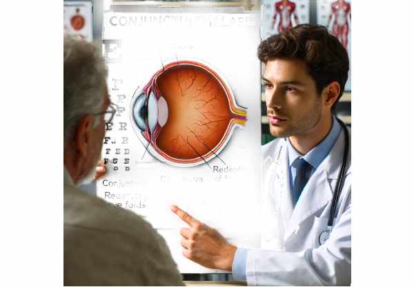
What is Conjunctivochalasis?
Conjunctivochalasis is an ocular condition marked by the presence of redundant, loose folds of conjunctival tissue, usually located between the globe of the eye and the lower eyelid. This condition can be extremely uncomfortable, with symptoms such as dryness, irritation, tearing, and a foreign body sensation. Conjunctivochalasis is commonly associated with aging, but it can also be caused by chronic inflammation, ocular surface disease, or mechanical factors like contact lens wear. Conjunctivochalasis, although frequently underdiagnosed, can have a significant impact on quality of life and visual function, necessitating a thorough understanding of its etiology and implications for effective management.
Conjunctivochalasis Insights
Conjunctivochalasis, derived from the Greek words “chalasis” meaning relaxation or slackening, is a condition affecting the conjunctiva, the thin, transparent membrane that covers the white part of the eye and lines the inside of the eyelids. In a healthy eye, the conjunctiva conforms smoothly to the eye’s contours. However, in conjunctivochalasis, this membrane relaxes and forms redundant folds, especially along the lower eyelid margin.
Etiology and Risk Factors
The precise cause of conjunctivochalasis is multifactorial and frequently idiopathic, but several contributing factors have been identified.
- Aging: Conjunctivochalasis is most commonly caused by aging. As people age, the conjunctiva loses elasticity, and the underlying Tenon’s capsule, which helps keep the conjunctiva in shape and position, atrophy, resulting in the formation of redundant tissue.
- Chronic Inflammation: Conditions such as chronic blepharitis, meibomian gland dysfunction, and allergic conjunctivitis can cause persistent inflammation of the conjunctiva, which contributes to laxity over time.
- Mechanical Factors: Prolonged contact lens wear, especially with poorly fitting lenses, can mechanically irritate the conjunctiva, resulting in conjunctivochalasis. Similarly, excessive eye rubbing or ocular surgery can have an impact.
- Tear Film Dysfunction: An unstable tear film, whether caused by dry eye syndrome or other factors, can exacerbate symptoms of conjunctivochalasis. The redundant conjunctival folds can disrupt normal tear flow and distribution, causing additional irritation and inflammation.
- Genetic Predisposition: Some people may have a genetic predisposition to conjunctivochalasis, though specific genetic markers have not been definitively identified.
Symptoms and Clinical Presentation
Conjunctivochalasis can cause a variety of symptoms, each with varying severity. Common symptoms include:
- Dryness: Patients frequently report a persistent sensation of dryness, despite normal or increased tear production. This paradoxical dryness occurs when redundant folds disrupt the tear film’s uniform distribution across the ocular surface.
- Foreign Body Sensation: Loose conjunctival folds can cause the sensation of having a foreign body in the eye. This discomfort can be ongoing and irritating.
- Tearing (Epiphora): The excess conjunctival tissue can obstruct the normal drainage of tears through the puncta, resulting in excessive tearing. This condition is especially bothersome in windy conditions or when doing activities that cause tear production.
- Blurred Vision: Although uncommon, some patients may experience intermittent blurred vision due to mechanical interference between the conjunctival folds and the corneal surface or tear film.
- Irritation and Redness: Chronic irritation of the conjunctival folds can cause persistent redness and a grittiness or burning sensation in the eyes.
Effects on Quality of Life
Conjunctivochalasis symptoms can have a significant impact on one’s quality of life. Persistent discomfort, dryness, and tearing can interfere with daily activities like reading, using digital devices, and going outside. The constant need to wipe away tears and manage discomfort can be distracting and frustrating in both personal and professional settings.
Pathophysiology
Recognizing the interplay of mechanical and inflammatory processes is essential for understanding conjunctivochalasis pathophysiology. The conjunctiva, a mucous membrane, is supported by Tenon’s capsule and episcleral tissue. With age or chronic inflammation, these supporting structures can weaken or atrophy, causing the conjunctiva to become redundant and fold.
The tear film, which is made up of three layers (lipid, aqueous, and mucin), is essential for keeping the ocular surface healthy. In conjunctivochalasis, redundant conjunctival folds can disrupt tear film stability by causing uneven tear distribution and interfering with normal tear drainage through the puncta. This disruption can worsen symptoms of dryness and irritation, resulting in a vicious cycle of inflammation and discomfort.
Differential Diagnosis
Several ocular conditions can cause symptoms similar to conjunctivochalasis, making differential diagnosis critical. These conditions include the following:
- Dry Eye Syndrome: While frequently associated with conjunctivochalasis, dry eye syndrome can cause symptoms such as dryness, irritation, and foreign body sensation. Diagnostic tests, such as tear break-up time (TBUT) and Schirmer’s test, can help distinguish between the two conditions.
- Blepharitis: Eyelid inflammation, like conjunctivochalasis, can cause redness, irritation, and tearing. Blepharitis, on the other hand, is often accompanied by crusting along the eyelid margins and dysfunction of the meibomian glands.
- Allergic Conjunctivitis: Allergic conjunctivitis causes redness, itching, and tearing, but it is typically accompanied by other allergic symptoms such as sneezing and nasal congestion. A detailed patient history and allergy testing can help distinguish these conditions.
- Punctal Stenosis: As with conjunctivochalasis, obstruction of the tear drainage system can cause excessive tearing. However, punctal stenosis is frequently detected during punctal examination and may necessitate specific treatments such as punctal dilation or surgical intervention.
Clinical Examination
A thorough clinical examination is required to diagnose conjunctivochalasis. The key components of the examination are:
- Visual Acuity Testing: Assessing visual acuity can help identify any effects on vision, though conjunctivochalasis rarely causes significant vision loss.
- Slit Lamp Examination: A slit lamp allows the ophthalmologist to see the conjunctiva and identify redundant folds, inflammation, and any associated ocular surface changes. Fluorescein staining can reveal areas of tear film instability and epithelial disruption.
- Tear Film Assessment: Evaluating the quantity and quality of the tear film using tests such as TBUT and Schirmer’s test provides insight into tear film dysfunction and aids in the diagnosis of concurrent dry eye syndrome.
- Eversion of the Eyelid: In some cases, eversion of the eyelid can reveal additional folds and improve visibility of the conjunctival redundancy.
How to Avoid Conjunctivochalasis
- Maintain Good Eye Hygiene: Clean your eyelids and eyelashes on a regular basis to prevent debris buildup and lower your risk of chronic inflammation, which can lead to conjunctivochalasis.
- Avoid Eye Rubbing: Avoid rubbing your eyes because it can aggravate mechanical irritation and contribute to the formation of redundant conjunctival folds.
- Use Properly Fitted Contact Lenses: To avoid mechanical irritation of the conjunctiva, ensure that contact lenses fit well and are used as directed.
- Effective Allergy Management: If you have allergies, control your symptoms and avoid known allergens to reduce the risk of chronic conjunctival inflammation.
- Stay Hydrated: Proper hydration promotes overall eye health and can support a more stable tear film.
- Protect Your Eyes: Wear sunglasses or protective eyewear in windy or dusty conditions to protect your eyes from irritants and reduce the risk of mechanical injury.
- Use Artificial Tears: Regular use of preservative-free artificial tears can help maintain tear film stability and alleviate dryness symptoms caused by conjunctivochalasis.
- Eat a Healthy Diet: A diet high in omega-3 fatty acids, antioxidants, and vitamins can help maintain ocular surface health and reduce inflammation.
- Regular Eye Exams: Schedule regular eye exams with an ophthalmologist to track your eye health, detect early signs of conjunctivochalasis, and effectively manage any underlying conditions.
- Reduce Screen Time: Limit your exposure to digital screens to avoid digital eye strain, which can exacerbate symptoms of dryness and discomfort.
Methods to Diagnose Conjunctivochalasis
Conjunctivochalasis is diagnosed through a comprehensive examination of the ocular surface, a detailed patient history, and specialized diagnostic tests. A thorough clinical examination is required to accurately diagnose this condition and distinguish it from other ocular surface diseases that present similar symptoms.
Clinical Examination
- Patient History: The diagnostic process starts with a thorough patient history to determine the onset, duration, and severity of symptoms. Questions about previous eye conditions, surgeries, contact lens use, and systemic diseases that may affect the eye are also relevant.
- Visual Acuity Testing: This standard procedure evaluates the patient’s vision to determine whether conjunctivochalasis affects visual acuity. Conjunctivochalasis rarely results in significant vision loss, but it can cause visual discomfort.
- Slit Lamp Examination: The ophthalmologist uses a slit lamp to examine the conjunctiva and eyelids. This examination detects the presence of redundant conjunctival folds, inflammation, and other abnormalities. Fluorescein staining can be used to highlight areas with tear film disruption and epithelial damage.
- Eyelid Eversion: In some cases, eversion of the eyelids may be performed to improve visibility of the conjunctival folds and determine the extent of the conjunctivochalasis.
Specialized Diagnostic Tests
- Tear Film Break-Up Time (TBUT): This test involves applying a fluorescein dye to the tear film and timing how long it takes for dry spots to appear on the cornea. A shortened TBUT suggests tear film instability, which is common in conjunctivochalasis patients.
- Schirmer’s Test: To measure tear production, place a small strip of filter paper under the lower eyelid. Reduced tear production can exacerbate the symptoms of conjunctivochalasis.
- Anterior Segment Optical Coherence Tomography (AS-OCT): AS-OCT is a sophisticated imaging technique that generates high-resolution cross-sectional images of the ocular surface. It can precisely measure conjunctival thickness and the presence of redundant folds, which helps with diagnosis and severity assessment of conjunctivochalasis.
- Impression Cytology: This minimally invasive procedure collects cells from the conjunctival surface with a cellulose acetate filter. The collected cells are then examined for signs of inflammation or other abnormalities, which provides additional information about the underlying pathology of conjunctivochalasis.
Emerging Diagnostic Techniques
- In Vivo Confocal Microscopy: This novel technique enables a detailed examination of the conjunctiva at the cellular level. It can assist in detecting microscopic changes in the conjunctival tissue that are not visible under a standard slit lamp examination.
- Tear Osmolarity Testing: Determining the osmolarity of tears can reveal information about tear film stability and ocular surface health. Elevated tear osmolarity is frequently associated with dry eye syndrome, which can coexist with or worsen conjunctivochalasis.
Conjunctivochalasis Treatment Options
Treating conjunctivochalasis entails addressing the underlying causes, relieving symptoms, and, in some cases, surgical intervention. The choice of treatment is determined by the severity of the condition and the patient’s specific symptoms.
Standard Treatment Options
- Lubricating Eye Drops: Preservative-free artificial tears are a popular first-line treatment for dryness and discomfort. These eye drops help to stabilize the tear film and relieve irritation caused by conjunctival folds.
- Anti-inflammatory Medications: Topical corticosteroids or non-steroidal anti-inflammatory drugs (NSAIDs) can be used to reduce inflammation and alleviate symptoms. To avoid potential side effects, these medications are typically only used for a short period of time.
- Warm Compresses and Lid Hygiene: Using warm compresses on a regular basis and practicing proper eyelid hygiene can help manage associated conditions such as blepharitis, which can lead to chronic inflammation and conjunctivochalasis.
- Punctal Plugs: For patients with severe tear film dysfunction, punctal plugs can be inserted into the tear ducts to reduce tear drainage and increase ocular surface moisture. This can help with symptoms of dryness and irritation.
Surgical Treatment Alternatives
- Conjunctival Resection: In severe cases where conservative treatments fail, surgical removal of the redundant conjunctival tissue may be required. This procedure removes excess conjunctiva to restore a smoother ocular surface and relieve symptoms.
- Amniotic Membrane Transplantation: This advanced surgical procedure involves applying an amniotic membrane graft to the affected area. The amniotic membrane promotes healing, reduces inflammation, and aids in restoring normal conjunctival architecture.
- Thermal Cautery: This procedure uses heat to shrink and tighten excess conjunctival tissue. It is an outpatient procedure that offers a minimally invasive treatment option for conjunctivochalasis.
Innovative and Emerging Therapies
- Autologous Serum Eye Drops: These eye drops are derived from the patient’s own blood serum and contain vital growth factors and nutrients that aid in healing and tear film stability. They are especially useful for patients who have severe dry eye symptoms caused by conjunctivochalasis.
- Biologic Agents: Research into biologic agents, such as cytokine inhibitors and growth factor modulators, is currently underway. These therapies aim to target specific pathways involved in inflammation and tissue remodeling, potentially opening up new treatment options for refractory conjunctivochalasis.
- Gene Therapy: Despite being in the experimental stage, gene therapy has the potential to treat ocular surface diseases by correcting genetic abnormalities that contribute to conditions such as conjunctivochalasis. This approach could provide long-term relief by addressing the disease’s underlying cause at the molecular level.
Trusted Resources
Books
- Ocular Surface Disease: Cornea, Conjunctiva and Tear Film by Edward J. Holland and Mark J. Mannis
- The Wills Eye Manual: Office and Emergency Room Diagnosis and Treatment of Eye Disease by Adam T. Gerstenblith and Michael P. Rabinowitz
- Conjunctival and Scleral Diseases: Diagnosis and Management by David G. Hwang


