What is Dacryocystitis?
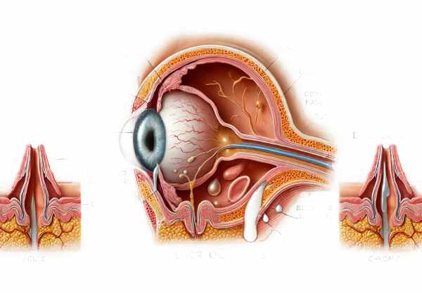
Dacryocystitis is an infection or inflammation of the lacrimal sac, a component of the eye’s tear drainage system. This condition is frequently caused by an obstruction in the nasolacrimal duct, resulting in the accumulation of tears and infection. It can affect people of all ages, but it is most common in infants and adults over the age of 40. Symptoms usually include pain, redness, and swelling around the inner corner of the eye, as well as excessive tearing. In more severe cases, pus may be present. Understanding the complexities of dacryocystitis is critical for identifying symptoms early and seeking appropriate medical treatment to avoid complications.
Comprehensive Dacryocystitis Overview
Dacryocystitis is a serious ocular condition characterized by inflammation of the lacrimal sac, usually caused by a blockage of the nasolacrimal duct. This obstruction prevents tears from draining properly, causing them to stagnate and create an environment ripe for infection. The condition can be acute or chronic, with each having unique clinical characteristics and implications for patient care.
Pathophysiology
The lacrimal apparatus consists of the lacrimal glands, which produce tears, and the nasolacrimal duct, which drains tears into the nasal cavities. When the nasolacrimal duct becomes obstructed, tears accumulate in the lacrimal sac, creating a breeding ground for bacteria. Staphylococcus aureus, Streptococcus pneumoniae, and Haemophilus influenzae are common causative organisms. Chronic cases frequently involve less virulent organisms, such as Staphylococcus epidermidis.
Epidemiology
Dacryocystitis affects people of all ages, but it is most common in newborns and adults over the age of 40. Neonatal dacryocystitis is caused by congenital nasolacrimal duct obstruction, which affects roughly 6% of infants. In adults, dacryocystitis is more common in women, most likely due to anatomical differences in the nasolacrimal duct.
Clinical Manifestations
Acute dacryocystitis is characterized by sudden onset of pain, redness, and swelling at the inner corner of the eye. Patients frequently report tenderness and may have a palpable mass over their lacrimal sac. Excessive tear production (epiphora) is common, and when pressure is applied to the puncta, purulent discharge may be released. Systemic symptoms such as fever and malaise may accompany severe infections.
Chronic dacryocystitis, on the other hand, presents with fewer severe symptoms. Patients usually have chronic epiphora and recurrent mild inflammation without much pain. The condition can cause persistent dacryocystocele (a cystic swelling in the lacrimal sac) or fistula formation.
Risk Factors
Several factors increase the likelihood of developing dacryocystitis. Congenital nasolacrimal duct obstruction is a major risk factor for infants. Adult risk factors include the following:
- Age and Gender: More common in women over 40.
- Nasal and Sinus Disease: Chronic sinusitis and nasal polyps can cause duct obstruction.
- Trauma or Surgery: Previous nasal or facial trauma, as well as surgery, can disrupt the nasolacrimal duct’s normal anatomy.
- Systemic Diseases: Sarcoidosis, Wegener’s granulomatosis, and certain cancers can all result in secondary obstruction.
- Medications: Prolonged use of topical medications, such as corticosteroids, may make people more susceptible to secondary infections.
Complications
If left untreated, dacryocystitis can cause a number of complications. Acute infections can lead to abscess formation, necessitating surgical intervention. Chronic cases may lead to the development of a dacryocystocele, fistula, or chronic sinusitis. In rare cases, the infection can spread to surrounding structures, resulting in orbital cellulitis, a serious and potentially blinding condition.
Prognosis
Dacryocystitis has a good prognosis if treated promptly and appropriately. Most patients recover completely and without long-term consequences. However, delayed treatment or recurrent infections can lead to complications that impair the patient’s quality of life and visual function.
Patient Education & Awareness
It is critical to educate patients about dacryocystitis symptoms and risk factors in order to detect and treat the condition early. Patients should be educated on the importance of seeking medical attention if they experience persistent tearing, swelling, or pain around their eyes. Healthcare providers should also closely monitor patients with known risk factors, such as chronic sinusitis or a history of nasolacrimal duct obstruction.
Prevention Tips
Dacryocystitis can be prevented by addressing the underlying causes and maintaining good ocular hygiene. Here are some key preventive measures:
- Maintain Ocular Hygiene: To avoid debris and bacteria buildup, clean the eyelids and eyelashes on a regular basis.
- Manage Nasal and Sinus Conditions: Treat chronic sinusitis and nasal polyps right away to reduce the risk of nasolacrimal duct obstruction.
- Avoid Trauma: Protect the facial area from trauma and seek immediate medical attention for any nasal or facial injuries.
- Use Medications Wisely: Avoid long-term use of topical corticosteroids and other medications that can increase the risk of infection.
- Monitor Systemic Diseases: Patients with systemic conditions such as sarcoidosis or Wegener’s granulomatosis should receive regular check-ups to detect any ocular involvement early on.
- Seek Early Treatment: If symptoms of dacryocystitis appear, seek medical attention right away to avoid complications and chronicity.
- Post-Surgical Care: After nasal or ocular surgery, carefully follow the post-operative care instructions to avoid infections and promote healing.
- Educate and Inform: Inform family members, particularly parents of newborns, about the symptoms of congenital nasolacrimal duct obstruction and the importance of prompt treatment.
Diagnostic methods
Dacryocystitis is diagnosed using a combination of clinical examination and imaging techniques to confirm the condition and assess the extent of the infection or obstruction.
Clinical Examination
A detailed patient history and physical examination are usually part of the initial evaluation. The clinician looks for pain, redness, and swelling around the lacrimal sac area, which is frequently accompanied by purulent discharge when the sac is pressed. Palpation can help detect a fluctuant mass that indicates an abscess.
The Fluorescein Dye Disappearance Test
The fluorescein dye disappearance test is a simple and non-invasive method for determining the drainage function of the nasolacrimal duct. A fluorescein dye drop is inserted into the conjunctival sac and its clearance is monitored. Delayed clearance indicates an obstruction in the tear drainage system.
Lacrimal Probing and Irrigation
Probing and irrigation are diagnostic procedures for confirming nasolacrimal duct obstruction. To detect blockages, a probe is gently inserted into the nasolacrimal duct through the puncta, followed by irrigation with saline. The ease or difficulty with which the fluid flows can aid in locating the source of obstruction.
Dacryocystography.
Dacryocystography is an imaging technique that involves injecting a contrast dye into the lacrimal sac, followed by X-ray imaging. This method aids in visualizing the anatomy of the tear drainage system, allowing for the identification of obstructions or structural anomalies.
Nasal Endoscopy
Nasal endoscopy allows for direct visualization of the nasal passages and nasolacrimal duct openings. It is especially useful when anatomical anomalies, such as nasal polyps or a deviated septum, are suspected of causing the obstruction.
Ultrasoundography
Ultrasound imaging of the lacrimal sac can aid in distinguishing dacryocystitis from other causes of periocular swelling, such as tumors or cyst. It allows for real-time imaging of the sac and surrounding tissues, revealing fluid collections and abscesses.
CT and MRI scans
Computed tomography (CT) and magnetic resonance imaging (MRI) are advanced imaging techniques used to evaluate deeper or more complex anatomical structures. These modalities provide detailed images of the lacrimal apparatus, surrounding structures, and potential infection spread.
Diagnostic innovations, such as high-resolution ultrasonography and advanced imaging techniques, continue to improve the accuracy and efficiency of dacryocystitis diagnosis, allowing for timely and appropriate treatment interventions.
Treatment for dacryocystitis varies based on the severity and duration of the infection. The goal is to alleviate symptoms, clear the infection, and restore proper tear drainage.
Standard Treatment Options
Antibiotics:
The first line of treatment for acute dacryocystitis is with systemic antibiotics. Oral antibiotics like amoxicillin-clavulanate and cephalexin are commonly prescribed. In cases of severe infection or abscess formation, intravenous antibiotics may be required.
Warm Compresses
Warm compresses applied to the affected area can promote drainage, thereby reducing pain and swelling. This simple home remedy is frequently recommended in addition to antibiotics.
Incisions and Drainage
If an abscess develops, surgical intervention may be necessary to drain the pus. This procedure, which is done under local anesthesia, involves making a small incision in the lacrimal sac to allow the infected material to exit.
Surgical Interventions
Dacryocystorhinostomy (DCR).
DCR is the standard surgical treatment for chronic dacryocystitis and recurring acute episodes. The procedure opens up a new drainage pathway between the lacrimal sac and the nasal cavity, bypassing the obstructed nasolacrimal duct. It can be performed externally or endoscopically, with endoscopic DCR being less invasive and resulting in faster recovery.
Balloon Dacryoplasty
This minimally invasive procedure involves inserting and inflating a balloon catheter into the nasolacrimal duct to clear the obstruction. Balloon dacryoplasty is especially effective in pediatric and adult cases where the obstruction is not severe.
Innovative and Emerging Therapies
Microendoscopic Surgery.
Microendoscopic techniques employ tiny endoscopes and instruments to carry out precise surgical procedures within the lacrimal drainage system. These advancements enable less invasive interventions, resulting in fewer postoperative complications and faster recovery times.
Laser-Assisted Procedures
Laser-assisted DCR is a new technique that employs laser energy to create a new drainage path. This method has the advantage of less bleeding, shorter operative times, and potentially lower recurrence rates.
Drug-Eluting Stents
Drug-eluting stents placed within the nasolacrimal duct can maintain patency while also delivering localized anti-inflammatory or antimicrobial therapy, lowering the risk of recurrence.
Continued research and technological advancements are improving the management of dacryocystitis, giving patients more effective and less invasive treatments.
Trusted Resources
Books
- “Diseases of the Lacrimal System” by Mohammad Javed Ali
- “Principles and Practice of Lacrimal Surgery” by Mohammad Javed Ali
- “The Lacrimal System: Diagnosis, Management, and Surgery” by Adam J. Cohen and Michael Mercandetti
Online Resources
- American Academy of Ophthalmology AAO
- National Eye Institute NEI
- Mayo Clinic Mayo Clinic
- MedlinePlus MedlinePlus


