What is Homonymous Hemianopia?
Homonymous hemianopia is a visual field defect that affects the same side of both eyes. This condition is caused by damage to the brain’s visual pathways, specifically the optic tract, optic radiation, and occipital cortex. Individuals with homonymous hemianopia lose half of their field of vision on the same side in both eyes, which significantly impairs their ability to read, drive, and recognize faces. Early diagnosis and appropriate management are critical for improving the quality of life for those affected.
Deep Dive into Homonymous Hemianopia
Homonymous hemianopia is a type of visual field loss in which both eyes lose the same half of their visual field. This condition is typically caused by lesions or damage to the visual pathways beyond the optic chiasm, specifically the optic tract, lateral geniculate nucleus, optic radiations, or occipital lobe.
Causes and Pathophysiology
The main causes of homonymous hemianopia are:
- Stroke: Cerebrovascular accidents are the leading cause, particularly those involving the posterior cerebral artery, which supplies the occipital lobe. Strokes can cause infarction or hemorrhage in the visual pathways.
- Traumatic Brain Injury (TBI): Severe head injuries can directly damage the brain structures that control vision, resulting in visual field deficits.
- Tumors: Brain tumors, especially those in the occipital, temporal, or parietal lobes, can compress or invade visual pathways, resulting in homonymous hemianopia.
- Infections and Inflammatory Conditions: Encephalitis, multiple sclerosis, and other demyelinating diseases can cause damage to the optic radiations and occipital cortex.
- Surgical Complications: Neurosurgical procedures involving the brain may inadvertently damage visual pathways, resulting in homonymous hemianopia.
- Congenital Causes: Although uncommon, congenital abnormalities can disrupt the visual pathways, resulting in homonymous hemianopia from birth.
Visual Pathway Anatomy
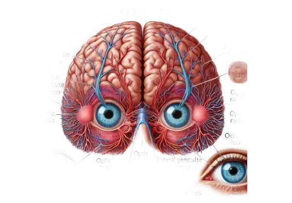
Understanding the anatomy of visual pathways is critical for understanding how homonymous hemianopia develops. The visual pathway contains:
- The Optic Nerve transports visual information from the retina to the optic chiasm.
- Optic Chiasm: The point at which the optic nerves partially intersect, with fibers from the nasal half of each retina crossing to the opposite side.
- Optic Tract: Runs from the optic chiasm to the lateral geniculate nucleus, containing fibers from the temporal half of the ipsilateral retina and the nasal half of the contralateral retina.
- Lateral Geniculate Nucleus (LGN): A relay center in the thalamus that processes visual input.
- Optic Radiations: Axons from the LGN branch out to the occipital lobe.
- Occipital Cortex: The primary visual processing center in the back of the brain.
Clinical Manifestations
Patients with homonymous hemianopia usually have:
- Visual Field Loss: Loss of one-half of the visual field on the same side in both eyes. For example, a lesion on the right side of the brain will cause both eyes to lose their left visual field (left homonymous hemianopia).
- Difficulty Reading: Reading can be difficult for patients because they may miss parts of the text due to an impaired visual field.
- Navigation Issues: Difficulty navigating through spaces because obstacles in the blind field are not detected.
- Facial Recognition Problems: Difficulty recognizing faces and interpreting visual scenes due to insufficient visual information.
Impact on Daily Life
Homonymous hemianopia impairs a person’s ability to perform daily tasks. Common challenges include:
- Driving: The loss of peripheral vision on one side makes it difficult to detect vehicles, pedestrians, and other hazards, posing serious safety risks.
- Reading: Patients frequently have reduced reading speed and comprehension because they miss words or lines in their blind field.
- Mobility: Increased risk of tripping or colliding with objects on the affected side, resulting in decreased confidence and independence.
- Social Interactions: Difficulties recognizing faces and interpreting social cues can impair social interactions and relationships.
Associated Conditions
Homonymous hemianopia can occur alone or in conjunction with other neurological deficits, depending on the location and extent of the brain lesion. Commonly related conditions include:
- Hemiparesis: Weakness on one side of the body, commonly associated with strokes that cause homonymous hemianopia.
- Aphasia: Language problems, especially if the lesion affects the left hemisphere in a right-handed person.
- Neglect Syndrome: A condition in which a patient is unaware of objects or their own body on one side, which is frequently associated with right hemisphere lesions.
Prognosis
The prognosis for homonymous hemianopia is dependent on the underlying cause, the severity of the lesion, and the patient’s overall health. Some patients may experience partial or complete recovery of their visual fields, especially after minor strokes or transient ischemic episodes. However, permanent visual field deficits are common, particularly when the brain suffers significant structural damage.
Rehabilitation
Rehabilitation is critical in helping patients adjust to their visual field loss. Strategies include:
- Visual Scanning Training: Teaching patients to compensate for field loss by actively scanning their surroundings, thereby increasing awareness of their blind side.
- Prism Glasses: Special glasses that alter the visual field, allowing patients to better detect objects in their blind field.
- Occupational Therapy: Helps patients develop strategies for managing daily activities and improving their quality of life.
Identifying Homonymous Hemianopia
To determine the underlying cause and extent of the visual field defect, homonymous hemianopia must be accurately diagnosed through a combination of clinical assessment, visual field testing, and neuroimaging.
Clinical Assessment
A thorough clinical assessment involves taking a detailed history of the patient’s symptoms, including their onset, duration, and progression. Information about recent head trauma, stroke, or other neurological conditions is critical.
- Visual Acuity Testing: Evaluates overall vision to rule out other ocular causes of vision loss.
- Ophthalmologic Examination: A thorough eye examination to look for any abnormalities in the anterior visual pathway that could lead to vision loss.
Visual Field Testing
Visual field testing is critical for detecting and characterizing homonymous hemianopia. Common methods include:
- Automated Perimetry is a computerized test in which patients respond to visual stimuli presented in various parts of their visual field. This test maps the visual field and detects areas of loss.
- Confrontation. Visual Field Testing is a preliminary, non-instrumental test performed by the clinician. The patient closes one eye and focuses on the examiner’s nose while identifying objects or fingers in different parts of the visual field.
Neuroimaging
Neuroimaging techniques are critical for determining the location and cause of the brain lesion causing homonymous hemianopia.
- Magnetic Resonance Imaging(MRI): MRI provides detailed images of brain structures, which aid in the diagnosis of strokes, tumors, traumatic injuries, and demyelinating diseases affecting visual pathways.
- Computed Tomography (CT) Scan: CT scans are useful for rapidly detecting acute hemorrhages, infarctions, and mass effects in the brain. It is commonly used in emergency situations where an MRI is not immediately available.
Treatment Options for Homonymous Hemianopia
The treatment of homonymous hemianopia focuses on identifying the underlying cause, managing symptoms, and improving the patient’s quality of life through rehabilitation strategies. While the primary goal is to treat the underlying cause, such as a stroke or tumor, a variety of rehabilitative approaches can help patients adjust to their visual impairments.
Standard Treatment Options:
- Medical Management: The most important aspect of treating homonymous hemianopia is to address the root cause. This may involve:
- Stroke: Using thrombolytic agents in the acute phase, managing blood pressure, controlling cholesterol levels, and using antiplatelet or anticoagulant medications to prevent future strokes.
- Brain Tumors: Depending on the type and location of the tumor, treatment options include surgical removal, chemotherapy, or radiotherapy.
- Inflammatory Conditions: Treating infections or autoimmune conditions with appropriate medications such as antibiotics, antivirals, or immunosuppressive agents.
- Traumatic Brain Injury: managing intracranial pressure, surgical interventions as needed, and rehabilitation.
- Visual Rehabilitation: Because homonymous hemianopia causes permanent visual field loss, rehabilitation aims to help patients adjust to their new visual reality.
- Visual Scanning Training: Teaching patients to consciously scan their surroundings to compensate for their lost visual field. To improve spatial awareness, they move their head and eyes to the blind side.
- Prism Glasses: Special lenses that shift the visual field and allow patients to see objects that would otherwise be in their blind spot.
- Vision Restoration Therapy (VRT): A computer-based therapy that uses repeated stimulation of the border area between the sighted and blind fields to improve visual field sensitivity.
- Occupational Therapy: Occupational therapists work with patients to develop strategies for managing daily activities. This may include:
- Adaptive Techniques: Making tasks easier and safer to complete, such as using contrasting colors to highlight steps or obstacles.
- Environmental Modifications: Changing home and work environments to reduce the risk of accidents, such as rearranging furniture or installing better lighting.
- Assistive Devices: Introducing tools and technologies that help with navigation and communication, such as GPS units or text-to-speech software.
Innovative and Emerging Therapies
1) Neuroplasticity-Based Approaches: Recent research has focused on using the brain’s neuroplasticity to improve visual function. Techniques like transcranial magnetic stimulation (TMS) and neurofeedback are designed to improve cortical reorganization and visual processing.
- Virtual Reality (VR) Rehabilitation: VR-based therapies create immersive environments that mimic real-world challenges, allowing patients to practice visual scanning and spatial navigation in a controlled, safe environment.
- Gene Therapy: For genetically based conditions, emerging gene therapies seek to correct underlying genetic defects, potentially restoring normal visual pathways.
- Stem Cell Therapy: Research into stem cell therapy investigates the potential to regenerate damaged neural tissues, providing hope for future treatments that could restore lost visual function.
By combining these treatment strategies, healthcare providers can assist patients with homonymous hemianopia in adapting to visual field loss, improving functional abilities, and improving quality of life.
Best Practices for Avoiding Homonymous Hemianopia
- Stroke Prevention: Implement lifestyle changes and medical interventions to lower the risk of stroke, which is a common cause of homonymous hemianopia. This includes eating a healthy diet, exercising regularly, managing hypertension, diabetes, and quitting smoking.
- Regular Health Check-Ups: Routine medical examinations can aid in the detection and management of risk factors for homonymous hemianopia, such as cardiovascular disease and hypertension.
- Protective Gear: Use appropriate protective equipment, such as helmets, to reduce the risk of traumatic brain injury, which can cause visual field defects.
- Monitor Vision Changes: Be aware of any sudden vision changes and seek immediate medical attention if they occur. Early intervention can prevent or reduce the severity of underlying conditions.
- Manage Chronic Conditions: Proper management of chronic conditions, such as multiple sclerosis or diabetes, can lower the risk of complications leading to homonymous hemianopia.
- Avoid Risky Behaviors: Limit activities that could result in head injuries, such as contact sports without proper safety equipment or driving under the influence of alcohol or drugs.
- Educate Yourself and Others: Learn about the symptoms and risk factors for conditions that can result in homonymous hemianopia, and educate family and friends to ensure timely intervention and support.
- Maintain a Healthy Lifestyle: Overall health and wellness can help prevent homonymous hemianopia. Balanced nutrition, regular physical activity, and stress management are all essential components.
Individuals who follow these preventive measures have a significantly lower risk of developing homonymous hemianopia and related complications.
Trusted Resources
Books
- “Neuro-Ophthalmology Illustrated” by Valerie Biousse and Nancy J. Newman
- “Clinical Neuro-Ophthalmology: A Practical Guide” by Ambar Chakravarty
- “The Neurology of Vision” by Jonathan D. Trobe


