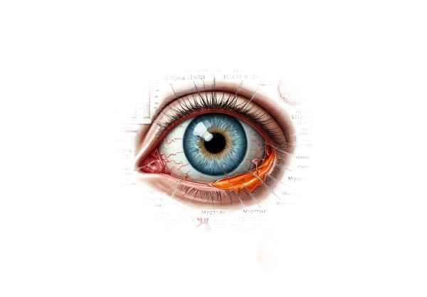
What is Marfan syndrome?
Marfan syndrome is a genetic disorder that affects the connective tissue that structures and supports the body’s organs and tissues. Mutations in the FBN1 gene cause this syndrome, which is characterized by defective fibrillin-1, a protein required for the formation of elastic fibers in connective tissue. Ocular manifestations of Marfan syndrome are severe and can impair vision, including lens dislocation (ectopia lentis), myopia, retinal detachment, and early-onset glaucoma. Understanding these ocular issues is critical for proper diagnosis and treatment.
In-Depth Look at Marfan Syndrome
Marfan syndrome is a systemic condition that affects many organs in the body, including the eyes. Ocular manifestations vary greatly and can have a significant impact on a patient’s quality of life. In this article, we will look at the key ocular features associated with Marfan syndrome, as well as their pathophysiology, clinical presentation, and complications.
Pathophysiology
Mutations in the FBN1 gene, which encodes fibrillin-1, cause Marfan syndrome. Fibrillin-1 is required for the structural integrity and proper function of connective tissues throughout the body, including the eyes. Defective fibrillin-1 weakens structural components, resulting in a variety of ocular complications.
Lens dislocation (Ectopia Lentis)
One of the distinguishing features of Marfan syndrome is lens dislocation, also known as ectopia lentis. This condition affects 60-80% of people with Marfan syndrome due to weakened zonular fibers that hold the lens in place. These fibrillin-based fibers fail to properly anchor the lens, resulting in displacement.
Clinical Presentation*:
- Visual Disturbance: Patients may experience blurred or double vision as a result of lens misalignments.
- Directional Displacement: The lens can be displaced upward, downward, or sideways, which affects vision depending on the direction and extent of the displacement.
- Refractive Errors: Lens dislocation frequently causes significant refractive errors, such as myopia or astigmatism.
Myopia
Myopia, or nearsightedness, is common in people with Marfan syndrome due to eyeball elongation. This elongation, when combined with lens dislocation, exacerbates visual disturbances. Myopia in Marfan syndrome ranges from mild to severe, and corrective lenses are typically required for optimal vision.
Retinal Detachment
Individuals with Marfan syndrome are significantly more likely than the general population to develop retinal detachment. The elongated eyeball and defective connective tissue make the retina more susceptible to tears and detachment.
Clinical Presentation*:
- Sudden Onset: Patients may notice floaters, flashes of light, or a shadow or curtain covering a portion of their visual field.
- Urgency: Retinal detachment is a medical emergency that requires immediate treatment to avoid permanent vision loss.
Glaucoma
Marfan syndrome can also cause early-onset glaucoma. The dislocated lens can obstruct the normal flow of aqueous humor, resulting in elevated intraocular pressure. This pressure can damage the optic nerve, resulting in vision loss.
Clinical Presentation*:
- Increased Intraocular Pressure: Patients may experience elevated intraocular pressure during routine eye exams.
- Optic Nerve Damage: If left untreated, progressive damage to the optic nerve can cause peripheral vision loss and, eventually, central vision loss.
Corneal Abnormalities
Individuals with Marfan syndrome may also have corneal abnormalities, such as increased curvature (keratoconus) and thinning of the cornea. These changes can cause additional refractive errors and visual disturbances.
Strabismus
Strabismus, or eye misalignment, can occur in people with Marfan syndrome. If not treated, this condition can progress to amblyopia (lazy eye) due to structural abnormalities in the eye.
Cataracts
Early-onset cataracts are more common in people with Marfan syndrome. Cataracts, or clouding of the lens, can worsen vision and necessitate surgical intervention.
Systemic Considerations
Because Marfan syndrome is a systemic disease, ocular manifestations frequently coexist with other symptoms. Cardiovascular issues, such as aortic root dilation and mitral valve prolapse, are especially concerning and necessitate ongoing monitoring. Skeletal abnormalities such as scoliosis and pectus excavatum are also common, adding to the disorder’s overall management challenges.
Effects on Quality of Life
The ocular manifestations of Marfan syndrome can have a significant impact on a patient’s quality of life. Visual disturbances, frequent medical appointments, and the possibility of serious complications all contribute to anxiety and decreased daily functioning. Early detection, regular monitoring, and appropriate interventions are critical for effectively managing these ocular issues and improving patient outcomes.
Current Research and Advances
Research into the ocular manifestations of Marfan syndrome is ongoing, with the goal of understanding the genetic basis and developing effective treatments. Advances in genetic testing enable earlier diagnosis and improved family counseling. Innovations in surgical techniques and intraocular lenses (IOLs) provide promising solutions for dealing with lens dislocation and other complications. Pharmacological approaches that target the underlying connective tissue defects are also being investigated to prevent or slow the progression of the disorder.
Diagnostic methods
Clinical evaluation, advanced imaging techniques, and genetic testing are all required for an accurate diagnosis of Marfan syndrome’s ocular manifestations. Early detection is critical for timely intervention and management.
Clinical Examination
- Visual Acuity Test: Measuring the patient’s visual acuity can help determine the severity of visual impairment caused by lens dislocation, myopia, or other refractive errors. This test assesses visual clarity and sharpness and can detect myopia, astigmatism, and other refractive errors.
- Slit-Lamp Examination: A slit-lamp examination provides a detailed view of the eye’s anterior segment, allowing the ophthalmologist to see the lens and detect any displacement or opacities. This examination can reveal the direction and extent of the lens dislocation, as well as the presence of any associated cataracts.
- Fundus Examination: A thorough examination of the retina and optic nerve is required to detect any signs of retinal detachment, glaucoma, or other retinal abnormalities. This examination helps to determine the overall health of the eye and to identify potential complications associated with lens dislocation.
Imaging Techniques
- Ultrasound Biomicroscopy (UBM) is a high-resolution imaging technique that produces detailed images of the eye’s anterior segment. It is especially useful for visualizing zonular fibers and determining the amount of lens displacement. UBM can also detect subtle changes that would not be visible under a slit lamp.
- Optical Coherence Tomography (OCT) is a non-invasive imaging technique that generates cross-sectional images of the retina and optic nerve. It is useful for determining the effect of lens dislocation on retinal structures and detecting secondary complications like glaucoma or macular edema.
- Anterior Segment Optical Coherence Tomography (AS-OCT): AS-OCT examines the anterior segment of the eye, which includes the cornea, iris, and lens. This imaging technique allows for the visualization of the lens’s position and the condition of the zonular fibers, which aids in the diagnosis and treatment of lens dislocation.
Genetic Testing
Genetic testing is an important part of diagnosing Marfan syndrome because it confirms the presence of mutations in the FBN1 gene. Identifying the specific mutation can provide critical information about the disorder’s severity and progression, guiding clinical management and genetic counseling.
- Molecular Genetic Testing: This test examines the patient’s DNA for mutations in the FBN1 gene. It can confirm the diagnosis of Marfan syndrome while also distinguishing it from other connective tissue disorders with similar characteristics.
- Family History and Genetic Counseling: A detailed family history can reveal inheritance patterns and help identify other family members who may be at risk. Genetic counseling informs patients and their families about the disorder’s genetic components, potential risks, and implications for future pregnancies.
Marfan Syndrome Ocular Manifestations Treatment
The treatment of ocular manifestations in Marfan syndrome focuses on symptom management, preventing complications, and improving visual function. The approach varies according to the ocular issues present, such as lens dislocation, myopia, retinal detachment, or glaucoma. Both non-surgical and surgical options are considered, with the treatment plan tailored to the patient’s specific needs.
Non-surgical Management
- Corrective Lenses: Prescription glasses or contact lenses are frequently the first line of treatment for refractive errors such as myopia and astigmatism caused by lens dislocation. Corrective lenses can significantly improve visual acuity while reducing eye strain.
- Medication: To lower intraocular pressure and protect the optic nerve, patients with early-onset glaucoma may be prescribed beta-blockers, prostaglandin analogs, or carbonic anhydrase inhibitors.
- Monitoring: Regular eye exams are essential for tracking the progression of ocular symptoms and detecting complications early on. Routine checks can improve the management of conditions such as glaucoma and retinal detachment.
Surgical Interventions
- Lens Removal (Lensectomy): If a dislocated lens significantly impairs vision or causes complications, surgical removal (lensectomy) may be required. This procedure involves removing the lens while keeping the capsular bag intact to support a future intraocular lens (IOL) implant.
- Intraocular Lens (IOL) Implantation: Following lens removal, an artificial intraocular lens (IOL) can be inserted to restore focusing power. Several techniques, including:
- Capsular Tension Ring (CTR): Inserting a CTR helps to stabilize the capsular bag and support the IOL, which is especially useful in patients with weak or damaged zonules.
- Scleral Fixation: If the capsular bag is not suitable, the IOL can be sutured to the sclera (white part of the eye).
- Vitrectomy: In some cases, a vitrectomy may be necessary to remove the vitreous gel and treat complications such as retinal detachment. This procedure, when combined with lensectomy and IOL implantation, can improve visual outcomes.
- Glaucoma Surgery: For patients with uncontrolled glaucoma, surgical procedures like trabeculectomy or glaucoma drainage implants may be required to reduce intraocular pressure and prevent further optic nerve damage.
Innovative and Emerging Therapies
- Minimally Invasive Techniques: Advances in minimally invasive surgical techniques, such as micro-incision cataract surgery (MICS), result in shorter recovery times and fewer complications. To remove the dislocated lens, these techniques make smaller incisions and use advanced phacoemulsification technology.
- Femtosecond Laser-Assisted Surgery: Femtosecond laser technology enables precise, bladeless incisions for lens removal and IOL implantation, thereby improving surgical accuracy and outcomes. This technique is especially useful in complex cases with weak zonules.
- Customizable IOLs: Research into customizable intraocular lenses seeks to provide tailored solutions for patients with specific visual requirements. Multifocal and toric IOLs can correct multiple refractive errors, resulting in improved overall visual quality.
- Pharmacological Advances: Researchers are looking into pharmacological agents that can strengthen zonular fibers or prevent their degradation, potentially reducing the need for surgical interventions. These treatments seek to address the underlying connective tissue defect in Marfan syndrome.
Effective management of ocular manifestations in Marfan syndrome necessitates a tailored approach that takes into account the severity of the condition, associated systemic manifestations, and patient preferences. Advances in surgical techniques and emerging therapies are improving patients’ outcomes and quality of life.
Effective Methods for Improving and Avoiding Marfan Syndrome Ocular Manifestations
- Regular Eye Examinations: Schedule regular eye exams to monitor your eyes’ health and detect early signs of lens dislocation or other complications. Early detection enables timely intervention and improved outcomes.
- Genetic Counseling: If you have a family history of Marfan syndrome, you should seek genetic counseling to determine your risk and discuss preventive measures. Genetic testing can provide useful information about the likelihood of passing the condition down to future generations.
- Maintain a Healthy Lifestyle: Eat a well-balanced diet high in vitamins and minerals that promote connective tissue health. Antioxidant-rich foods, such as fruits and vegetables, can help keep your eyes healthy.
- Avoid High-Risk Activities: Avoid activities that could cause eye trauma, such as contact sports, as these can exacerbate lens dislocation. If you’re doing anything that could cause an eye injury, wear protective eyewear.
- Manage Systemic Conditions: Collaborate with your healthcare provider to address systemic conditions associated with Marfan syndrome, such as cardiovascular problems. Controlling these conditions can help to reduce the overall disease burden and prevent complications.
- Educate Yourself: Understand the symptoms and potential complications of Marfan syndrome and lens dislocation. Being informed enables you to recognize changes in your vision and seek immediate medical attention.
- Use Corrective Lenses: If prescribed, always wear glasses or contact lenses to correct refractive errors caused by lens dislocation. This can improve visual acuity and reduce eye strain.
- Stay Hydrated: Drink plenty of water to keep your skin and eyes hydrated. Proper hydration promotes overall eye health and helps the ocular structures function properly.
- Protect Your Eyes from UV Radiation: To protect your eyes from harmful ultraviolet rays, wear UV-protective sunglasses. UV exposure can worsen ocular conditions and raise the risk of complications.
- Follow Medical Advice: Stick to the treatment plan and seek advice from your healthcare providers. Regular follow-ups and adherence to prescribed treatments are essential for managing Marfan syndrome and avoiding complications.
Implementing these preventive measures and lifestyle changes can help Marfan syndrome patients manage symptoms, reduce the risk of complications, and improve their overall quality of life.
Trusted Resources
Books
- “The Marfan Syndrome: A Primer for Clinicians and Scientists” by Peter N. Robinson and Maurice Godfrey
- “Marfan Syndrome: A Multidisciplinary Approach” by Alan C. Braverman and Harry C. Dietz
- “The Marfan Handbook: Everything You Need to Know” by Carolyn B. Munch and Peter K. Smith
Online Resources
- Marfan Foundation – marfan.org
- National Marfan Foundation – marfan.org
- Genetics Home Reference – Marfan Syndrome – ghr.nlm.nih.gov
- National Eye Institute (NEI) – nei.nih.gov


