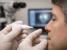
Chronic lacrimal canaliculitis is a persistent infection of the lacrimal canaliculi—tiny channels near the inner corner of the eyelid that drain tears from the eye. Often mistaken for conjunctivitis or “dry eye,” this underdiagnosed condition can cause tearing, discharge, and irritation that do not resolve with typical treatments. If left unmanaged, it can lead to chronic discomfort, recurrent infections, or even obstruction of the tear duct. This comprehensive guide explores the condition’s key features, evidence-based therapies, innovative surgical approaches, and the latest breakthroughs—empowering you with the knowledge to seek optimal care for lasting relief and ocular health.
Table of Contents
- Condition Overview and Epidemiology
- Conventional and Pharmacological Therapies
- Surgical and Interventional Procedures
- Emerging Innovations and Advanced Technologies
- Clinical Trials and Future Directions
- Frequently Asked Questions
- Disclaimer
Condition Overview and Epidemiology
Chronic lacrimal canaliculitis is a long-standing infection of the canaliculi, the tiny ducts that channel tears from the eye’s surface to the nose. Often underrecognized, this condition is commonly misdiagnosed as conjunctivitis or dacryocystitis.
Defining Features and Pathophysiology:
- Canaliculitis typically results from microbial infection—most often by Actinomyces species, but also by bacteria like Staphylococcus, Streptococcus, or fungi.
- The chronic nature stems from the formation of “sulfur granules” or concretions—yellowish, gritty debris that harbors infection in the canaliculus.
Epidemiology and Demographics:
- While exact prevalence is unclear, canaliculitis is relatively rare but likely underdiagnosed due to its subtle presentation.
- Middle-aged to elderly adults are most frequently affected.
- No significant sex predilection has been consistently observed.
Risk Factors:
- Advanced age (tissue changes and decreased immune response)
- History of chronic conjunctivitis or blepharitis
- Presence of punctal plugs or foreign bodies
- Previous ocular surgeries, especially those involving the tear drainage system
- Poor eyelid hygiene or chronic eye rubbing
Symptoms and Clinical Clues:
- Chronic tearing (epiphora)
- Mucopurulent discharge, often expressed with pressure over the inner eyelid
- Redness, swelling, and tenderness near the inner canthus
- Pouting or swelling of the punctum (tear duct opening)
- Failure to respond to standard topical antibiotics
Diagnosis:
- Careful clinical examination is key, often including gentle pressure over the canaliculus to express discharge.
- Microbiological cultures or histopathology of concretions may confirm the diagnosis.
- Imaging is rarely needed except for atypical or recurrent cases.
Practical Advice:
If you have chronic eye irritation, tearing, or persistent discharge that doesn’t improve with regular eye drops, ask your eye doctor about canaliculitis—a thorough exam may reveal this often-missed culprit.
Conventional and Pharmacological Therapies
Medical therapy remains the first line for many patients with early or mild chronic canaliculitis, and is also an essential adjunct to surgical care. However, the unique challenges of biofilm and concretions make eradication with medication alone difficult.
First-Line Medical Approaches:
- Topical Antibiotics:
- Broad-spectrum agents such as ciprofloxacin, moxifloxacin, or tobramycin are commonly prescribed.
- Drops or ointments are usually applied several times daily for several weeks.
- Topical antifungal or antiviral agents may be added if culture suggests unusual organisms.
- Systemic Antibiotics:
- Oral penicillin or amoxicillin may be used for suspected Actinomyces infection, often in combination with topical therapy.
- Doxycycline is sometimes selected for its anti-inflammatory and antimicrobial effects.
- Irrigation and Local Debridement:
- In-office irrigation of the canaliculus with antibiotic solution can reduce superficial infection.
- Gentle probing may help express concretions and biofilm, increasing medication effectiveness.
Supportive Measures:
- Warm compresses applied 2–3 times daily can promote drainage and reduce discomfort.
- Lid hygiene: Regular cleansing of the eyelids and lashes with diluted baby shampoo or commercial lid scrubs.
Limitations of Medical Therapy:
- Concretions often act as a reservoir for persistent infection, making complete cure rare without physical removal.
- Symptoms may improve temporarily but often recur after stopping treatment.
Practical Advice:
Adherence is key—use all medications as directed, complete the full course, and attend all follow-up appointments. If symptoms return quickly or do not improve after a few weeks, surgical intervention may be necessary.
Surgical and Interventional Procedures
When conservative management fails, surgical intervention is the standard of care for chronic lacrimal canaliculitis. The goal is complete removal of concretions and infected tissue, restoring normal tear drainage and resolving infection.
Surgical Strategies:
- Canaliculotomy:
- The primary procedure for chronic canaliculitis.
- Under local anesthesia, a small incision is made along the canaliculus to access and remove concretions, debris, and infected lining.
- The canaliculus is irrigated with antibiotic solution.
- The incision is often left to heal naturally (secondary intention) or may be closed with fine sutures.
- Curettage and Debridement:
- Curettes or small forceps are used to physically remove all visible granules and biofilm.
- Repeated irrigation ensures thorough cleansing.
- Punctoplasty or Punctal Reconstruction:
- In cases with significant scarring or obstruction, the punctum (tear duct opening) may be widened or reconstructed.
- Adjunctive Treatments:
- Intraoperative or postoperative antibiotic irrigation.
- Silicone stent insertion in select cases to maintain patency.
Minimally Invasive Innovations:
- Micro-incision canaliculotomy techniques are being developed to reduce trauma and speed recovery.
- Office-based, endoscope-assisted removal is under research in specialized centers.
Recovery and Postoperative Care:
- Mild soreness or swelling is expected for a few days.
- Topical antibiotics and anti-inflammatories are often prescribed postoperatively.
- Most patients experience rapid and lasting relief after surgery.
Practical Advice:
Ask your surgeon to explain what to expect before, during, and after surgery—including potential risks, recovery timeline, and self-care tips for optimal healing.
Emerging Innovations and Advanced Technologies
Advances in technology and a better understanding of biofilm-driven infections are leading to improved outcomes in chronic lacrimal canaliculitis care.
Novel Diagnostic Tools:
- High-resolution imaging (anterior segment optical coherence tomography, or AS-OCT) may help visualize canalicular changes and guide treatment in ambiguous cases.
- Polymerase chain reaction (PCR) and molecular diagnostic panels can rapidly identify causative microbes when standard cultures are negative.
Biofilm-Targeted Therapies:
- Enzyme-based irrigation solutions are being developed to break down biofilm and concretions, enhancing antibiotic penetration.
- Research on antimicrobial peptides and topical agents that disrupt microbial biofilm is ongoing.
Innovative Surgical Devices:
- Micro-endoscopes and miniaturized curettes allow for more targeted, minimally invasive removal of concretions.
- Biodegradable stents are under study to maintain canalicular patency during healing.
Postoperative Advances:
- Sustained-release antibiotic and anti-inflammatory implants are being explored to minimize recurrence and reduce the need for daily eye drops.
AI and Digital Monitoring:
- Artificial intelligence tools can help flag subtle, recurrent symptoms through patient-reported outcome apps, prompting earlier intervention.
Practical Advice:
Inquire about emerging technologies if you have persistent or recurrent symptoms, or if you’ve had multiple unsuccessful treatments. Some centers offer participation in pilot studies for advanced therapies.
Clinical Trials and Future Directions
Research is expanding our understanding and management of chronic lacrimal canaliculitis, aiming for faster diagnosis, less invasive treatment, and better patient outcomes.
Key Focus Areas:
- Comparative trials of new enzyme-based and biofilm-targeted irrigants versus traditional antibiotics.
- Studies of minimally invasive surgical techniques and the role of micro-endoscopy.
- Development and evaluation of sustained-release or implantable drug devices for the canalicular system.
- Exploration of patient-friendly, digital monitoring tools for early detection of relapse.
Pipeline of Future Advances:
- Identification of genetic or immunological markers for susceptibility.
- Personalized medicine approaches tailoring treatment to the specific microbial profile.
- Partnerships with device manufacturers to produce more effective, minimally invasive surgical tools.
Patient Participation and Access:
- Some clinical trials are open for enrollment, particularly at tertiary ophthalmology centers or research hospitals.
- Benefits may include access to novel therapies, expert follow-up, and contributing to the improvement of care for future patients.
What to Expect Next:
- As diagnostic and therapeutic options grow, more patients will receive timely, definitive treatment—reducing chronic discomfort and risk of complications.
Practical Advice:
If you have chronic symptoms, consider discussing research participation with your provider. ClinicalTrials.gov and major eye hospital websites list open studies; advocacy groups may offer additional support and information.
Frequently Asked Questions
What is chronic lacrimal canaliculitis and what causes it?
Chronic lacrimal canaliculitis is a persistent infection of the canaliculus (tear drainage duct), most often caused by Actinomyces bacteria or other microbes. It leads to ongoing tearing, discharge, and eye irritation.
How is chronic lacrimal canaliculitis diagnosed?
Diagnosis is usually made by examining the eyelid and canaliculus for swelling, discharge, and “sulfur granules.” Cultures or microscopic analysis of debris may confirm the infection.
What are the most effective treatments for chronic lacrimal canaliculitis?
Surgical removal of concretions and infected tissue (canaliculotomy) is the most effective cure. Antibiotics alone rarely eradicate the infection due to biofilm formation within the canaliculus.
Can chronic lacrimal canaliculitis recur after treatment?
Yes, recurrence is possible if all concretions are not removed or if the infection returns. Proper surgical technique and postoperative care reduce this risk.
What are the risks of not treating chronic lacrimal canaliculitis?
Untreated canaliculitis can cause ongoing discomfort, persistent discharge, blockage of the tear duct, and, in rare cases, spread of infection to nearby tissues.
How can I prevent chronic lacrimal canaliculitis?
Practicing good eyelid hygiene, avoiding unnecessary punctal plugs, and treating eye infections promptly may reduce the risk. There’s no guaranteed prevention, but early recognition helps.
Are there any new treatments or clinical trials for chronic lacrimal canaliculitis?
Yes, clinical trials are investigating enzyme-based irrigants, minimally invasive surgeries, and sustained-release antibiotics. Ask your eye care provider or check ClinicalTrials.gov for the latest options.
Disclaimer
The information presented in this article is intended for educational purposes only and does not substitute for professional medical advice, diagnosis, or treatment. Always consult your physician or a qualified eye care specialist regarding any questions about your health or treatment.
If you found this article helpful, please consider sharing it on Facebook, X (formerly Twitter), or any platform you prefer. Your support helps us continue producing accessible, high-quality health content. Don’t forget to follow us on social media for the latest updates and insights—your involvement truly makes a difference!


