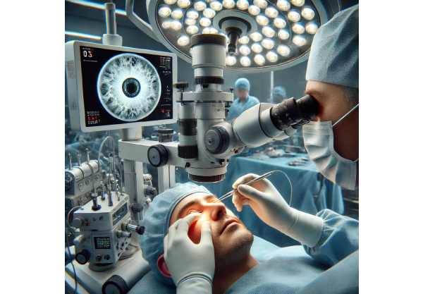
Iris dialysis is a relatively rare but potentially vision-threatening eye injury, often resulting from trauma or surgical complications, where the iris becomes partially or completely detached from its attachment at the ciliary body. This disruption can lead to noticeable visual changes, discomfort, and cosmetic concerns, while sometimes causing secondary issues such as glare or light sensitivity. Fortunately, advances in both medical and surgical management now provide more options for restoring eye function and appearance. In this comprehensive guide, we will examine the causes, therapies, innovative procedures, and latest breakthroughs for iris dialysis, equipping you to make informed decisions about your care or support someone navigating this condition.
Table of Contents
- Condition Overview and Epidemiology
- Conventional and Pharmacological Therapies
- Surgical and Interventional Procedures
- Emerging Innovations and Advanced Technologies
- Clinical Trials and Future Directions
- Frequently Asked Questions
Condition Overview and Epidemiology
Iris dialysis refers to the detachment of the iris—the colored part of the eye—from its base at the ciliary body. This detachment is typically the result of blunt or penetrating ocular trauma but can occasionally occur during intraocular surgery or, very rarely, due to certain eye diseases or congenital conditions.
Definition and Pathophysiology
- Iris dialysis is characterized by a separation (often partial, but sometimes complete) of the iris root from its normal attachment.
- The injury disrupts the normal circular structure of the iris, causing a gap at the periphery, which can be visible on close examination.
Prevalence and Causes
- Most commonly follows direct eye trauma (sports injuries, accidents, assaults).
- Can result from iatrogenic causes (complications during cataract or glaucoma surgery).
- Rarely associated with certain eye diseases or congenital abnormalities.
Risk Factors
- Participation in contact sports without protective eyewear
- History of eye trauma or surgery
- Pre-existing eye conditions such as angle recession or glaucoma
Clinical Presentation
- Visible irregularity or notch at the iris margin (best seen with slit-lamp examination)
- Photophobia (light sensitivity)
- Monocular diplopia (double vision in one eye)
- Glare and decreased contrast sensitivity
- Cosmetic concerns due to abnormal pupil shape or iris appearance
Associated Complications
- Secondary glaucoma (due to altered aqueous flow)
- Hyphema (bleeding in the anterior chamber)
- Traumatic cataract
- Corneal edema or scarring in severe cases
Diagnosis
- Based on history, symptoms, and slit-lamp examination.
- Gonioscopy and anterior segment optical coherence tomography (AS-OCT) may aid in assessing the extent and effects of the detachment.
Practical Tips
- Always seek immediate ophthalmic evaluation after any significant eye trauma.
- Use appropriate eye protection during high-risk activities.
- If you notice visual changes or abnormal iris appearance, consult an eye specialist promptly.
Conventional and Pharmacological Therapies
While many cases of iris dialysis may ultimately require surgical intervention, there are several initial medical therapies and non-surgical management strategies that can minimize symptoms, prevent complications, and improve quality of life—especially when the dialysis is small or not visually significant.
Observation and Monitoring
- Small, stable detachments with no significant symptoms may be safely monitored.
- Regular eye exams are essential to detect changes in intraocular pressure or the development of secondary glaucoma.
Symptom Relief and Eye Protection
- Photochromic and Tinted Lenses:
- Glasses with tints or light-adaptive lenses can reduce glare and light sensitivity.
- Mydriatic or Miotic Drops:
- In select cases, eye drops may be prescribed to control pupil size and minimize symptoms.
- Caution: Not all patients are candidates, and these should only be used under ophthalmic guidance.
Medical Management of Complications
- Intraocular Pressure Control:
- If secondary glaucoma develops, pressure-lowering eye drops may be initiated (e.g., prostaglandin analogs, beta-blockers).
- Anti-inflammatory Agents:
- Topical steroids or nonsteroidal anti-inflammatory drops may be prescribed if inflammation or hyphema is present.
Practical Lifestyle Advice
- Wear sunglasses outdoors or in bright conditions to reduce discomfort.
- Avoid eye rubbing or trauma to the affected eye.
- Schedule regular follow-up visits for eye pressure and vision monitoring.
Limitations of Medical Management
- Non-surgical therapies primarily address symptoms and secondary issues rather than the underlying detachment.
- If symptoms persist or worsen, referral for surgical evaluation is indicated.
Surgical and Interventional Procedures
When iris dialysis results in significant visual disturbance, cosmetic problems, or the risk of further complications, surgical repair is the primary treatment. Advances in microsurgical techniques now enable precise, safe, and cosmetically pleasing outcomes.
Indications for Surgery
- Large or symptomatic detachment
- Persistent glare, diplopia, or photophobia
- Secondary glaucoma unresponsive to medical therapy
- Aesthetic concerns affecting quality of life
Key Surgical Approaches
- Iris Root Suturing (Iridoplasty/Iridorrhaphy):
- The standard technique for reattaching the iris root to the ciliary body or scleral spur.
- Uses fine, non-absorbable sutures under a microscope.
- May involve ab externo (from outside) or ab interno (from inside the eye) methods.
- Often combined with other procedures (e.g., cataract extraction).
- Segmental Iris Prosthesis or Artificial Iris Implant:
- For extensive loss or defects, a customized artificial iris or segment may be implanted.
- Provides both functional and cosmetic restoration.
- Anterior Chamber Reconstruction:
- When iris dialysis is accompanied by other injuries (e.g., lens or cornea trauma), simultaneous repair of multiple structures may be performed.
Surgical Innovations and Minimally Invasive Techniques
- Microincisional and sutureless procedures for selected cases.
- Advanced visualization (intraoperative OCT, digital microscopy) for improved precision.
Postoperative Care and Recovery
- Use of anti-inflammatory and antibiotic eye drops.
- Protective eye shield as advised.
- Follow-up visits for suture removal (if non-buried) and to monitor healing.
Risks and Complications
- Infection (endophthalmitis)
- Recurrence or incomplete repair
- Intraocular pressure changes
Practical Guidance for Patients
- Carefully follow pre- and post-surgery instructions.
- Avoid strenuous activities, bending, or heavy lifting during initial healing.
- Contact your surgeon promptly if you experience pain, vision loss, or redness.
Surgical intervention offers substantial benefits for both visual function and cosmetic appearance, with most patients enjoying significant improvements.
Emerging Innovations and Advanced Technologies
Ophthalmic surgery and diagnostics have witnessed a wave of innovation, bringing exciting new options for managing iris dialysis—especially for complex or recurrent cases.
Novel Iris Repair Techniques
- Femtosecond Laser-Assisted Procedures:
- These ultra-precise lasers are now being used to assist with iris repair, improving accuracy and minimizing trauma.
- Advanced Suture Materials:
- New, more biocompatible suture materials reduce inflammation and improve long-term outcomes.
Custom Artificial Iris Technology
- 3D-Printed Implants:
- Personalized artificial irises designed with advanced imaging and 3D printing technology can precisely match the patient’s natural iris color and structure.
- Foldable Artificial Iris Devices:
- Allow minimally invasive implantation through tiny incisions, reducing recovery time.
Imaging and Visualization Breakthroughs
- High-Definition Anterior Segment OCT:
- Enhances preoperative planning and postoperative monitoring.
- Digital Surgical Microscopy:
- Offers unprecedented visualization during delicate repairs.
Adjunctive Technologies
- Anti-Scarring Agents and Drug-Eluting Implants:
- Under investigation for reducing post-surgical fibrosis and improving surgical success.
Teleophthalmology and Remote Care
- Secure video follow-ups and remote vision assessments are making specialist care more accessible, especially for rural or mobility-limited patients.
Patient Empowerment Tools
- Mobile apps to track recovery, vision changes, and medication adherence.
- Supportive virtual communities for sharing experiences and resources.
Practical Patient Advice
- Ask your surgeon about new technologies and custom implant options.
- Participate in research or registry programs if eligible, as these often offer access to leading-edge care.
These innovations are not just refining surgical outcomes—they’re helping patients achieve optimal visual rehabilitation and cosmetic satisfaction.
Clinical Trials and Future Directions
As the understanding of iris dialysis deepens, ongoing research is paving the way for next-generation therapies, devices, and techniques to further enhance patient care and outcomes.
Key Research Frontiers
- Next-Generation Biomaterials
- Trials are underway for advanced suture and implant materials designed to improve healing and reduce complications.
- Regenerative Medicine and Tissue Engineering
- Experimental work with stem cells and bioengineered tissues aims to restore natural iris structure and function for patients with extensive loss.
- Gene Therapy Approaches
- Investigational gene therapies could, in the future, stimulate endogenous repair pathways or prevent secondary complications like glaucoma.
- AI-Assisted Surgical Planning
- Artificial intelligence is being harnessed to optimize surgical techniques and personalize implant design, potentially raising the bar for results.
- Virtual and Augmented Reality in Surgery
- Trials are exploring immersive technologies to train surgeons and support intraoperative decision-making for complex repairs.
- Remote Rehabilitation and Vision Monitoring
- Digital programs that guide patients through post-op exercises and track long-term vision stability are in development.
Finding Clinical Trials
- Search clinicaltrials.gov or academic medical center websites for studies on iris repair, ocular trauma, or anterior segment surgery.
- Discuss trial eligibility and safety with your care team.
Patient Participation Advice
- Prepare a list of questions for research coordinators.
- Keep records of your symptoms and surgical history to streamline screening.
- Remain in close communication with both your regular ophthalmologist and the research team throughout participation.
These research efforts hold great promise for the future—expanding choices, improving precision, and restoring both function and appearance for people affected by iris dialysis.
Frequently Asked Questions
What causes iris dialysis and who is at risk?
Iris dialysis is most often caused by blunt or penetrating eye trauma, but can also result from eye surgery complications. Those at higher risk include contact sports participants and people with prior eye injuries or surgeries.
How is iris dialysis diagnosed?
Diagnosis is made through a detailed eye exam with slit-lamp biomicroscopy, sometimes supplemented by imaging like anterior segment OCT. Your eye doctor looks for a gap or notch at the iris root and assesses related damage.
Is surgery always required for iris dialysis?
Not all cases require surgery. Small, asymptomatic detachments may only need observation. Surgery is recommended for significant vision problems, photophobia, or cosmetic issues.
What are the best surgical options for iris dialysis?
Microsurgical iris repair (iridoplasty or iridorrhaphy) is the main approach. For extensive defects, artificial iris implants offer both cosmetic and functional restoration. Your surgeon will recommend the best method for your situation.
Can iris dialysis lead to other eye problems?
Yes, complications can include secondary glaucoma, traumatic cataract, or corneal damage. Regular follow-up and prompt treatment help prevent and manage these risks.
How long does it take to recover from iris dialysis surgery?
Recovery times vary, but most people experience significant improvement within a few weeks. You’ll need follow-up visits to monitor healing and vision. Carefully following post-op instructions is key to a good result.
Are there new technologies or treatments for iris dialysis?
Yes, advances include 3D-printed artificial irises, femtosecond laser repair, and minimally invasive techniques. Participation in clinical trials may give access to innovative options not widely available.
Disclaimer:
This guide is for informational purposes only and should not be considered a substitute for professional medical advice, diagnosis, or treatment. Always consult with a qualified eye care provider for advice tailored to your situation.
If this article was helpful, please consider sharing it on Facebook, X (formerly Twitter), or your favorite social media. Your support helps us keep producing in-depth, accessible health resources for all—thank you for helping spread awareness and knowledge!


