An iris nevus is a benign, pigmented growth found on the colored part of the eye. While most iris nevi remain harmless throughout life, their resemblance to more serious lesions like iris melanoma means they require careful monitoring and, occasionally, intervention. With ongoing advances in eye imaging, medical management, and surgical options, today’s patients and clinicians are better equipped than ever to distinguish, track, and treat iris nevi. In this comprehensive guide, we’ll explore the nature of iris nevus, available therapies, surgical techniques, the latest technological breakthroughs, and what the future holds for safe, effective management of this common ocular finding.
Table of Contents
- Condition Overview and Epidemiology
- Conventional and Pharmacological Therapies
- Surgical and Interventional Procedures
- Emerging Innovations and Advanced Technologies
- Clinical Trials and Future Directions
- Frequently Asked Questions
Condition Overview and Epidemiology
An iris nevus is a benign (non-cancerous) growth of melanocytes—the pigment-producing cells—within the iris. These small, flat, or slightly raised spots are often found incidentally during a routine eye exam, but can sometimes cause cosmetic concern or anxiety due to their resemblance to more serious eye conditions.
Definition and Pathophysiology
- Iris nevi appear as localized, pigmented areas (brown, tan, gray, or rarely blue/black) on the surface or within the substance of the iris.
- Unlike iris freckles, which are superficial and typically multiple, iris nevi are deeper and usually solitary.
Prevalence and Demographics
- Iris nevi are detected in up to 6% of the general population.
- They are more common in adults but can occur at any age.
- There is no strong sex predilection; nevi may be slightly more frequent in people with lighter-colored irides.
Risk Factors and Causes
- Genetics play a role; people with fair skin or lighter irises may be more susceptible.
- Sun exposure has not been definitively linked, but UV light is a known risk factor for other ocular melanocytic lesions.
- History of trauma or inflammation does not typically influence the risk of developing a nevus.
Clinical Presentation
- Usually asymptomatic and discovered incidentally.
- Rarely, a nevus may be associated with mild visual disturbance if it encroaches upon the pupil or causes irregularity.
- Some nevi may be vascularized, have a cystic component, or demonstrate subtle growth over time.
Risk of Malignant Transformation
- The vast majority of iris nevi remain benign.
- The lifetime risk of an iris nevus turning into iris melanoma is estimated at less than 1%.
- Features raising suspicion for malignancy include documented growth, increased vascularity, secondary glaucoma, or local extension.
Diagnosis
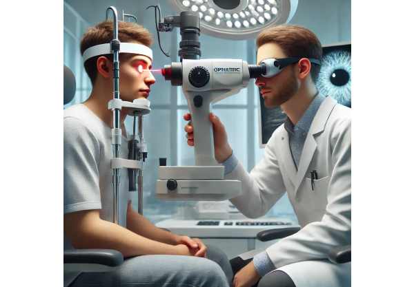
- Clinical examination with slit-lamp biomicroscopy is the gold standard.
- Additional imaging (anterior segment OCT, ultrasound biomicroscopy, or photography) is used to document size, thickness, and features over time.
Practical Tips
- Schedule regular eye exams to track any changes in size, shape, or color of the lesion.
- Report new symptoms such as pain, vision changes, or iris distortion to your eye care provider promptly.
- Photograph the eye (with flash off) periodically for home monitoring between visits.
Conventional and Pharmacological Therapies
Most iris nevi do not require treatment, only periodic observation to ensure stability. However, medical management may play a role in certain situations—particularly for symptoms, cosmetic issues, or if complications arise.
Observation and Surveillance
- Primary approach:
- The majority of iris nevi are monitored every 6–12 months with eye exams and photographic documentation.
- Growth, changes in shape, or the development of abnormal blood vessels are closely watched.
Pharmacological Management
- Symptomatic Management:
- If a nevus leads to minor irritation or local inflammation (rare), lubricating drops or anti-inflammatory medications may be used under professional supervision.
- Intraocular Pressure Control:
- Occasionally, nevi can cause secondary glaucoma. Standard glaucoma drops (e.g., prostaglandin analogs, beta-blockers) may be prescribed as needed.
Cosmetic and Comfort Measures
- Colored Contact Lenses:
- Specially tinted soft contact lenses can mask the appearance of a visible iris nevus for cosmetic comfort.
- These should be fitted by an eye care professional to avoid complications.
Patient Empowerment & Lifestyle Guidance
- Wear sunglasses with UV protection to safeguard overall ocular health.
- Avoid eye rubbing or trauma to reduce risk of complications.
- If you have a family history of eye cancer, share this information with your eye doctor for personalized surveillance.
Limitations
- No medication can shrink or eliminate a benign nevus.
- Medical therapy does not reduce the small risk of malignant transformation; only vigilant monitoring can address this concern.
Surgical and Interventional Procedures
Though uncommon, surgery or laser intervention may be indicated for an iris nevus if there are significant symptoms, cosmetic concerns, or signs suggestive of malignancy.
Indications for Surgical or Laser Treatment
- Documented growth of the lesion over time
- Secondary glaucoma or significant vision impairment
- Local irritation unresponsive to medical therapy
- Suspicion of malignant transformation (changing color, bleeding, extension)
Surgical Approaches
- Sector Iridectomy
- Surgical removal of the nevus-containing segment of iris.
- Performed under local or general anesthesia, using fine microsurgical techniques.
- Tissue is sent for pathological examination to rule out melanoma.
- Laser Therapy
- Laser photocoagulation can be used in rare cases for superficial nevi, particularly those with visible vascularity.
- Less invasive but not widely performed due to risk of scarring and pigment dispersion.
- Anterior Segment Reconstruction
- For larger nevi or those causing secondary structural damage, surgery may include iris reconstruction using sutures or artificial iris segments.
Risks and Considerations
- Infection, bleeding, or inflammation
- Potential for iris deformity, glare, or photophobia
- Rare but serious risk of intraocular spread if the lesion is actually melanoma
Recovery and Postoperative Care
- Use of topical antibiotics and steroids to reduce inflammation
- Shielding the eye for several days post-surgery
- Follow-up visits for wound check and to monitor healing
Practical Advice
- Always choose an experienced ocular oncologist or anterior segment surgeon for iris lesion surgery.
- Surgery is not recommended for purely cosmetic reasons if the lesion is stable and benign.
- Keep detailed records of your eye exams and surgical history.
Emerging Innovations and Advanced Technologies
Recent years have brought new tools and techniques to the detection, analysis, and management of iris nevi—enabling more personalized, precise, and less invasive care.
Advanced Imaging Modalities
- High-Resolution Anterior Segment OCT:
- Provides detailed cross-sectional images to track nevus thickness and structure over time.
- Ultrasound Biomicroscopy (UBM):
- Enables in-depth assessment of lesions hidden beneath the iris surface.
- Automated Digital Photography:
- Facilitates home-based monitoring and remote consultation, supporting teleophthalmology.
Artificial Intelligence (AI) and Predictive Analytics
- AI-Driven Image Analysis:
- Algorithms can identify subtle features or early signs of malignancy missed by the human eye, aiding in timely referral and management.
- Risk Stratification Tools:
- New scoring systems combine imaging and patient data to assess risk and personalize follow-up intervals.
Minimally Invasive Techniques
- Microincisional Iridectomy:
- Tiny surgical instruments enable removal of small lesions with minimal tissue disruption and fast recovery.
- Femtosecond Laser-Assisted Procedures:
- Improve surgical precision for selected cases, reducing the risk of collateral damage.
Customized Cosmetic Solutions
- 3D-Printed Artificial Iris Segments:
- For select cases requiring reconstruction, highly individualized implants can now restore both appearance and function.
Genetic and Molecular Profiling
- Investigational studies are exploring genetic markers to better predict the risk of nevus transformation to melanoma.
Digital Health and Patient Engagement
- Smartphone apps and patient portals now let individuals track changes, schedule reminders, and communicate concerns quickly.
Practical Tips
- Ask your doctor about digital imaging at each visit.
- If you’re part of a high-risk group, inquire about AI-based analysis or genetic counseling.
- Share any changes or new symptoms with your provider, even between visits.
Clinical Trials and Future Directions
Research into iris nevi is evolving, with scientists seeking to refine diagnostic accuracy, reduce unnecessary interventions, and better predict which lesions are at risk of becoming cancerous.
Key Research Themes
- Longitudinal Imaging Studies
- Ongoing trials use serial high-resolution imaging and AI to determine growth patterns and risk factors for malignant change.
- Noninvasive Biomarker Discovery
- Studies are searching for tear fluid, blood, or tissue biomarkers that can distinguish benign from malignant lesions without surgery.
- Genetic Profiling and Personalized Risk
- Genomic research may soon allow individual risk scoring for each patient, enabling tailored follow-up and intervention.
- Targeted Therapies for Early Melanoma
- New medications, immunotherapies, or focal laser therapies under investigation could treat early malignant transformation before spread occurs.
- Patient-Driven Monitoring Programs
- Digital health platforms and remote home-based imaging are being trialed to make surveillance more accessible and effective.
Participating in Clinical Research
- Major eye centers and academic hospitals frequently enroll patients for studies.
- Discuss eligibility, risks, and potential benefits with your care team.
Future Outlook
- Advances are moving toward personalized, noninvasive surveillance and interventions that are safer, faster, and more comfortable for patients.
Advice for Patients
- Stay up to date by joining patient registries or advocacy organizations.
- Participate in research or long-term studies if eligible, to help improve future care for others.
Frequently Asked Questions
What is an iris nevus and how can I tell if it’s dangerous?
An iris nevus is a benign pigmented spot on the iris, often harmless. Most do not become cancerous. However, if you notice growth, color change, pain, or blurred vision, see your eye doctor for prompt evaluation.
How often should an iris nevus be checked?
Most nevi require monitoring every 6–12 months, especially for the first few years after discovery. Your eye doctor may increase frequency if there are suspicious features or a family history of eye cancer.
Can an iris nevus turn into cancer?
The risk is very low—less than 1%. Features like documented growth, new blood vessels, or secondary glaucoma increase the risk. Regular follow-up is key for early detection of any malignant change.
Is treatment required for iris nevus?
Most cases do not need treatment, only observation. Surgery or laser therapy is reserved for rare cases with symptoms, vision problems, or signs of malignant transformation.
What are the signs that an iris nevus might be melanoma?
Warning signs include rapid growth, irregular borders, bleeding, development of abnormal vessels, and secondary glaucoma. Any of these should prompt urgent assessment by an eye specialist.
Are there cosmetic solutions for visible iris nevi?
Yes, colored contact lenses can help mask a visible nevus for cosmetic comfort. In rare cases, surgical reconstruction with artificial iris segments is considered.
Are there new technologies to monitor or treat iris nevi?
Yes, advanced imaging, AI risk analysis, and even minimally invasive procedures or custom artificial irises are now available at leading eye centers.
Disclaimer:
This article is for educational purposes only and should not be considered medical advice. Always consult a qualified eye care professional about your personal health or any changes in vision.
If you found this guide helpful, please share it with friends, family, or your network on Facebook, X (formerly Twitter), or your favorite social media platform. Your support helps us continue producing quality, accessible health resources for all—thank you!


