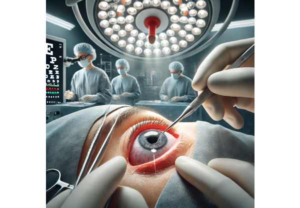
Lacrimal fistula is a rare yet significant condition where an abnormal channel forms between the lacrimal system and the skin, most often resulting in persistent tearing and discharge from an opening near the inner corner of the eye. This guide explores the underlying mechanisms, epidemiology, and risk factors, and provides a thorough overview of the latest therapeutic approaches—including both established medical treatments and the most advanced surgical and technological innovations. Whether you’re a patient, caregiver, or health professional, this resource is designed to help you navigate the evolving landscape of lacrimal fistula treatment and management.
Table of Contents
- Condition Overview and Epidemiology
- Conventional and Pharmacological Therapies
- Surgical and Interventional Procedures
- Emerging Innovations and Advanced Technologies
- Clinical Trials and Future Directions
- Frequently Asked Questions
Condition Overview and Epidemiology
Lacrimal fistula, sometimes called congenital lacrimal fistula or acquired lacrimal fistula, is an abnormal tract that allows tears to bypass their normal drainage path and exit onto the skin. Most commonly found near the inner canthus, it may be present at birth or develop later due to trauma, infection, surgery, or chronic inflammation. This direct connection between the lacrimal sac or duct and the skin leads to persistent tearing and, in some cases, mucous or purulent discharge.
Definition and Pathophysiology:
- Lacrimal fistula is an epithelial-lined tract connecting the lacrimal system (puncta, canaliculi, sac, or duct) to the skin.
- Congenital cases result from incomplete embryological development.
- Acquired forms can develop from trauma, infection, chronic dacryocystitis, or complications from previous surgery.
Prevalence and Epidemiology:
- Congenital lacrimal fistula is very rare, occurring in approximately 1 in 2,000 to 1 in 10,000 live births.
- Slight female preponderance has been noted.
- Acquired fistulas are less common, with most cases seen in adults following trauma, chronic infection, or after nasolacrimal duct surgery.
Risk Factors:
- Family history of congenital lacrimal anomalies.
- History of chronic dacryocystitis or lacrimal duct obstruction.
- Facial trauma or previous eye/nasal surgeries.
- Autoimmune conditions or granulomatous diseases affecting the lacrimal system.
Clinical Presentation:
- Constant tearing (epiphora), especially when crying or exposed to wind.
- Intermittent or persistent watery or mucopurulent discharge from a pinpoint skin opening near the inner canthus.
- Recurrent periocular skin irritation or infection.
- Redness, swelling, or tenderness around the fistula site in cases of superimposed infection.
Diagnosis:
- Clinical inspection: visualizing the external opening.
- Gentle pressure over the lacrimal sac may express tears or pus through the fistula.
- Fluorescein dye tests and probing can help delineate the tract’s anatomy.
- Imaging (dacryocystography, CT, or MRI) may be used in complex or recurrent cases.
Practical Advice:
- Avoid manipulating or squeezing the fistula, as this can worsen irritation or introduce infection.
- Persistent tearing or discharge should prompt evaluation by an ophthalmologist, especially if associated with skin changes or redness.
Conventional and Pharmacological Therapies
Non-surgical management of lacrimal fistula may be effective in select patients, especially those with minimal symptoms or without recurrent infections. The approach is tailored by age, underlying cause, and presence of complications.
Conservative Management:
- Observation: In rare cases, especially in infants with no infection and minimal symptoms, simple observation may be reasonable.
- Eyelid hygiene: Regular cleansing of the periocular skin with gentle, non-irritating solutions to prevent secondary infection.
- Warm compresses: Useful for mild inflammation or to encourage spontaneous closure in infants (rare).
Pharmacological Approaches:
- Topical antibiotics: Prescribed when discharge is present, especially if mucopurulent, to control superficial bacterial growth.
- Oral antibiotics: Reserved for episodes of cellulitis, dacryocystitis, or deeper soft tissue infection.
- Anti-inflammatory drops: In select cases, to reduce peri-fistula irritation and swelling.
Management of Associated Lacrimal Obstruction:
- Crigler massage: For infants with coexisting nasolacrimal duct obstruction, massage can improve symptoms and possibly reduce fistula output.
- Saline irrigation: Occasionally used to flush the lacrimal system and maintain patency, particularly in congenital cases.
When Conservative Management Is Not Enough:
- Persistent symptoms, skin irritation, or recurrent infections usually require procedural intervention.
- Adults with acquired fistulas rarely benefit from conservative measures alone.
Practical Tips:
- Practice gentle cleansing and avoid harsh soaps near the fistula.
- Use all medications as directed; overuse of antibiotics can lead to resistance or skin irritation.
- Keep a log of symptoms to aid your provider in decision-making.
Surgical and Interventional Procedures
Surgical management remains the mainstay for symptomatic or complicated lacrimal fistulas. The choice of technique is based on fistula anatomy, patient age, coexisting conditions, and previous interventions.
Fistulectomy (Excision of Fistula):
- The fistula tract is identified, dissected, and completely excised down to its connection with the lacrimal system.
- The lacrimal system is repaired, and the skin is closed.
- In congenital cases, may be combined with probing or stenting of the nasolacrimal duct.
Dacryocystorhinostomy (DCR):
- Indicated for patients with both lacrimal duct obstruction and fistula.
- A new drainage pathway is created from the lacrimal sac to the nasal cavity, often performed via endoscopic or external approach.
- Fistulectomy is performed concurrently, and the skin defect is repaired.
Canaliculodacryocystorhinostomy:
- For fistulas involving the canaliculi (closer to the eyelid margin), this more complex procedure creates a new channel for tear drainage.
Silicone Intubation/Stenting:
- After tract excision or duct repair, silicone tubes may be placed to keep the passage open and reduce risk of restenosis.
- Stents are typically removed after several weeks.
Laser-Assisted Fistula Closure:
- Lasers (CO2 or diode) can ablate the fistula tract with minimal bleeding and promote healing.
- Sometimes used as adjuncts to surgical repair.
Postoperative Care:
- Topical and oral antibiotics to prevent infection.
- Cold compresses and anti-inflammatory drops to reduce swelling.
- Instructions to avoid nose blowing, heavy lifting, or direct trauma to the area during healing.
Complications and Revision:
- Recurrence of fistula, infection, or scar formation may require repeat intervention.
- Early recognition of signs of infection—such as redness, pain, or discharge—is critical for prompt treatment.
Practical Advice:
- Discuss anesthesia options and likely downtime with your surgeon.
- Maintain follow-up visits to monitor for recurrence or healing issues.
- Protect the surgical site from trauma, especially in the early weeks after surgery.
Emerging Innovations and Advanced Technologies
The management of lacrimal fistula is evolving, with several technological and procedural innovations enhancing both patient outcomes and comfort.
Endoscopic and Minimally Invasive Techniques:
- High-resolution endoscopes enable direct visualization of the lacrimal system and fistula tract for precise, targeted repair.
- Minimally invasive approaches reduce scarring, postoperative discomfort, and recovery time.
Bioengineered and Absorbable Stents:
- New stents made of bioabsorbable materials keep the repaired duct open and dissolve naturally, reducing the need for secondary procedures.
- Drug-eluting stents release antibiotics or anti-scarring agents, further lowering risk of recurrence.
Laser and Radiofrequency Ablation:
- Latest-generation lasers and radiofrequency tools offer non-contact, bloodless excision or ablation of the fistula, minimizing tissue damage.
3D Imaging and Surgical Planning:
- Preoperative imaging with 3D reconstruction aids in mapping complex fistulas, leading to more accurate repairs and fewer complications.
Tissue Engineering and Regenerative Medicine:
- Early research into stem cell–based therapies and tissue scaffolds for lacrimal duct reconstruction offers hope for severe or recurrent cases.
Telemedicine and Patient Monitoring:
- Remote follow-up with video consultations enables early detection of postoperative issues and supports patients unable to travel frequently.
Patient-Focused Innovation:
- Digital tools help patients track symptoms and recovery, ensuring early intervention if recurrence occurs.
- Educational platforms provide video demonstrations of postoperative care, improving adherence and outcomes.
Practical Tips:
- Ask your care team if you may benefit from newer, minimally invasive or laser-assisted options.
- Use digital symptom trackers to log recovery and share updates with your provider.
Clinical Trials and Future Directions
Ongoing research is driving improvements in both surgical and non-surgical management of lacrimal fistulas, with a focus on less invasive techniques and better quality of life.
Current Research Priorities:
- Comparison of endoscopic versus open surgical approaches for fistula closure.
- Evaluation of bioabsorbable and drug-eluting stents in maintaining duct patency after repair.
- Use of autologous tissue grafts or stem cell therapy for complex or recurrent fistulas.
- Clinical outcomes in pediatric versus adult populations following innovative repair techniques.
Active and Upcoming Clinical Trials:
- Several multicenter trials are underway investigating new materials for stents and novel ablation techniques.
- Patient-reported outcomes—such as comfort, cosmetic appearance, and recurrence rates—are increasingly used as primary endpoints.
Research Pipeline and Anticipated Advancements:
- Personalized surgical planning using AI-driven imaging is expected to improve outcomes in complex cases.
- Development of topical agents that promote tract closure or reduce inflammation post-surgery.
- Remote monitoring platforms and mobile health technologies for early detection of complications.
Patient Advocacy and Involvement:
- Patients interested in participating in clinical trials should speak with their provider or explore national clinical trial registries.
- Support groups and online communities can provide valuable information and emotional support during recovery or decision-making.
Practical Advice for Patients:
- Stay informed about ongoing research—some clinical trials offer early access to breakthrough treatments.
- If you have a recurrent or complex fistula, ask about experimental therapies or trial enrollment.
- Engage with patient advocacy organizations for resources and community support.
Frequently Asked Questions
What is a lacrimal fistula and how is it diagnosed?
A lacrimal fistula is an abnormal channel connecting the tear duct system to the skin, often causing persistent tearing or discharge. Diagnosis is made through a detailed eye exam, sometimes aided by dye tests or imaging.
Can a lacrimal fistula go away on its own?
Rarely, mild congenital fistulas may close spontaneously in infants. Most require medical or surgical treatment, especially if symptoms are persistent or infections occur.
What are the common treatments for lacrimal fistula?
Conservative measures include hygiene and antibiotics, but most cases—especially those with recurrent symptoms—are best treated with surgical excision of the fistula, often combined with repair of the tear duct system.
How safe is surgery for a lacrimal fistula?
Surgery is generally safe, especially with modern minimally invasive and endoscopic techniques. Risks are low, but can include infection, scarring, or recurrence.
Are there innovative or minimally invasive options for fistula treatment?
Yes. Laser-assisted procedures, endoscopic surgery, and bioabsorbable stents are among the latest innovations designed to improve results and minimize recovery time.
When should I see a specialist about a lacrimal fistula?
See an ophthalmologist or oculoplastic surgeon if you have constant tearing, a skin opening near the eye, or repeated infections. Early evaluation improves outcomes and can prevent complications.
Disclaimer:
This article is for educational purposes only and does not substitute for professional medical advice, diagnosis, or treatment. For any symptoms or questions related to lacrimal fistula, please consult an eye care specialist.
If you found this information helpful, please consider sharing it on Facebook, X (formerly Twitter), or your favorite platform. Your support empowers us to keep providing trusted, up-to-date resources for the community. Thank you!


