What is Punctal Stenosis?
Punctal stenosis is an ocular condition defined by the narrowing or complete blockage of the lacrimal puncta, which are small openings on the eyelid margins near the nose that allow tears to drain from the eye’s surface into the nasal cavity. This condition causes poor tear drainage, resulting in persistent tearing (epiphora), irritation, and discomfort. Punctal stenosis can affect one or both eyes and have a significant impact on quality of life, resulting in chronic tearing, blurred vision, and frequent eye infections.
The condition may be congenital or acquired. Congenital punctal stenosis is present from birth and is frequently associated with other lacrimal system abnormalities. Chronic blepharitis (eyelid inflammation), conjunctivitis, long-term use of certain medications, trauma, or age-related changes can all contribute to acquired punctal stenosis, which is more common in adults. Chronic inflammation and scarring caused by conditions such as Stevens-Johnson syndrome or ocular cicatricial pemphigoid can both contribute to the development of punctal stenosis.
Early detection and treatment are critical in managing punctal stenosis to avoid complications and improve patient comfort. Understanding the underlying causes and symptoms of punctal stenosis is critical for timely intervention and effective management of this frequently debilitating condition.
Punctal Stenosis: Management and Treatment Strategies
Managing and treating punctal stenosis requires a combination of non-surgical and surgical approaches, depending on the severity and underlying cause of the disease. The primary goals of treatment are to restore proper tear drainage, relieve symptoms, and avoid recurring infections and complications.
Non-surgical Treatments
For mild cases of punctal stenosis or as an initial approach, non-surgical treatments may be recommended:
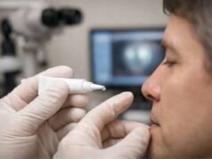
- Lubricating Eye Drops: Artificial tears or lubricating eye drops can help relieve the symptoms of dryness and irritation caused by punctal stenosis. These drops provide temporary relief, but they do not treat the underlying blockage.
- Topical Steroids: If inflammation is a contributing factor, topical corticosteroids may be used to reduce swelling and inflammation around the puncta, potentially improving tear drainage.
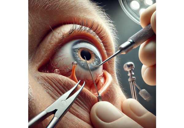
- Punctal Probing and Dilation: This procedure involves inserting a thin instrument into the punctum to dilate and open the obstructed tear duct. Probing can be performed in a clinical setting using local anesthesia and is frequently combined with irrigation to flush out any debris or blockages in the tear drainage system.
Surgical Treatments
Surgical intervention is frequently required for more severe cases of punctal stenosis or when nonsurgical treatments fail to provide relief. There are several surgical techniques available for restoring tear drainage.
- Punctoplasty: This procedure entails making small incisions to widen the punctal opening. Simple punctal dilation, three-snip punctoplasty, and laser punctoplasty are some of the techniques available. Punctoplasty is usually done under local anesthesia and has a high success rate in relieving symptoms.
- ** Punctal Stents**: In cases where punctoplasty alone is insufficient, small silicone or thermoplastic stents can be inserted to keep the puncta open. These stents promote tear drainage and prevent recurrent stenosis. The stents are typically left in place for several months to allow the puncta to heal in an open position.
- Conjunctivodacryocystorhinostomy (CDCR): This more difficult procedure is reserved for severe cases in which other treatments have failed. CDCR involves creating a new tear drainage pathway by bypassing the blocked puncta and connecting the conjunctiva directly to the nasal cavity with a small glass tube (Jones tube). This surgery is performed under general anesthesia and requires meticulous postoperative care.
Post-operative Care
Postoperative care is critical for achieving successful outcomes after punctal stenosis surgery. Patients are usually given antibiotic and anti-inflammatory eye drops to prevent infection and inflammation. Regular follow-up visits are required to monitor healing, assess the effectiveness of the procedure, and manage any complications. Additional follow-up is required to monitor the position and function of stents or tubes.
Latest Innovations in Punctal Stenosis Treatment
Recent advances in punctal stenosis treatment have resulted in the development of novel techniques and technologies that improve the efficacy and outcomes of both non-surgical and surgical procedures. These cutting-edge innovations are transforming the treatment of punctal stenosis, giving patients new hope.
Minimal Invasive Techniques
Minimally invasive approaches are gaining popularity in the treatment of punctal stenosis due to their shorter recovery times, lower risk of complications, and improved patient comfort.
- Balloon Dilation: Balloon catheter dilation is a minimally invasive procedure that employs a tiny balloon to gently expand the narrowed punctum. The balloon is inserted into the punctum and inflated to widen the tear duct, which improves tear drainage. This technique uses local anesthesia and has shown promising results in restoring tear flow with minimal discomfort.
- Microdrill Canaliculoplasty: This novel technique uses a microdrill to gently remove obstructions and expand the punctal opening. The microdrill causes precise, controlled enlargements of the puncta, which improves tear drainage while minimizing tissue damage. Microdrill canaliculoplasty is a quick outpatient procedure with a high success rate and minimal complications.
Advanced Stent Technologies
New stent technologies have significantly improved surgical outcomes for punctal stenosis. These advanced stents provide improved biocompatibility, durability, and ease of insertion.
- Biodegradable Stents: Biodegradable stents are intended to provide temporary support for the puncta before dissolving over time. These stents eliminate the need for a second procedure to remove them and lower the risk of long-term complications that come with permanent stents.
- Smart Stents: Smart stents use cutting-edge materials and technologies like shape-memory alloys and drug-eluting coatings. Shape-memory stents can change shape in response to temperature changes, ensuring a good fit and punctal patency. Drug-eluting stents gradually release anti-inflammatory or antiscarring medications, lowering the risk of restenosis and promoting healing.
Laser-Assisted Procedures
Laser technology is increasingly being used to treat punctal stenosis, providing greater precision and results.
- Laser Punctoplasty: Laser-assisted punctoplasty uses concentrated laser energy to make small incisions and widen the punctal opening. This technique provides precise control with minimal tissue damage, resulting in faster recovery and less postoperative discomfort. Laser punctoplasty is especially useful for patients with recurrent stenosis or who have not responded to conventional surgical methods.
- Laser-Assisted Canaliculotomy: This procedure uses a laser to precisely incise and open blocked canaliculi (tear ducts). Laser-assisted canaliculotomy is less invasive than traditional surgical methods, providing greater accuracy and shorter recovery times. It is an effective treatment option for patients with complex or multiple-level obstructions.
Regenerative Medicine and Tissue Engineering
The use of regenerative medicine and tissue engineering in the treatment of punctal stenosis is a growing field with enormous potential.
- Stem Cell Therapy: Stem cell therapy attempts to regenerate damaged or scarred punctal tissues, thereby restoring normal tear drainage function. Stem cell-based treatments that promote tissue healing and reduce the need for surgical intervention are currently under development. Early animal studies have yielded promising results, and clinical trials are currently underway to determine the safety and efficacy of stem cell therapy for punctal stenosis.
- Tissue-Engineered Grafts: Tissue engineering is the process of creating bioengineered grafts to replace damaged or stenotic punctal tissues. These grafts are intended to mimic the structure and function of natural tissues, thereby encouraging integration and healing. Tissue-engineered grafts could be a viable option for patients with severe or recurring punctal stenosis who require reconstructive surgery.
Personalized Treatment Approaches
The rise of personalized medicine is also having an impact on punctal stenosis treatment. Tailoring treatments to individual patient characteristics and needs improves outcomes and satisfaction.
- Customizable Stents: Personalized stents are being developed to accommodate each patient’s unique anatomy and needs. Stents are created using advanced imaging techniques such as 3D printing and computer-aided design to ensure a precise fit and optimal support. Customizable stents improve treatment efficacy while lowering the risk of complications.
- Genetic Profiling: Genetic profiling is being investigated in order to identify patients with a higher risk of developing punctal stenosis and tailor preventive and therapeutic interventions accordingly. Understanding the genetic factors that cause punctal stenosis can aid in the development of targeted treatments and better patient outcomes.


