Traumatic endophthalmitis is a severe, sight-threatening intraocular infection that occurs after an open-globe injury that compromises the eye’s integrity. This condition is characterized by inflammation of the intraocular tissues, including the vitreous and/or aqueous humor, as a result of microbial contamination during injury. Traumatic endophthalmitis is considered an ophthalmic emergency, necessitating immediate medical attention to avoid permanent vision loss or even loss of the eye.
Etiology and Pathogenesis
The primary cause of traumatic endophthalmitis is microorganisms entering the eye during an open-globe injury. This can happen when a foreign body enters the eye or when the eye comes into contact with contaminated objects or environments. Bacteria are the most common pathogens responsible for traumatic endophthalmitis, but fungi and, in rare cases, viruses or parasites can also be involved. The etiology of the infection is largely determined by the environment in which the injury occurred, the nature of the object that caused the injury, and the time passed before medical treatment is initiated.
Bacterial Infections
Bacterial infections are the leading cause of traumatic endophthalmitis, with the most commonly isolated organisms being Staphylococcus aureus, Staphylococcus epidermidis, and Streptococcus species. Gram-negative bacteria, such as Pseudomonas aeruginosa, Enterobacteriaceae, and Proteus species, are also important pathogens, especially in agricultural settings or cases of soil contamination.
When a foreign body enters the eye, it frequently transports bacteria from the surface or environment into the sterile interior. Once inside the eye, these bacteria can quickly multiply in the nutrient-rich environment of the vitreous and aqueous humor, causing a severe inflammatory reaction. In response to the infection, the body’s immune system releases inflammatory mediators and recruits immune cells to the site, exacerbating the damage to ocular tissues.
Fungal Infections
Fungal endophthalmitis is less common than bacterial endophthalmitis, but it is a major concern, especially in tropical and subtropical regions where filamentous fungi like Aspergillus and Fusarium species thrive. Fungal endophthalmitis is frequently associated with injuries to organic matter, such as plant material, which can introduce fungal spores into the eye. Fungal infections are more indolent than bacterial infections, which means they progress more slowly but are more difficult to treat and eradicate.
Endophthalmitis is caused by fungal pathogens that germinate and proliferate within the eye, resulting in the formation of inflammatory granulomas and potentially severe damage to ocular structures. The chronic nature of fungal infections can cause delays in diagnosis and treatment, resulting in poorer visual outcomes than bacterial endophthalmitis.
Other pathogens
Viral or parasitic infections can also cause traumatic endophthalmitis, though this is rare. Viral endophthalmitis is uncommon and usually affects immunocompromised people. Herpes simplex virus (HSV) and cytomegalovirus (CMV) are the most frequently implicated viral agents. Parasitic infections, such as those caused by Toxoplasma gondii, are extremely uncommon, but they can occur in specific endemic areas or under certain conditions.
Risk Factors
Several risk factors raise the possibility of developing traumatic endophthalmitis after an eye injury. The most significant risk factors are:
- Nature of the Injury: Penetrating injuries involving contaminated objects, such as those in agricultural settings or involving organic material, are more likely to introduce pathogenic organisms into the eye. High-velocity foreign body injuries, such as those involving metal or glass fragments, are also more likely to cause endophthalmitis.
- Delay in Treatment: The time between the injury and the start of medical treatment is an important factor in the development of traumatic endophthalmitis. Delayed treatment gives pathogens more time to spread and cause significant damage before they can be effectively controlled.
- Foreign Body Retention: The presence of an intraocular foreign body (IOFB) raises the risk of developing endophthalmitis. Foreign bodies, particularly those made of organic material or heavily contaminated, can act as nidus for infection.
- Poor Wound Closure: Inadequate or delayed closure of the ocular wound can allow microorganisms to enter the eye and cause infection. Proper wound closure is required to prevent pathogen infiltration and maintain the integrity of the ocular barrier.
- Immune Status: People with weakened immune systems, whether due to underlying medical conditions or immunosuppressive treatments, are more likely to develop severe infections, such as endophthalmitis.
Clinical Presentation
The clinical presentation of traumatic endophthalmitis differs depending on the causative pathogen, the severity of the infection, and the time since injury. The condition usually manifests acutely, with symptoms appearing within hours to days of the trauma. In cases of fungal or indolent bacterial infections, symptoms may appear gradually over several days to weeks.
Common signs and symptoms of traumatic endophthalmitis are:
- Severe Ocular Pain: Patients frequently describe intense eye pain that is accompanied by a deep, throbbing sensation. Eye movement usually exacerbates the pain, and it is often one of the first symptoms to appear.
- Decreased Vision: Traumatic endophthalmitis is characterized by rapid and significant vision loss. Patients may have blurred vision, floaters, or the sensation that a curtain is being drawn over their visual field. In severe cases, vision may be reduced to the perception of light or even the absence of light perception.
- Redness and Swelling: The affected eye is typically red and swollen, with significant conjunctival injection (redness of the white part of the eye) and chemosis. The eyelids may also swell, making it difficult to open the eyes.
- Hypopyon: Hypopyon, or the accumulation of pus in the anterior chamber of the eye, is a common sign of endophthalmitis. It appears as a white or yellowish layer of inflammatory cells in the lower part of the anterior chamber, indicating intraocular inflammation.
- Photophobia: Patients with traumatic endophthalmitis frequently experience photophobia, or light sensitivity, as a result of intraocular inflammation. Bright light can cause discomfort and worsen pain.
- Floaters and Vitreous Opacities: The presence of floaters, which are small, dark shapes that drift across the visual field, is a common sign of vitreous involvement in endophthalmitis. The inflammatory debris in the vitreous humor, the gel-like substance that fills the eye, is what causes these floaters.
- Corneal Edema: In some cases, inflammation and increased intraocular pressure can cause the cornea to swell. Corneal edema can make the cornea appear cloudy or hazy, which contributes to vision loss.
- Systemic Symptoms: Although less common, systemic symptoms such as fever, malaise, or a general sense of ill health can occur in severe or systemic infections.
Complications
Traumatic endophthalmitis carries a high risk of complications, especially if not treated promptly and effectively. Some of the most serious complications are:
- Retinal Detachment: Inflammation and infection can weaken the retina’s attachments, causing retinal detachment. This is a serious complication that can lead to permanent vision loss if not treated promptly.
- Phthisis Bulbi: This is a condition in which the eye becomes shrunken and non-functional as a result of extensive inflammation and scarring. Phthisis bulbi is a common complication of untreated or poorly managed endophthalmitis that results in eye loss.
- Glaucoma: Increased intraocular pressure due to inflammation or scar tissue formation can cause secondary glaucoma, which can further damage the optic nerve and worsen vision loss.
- Corneal Scarring: Severe inflammation and infection can cause corneal scarring, resulting in significant vision impairment. Corneal scarring may necessitate surgical treatment, such as corneal transplantation, to restore vision.
- Infection Spread: In rare cases, the infection can spread beyond the eye and into the surrounding tissues (orbital cellulitis) or even into the bloodstream, resulting in systemic sepsis, a potentially fatal condition.
Diagnostic methods
To diagnose traumatic endophthalmitis, a clinical examination, laboratory testing, and imaging studies are all required. Prompt and accurate diagnosis is critical for initiating appropriate treatment and increasing the likelihood of preserving vision.
Clinical Examination
The clinical examination is the initial step in diagnosing traumatic endophthalmitis. An ophthalmologist will perform a thorough examination of the eye, looking for signs of infection and inflammation.
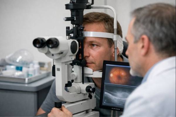
- Slit-Lamp Biomicroscopy: This technique allows an ophthalmologist to closely examine the anterior segment of the eye. The slit lamp can reveal signs such as conjunctival injection, hypopyon, corneal edema, and anterior chamber inflammation. The presence of these signs, combined with a history of trauma, strongly suggests endophthalmitis.
- Fundus Examination: A thorough examination of the posterior segment of the eye, including the retina and vitreous, is required. The ophthalmologist uses an ophthalmoscope or indirect ophthalmoscopy to look for vitreous opacities, retinal detachment, and other signs of posterior segment involvement. In cases where dense media opacities or inflammation obscure the view, additional diagnostic tools may be required to fully evaluate the posterior segment.
Laboratory Testing
Laboratory testing is critical for confirming the diagnosis of traumatic endophthalmitis and determining the causative pathogen. Common methods include the following:
- Aqueous and Vitreous Taps: Fine-needle aspiration is used to collect samples of aqueous humor (from the anterior chamber) and vitreous humor. These samples are then microbiologically analyzed, including Gram staining and culture, to determine the causative organism. Gram staining can provide quick preliminary information on the type of bacteria or fungi involved, whereas cultures can identify and test specific pathogens to guide antibiotic or antifungal therapy.
- Polymerase Chain Reaction (PCR) is a highly sensitive molecular technique for detecting the genetic material of bacteria, fungi, or viruses in aqueous or vitreous samples. PCR is especially useful when traditional cultures are negative or rapid pathogen identification is required. It is particularly useful in cases of fungal or atypical bacterial infections.
- Blood Culture: In cases of suspected systemic involvement or sepsis, a blood culture may be used to identify the pathogen in the bloodstream. This is critical for tailoring systemic antibiotic therapy and managing any associated complications.
Imaging Studies
Imaging studies are frequently required to determine the extent of the infection, particularly when the clinical examination is limited due to media opacities or other conditions.
- B-Scan Ultrasonography: B-scan ultrasound is a non-invasive imaging technique that produces cross-sectional images of the eye, allowing for visualization of the posterior segment, which includes the vitreous, retina, and choroid. It is especially useful when corneal opacity, hypopyon, or dense vitreous opacities obscure the retinal view. B-scans can detect vitreous debris, retinal detachment, and choroidal thickening, all of which are signs of endophthalmitis.
- Optical Coherence Tomography (OCT): OCT produces high-resolution images of the retina and optic nerve, allowing for a thorough examination of retinal structure. While not commonly used in the acute setting, OCT can be useful in determining the extent of retinal damage and monitoring treatment response during the follow-up period.
- Fluorescein Angiography (FA) is used to assess retinal and choroidal circulation, especially when retinal ischemia or choroidal involvement is suspected. It entails injecting fluorescein dye into the bloodstream and taking images of the retina as it circulates. This test can detect areas of retinal non-perfusion, leakage, or neovascularization, which may indicate severe endophthalmitis.
Effective Strategies for Traumatic Endophthalmitis Management
Traumatic endophthalmitis is a medical emergency that necessitates immediate treatment to avoid irreversible vision loss and potential eye loss. The treatment plan usually includes a combination of systemic and local antimicrobial therapy, surgical intervention, and supportive care. Early and aggressive treatment is critical for improving the prognosis and visual outcomes for patients with this condition.
Antimicrobial Therapy
The early initiation of broad-spectrum antimicrobial therapy to eliminate the infecting organisms is critical in the treatment of traumatic endophthalmitis. This includes both intravitreal injections and systemic administration of antibiotics or antifungals.
- Intravitreal Injections: Direct injections of antibiotics or antifungal agents into the vitreous cavity are required to achieve high drug concentrations at the site of infection. The suspected or identified pathogens guide the choice of antimicrobial agents. Intravitreal injections of vancomycin (effective against Gram-positive bacteria) and ceftazidime or amikacin (effective against Gram-negative bacteria) are common treatments for bacterial endophthalmitis. In cases of fungal endophthalmitis, intravitreal amphotericin B or voriconazole are given. These agents are chosen for their ability to penetrate the vitreous and their effectiveness against the suspected organisms.
- Systemic Antibiotics: Systemic antibiotics are used in conjunction with intravitreal injections to provide additional protection and treat any potential systemic involvement. The pathogen identified or suspected, as well as the patient’s clinical condition, influence antibiotic selection. For example, broad-spectrum antibiotics like fluoroquinolones or third-generation cephalosporins may be used initially, with adjustments based on culture and sensitivity results.
- Topical and Subconjunctival Antibiotics: In addition to intravitreal and systemic therapy, topical antibiotic eye drops and subconjunctival injections are frequently used to manage the infection and reduce the risk of corneal involvement. These may include fortified antibiotics such as vancomycin and tobramycin, which are given frequently to maintain high drug concentrations on the ocular surface.
Surgical Intervention
In cases of severe or refractory endophthalmitis, surgical intervention, specifically pars plana vitrectomy (PPV), is frequently required. Vitrectomy removes the infected or inflammatory vitreous humor, allowing for direct access to the posterior segment for more effective treatment.
- Pars Plana Vitrectomy (PPV) is a procedure that removes infected vitreous material, reduces microbial load, and clears inflammatory exudates from the vitreous cavity. This procedure also allows for the administration of additional intravitreal antibiotics during surgery, improving drug penetration into the posterior segment. Vitrectomy is recommended in cases of dense vitreous opacity, retinal detachment, or no significant improvement with initial medical therapy. The timing of vitrectomy is critical, and early intervention is associated with improved visual outcomes.
- Foreign Body Removal: When an intraocular foreign body is present, its removal is critical because it can act as a nidus for ongoing infection. The foreign body can be removed during the vitrectomy procedure or as a separate surgical intervention, depending on its nature and location within the eye.
- Wound Revision: Proper wound closure is required to prevent further pathogen infiltration and to restore the structural integrity of the eye. Surgical revision of the wound may be required, particularly if the initial closure was inadequate or if intraocular fluid leakage persists.
Supportive Care
Supportive care is essential in the overall management of traumatic endophthalmitis and includes measures to reduce inflammation, manage intraocular pressure, and relieve pain.
- Corticosteroids: Corticosteroids are frequently used to reduce inflammation and prevent tissue damage caused by the inflammatory response. Depending on the severity of the inflammation, these can be administered systemically, intravenously, or topically. However, the use of corticosteroids must be carefully balanced with the need to control the infection, as they have the potential to suppress immune response.
- Intraocular Pressure Control: Inflammation and blockage of the trabecular meshwork cause elevated intraocular pressure (IOP), a common complication of endophthalmitis. To manage ocular hypertension and prevent additional optic nerve damage, IOP-lowering medications such as topical beta-blockers, carbonic anhydrase inhibitors, or oral acetazolamide may be required.
- Pain Management: Traumatic endophthalmitis causes significant pain, which can be treated with systemic analgesics or topical anesthetics. Adequate pain relief is critical for patient comfort and compliance with treatment.
Prognosis and Follow-up
The prognosis for traumatic endophthalmitis is determined by several factors, including the causative organism, the severity of the infection, the timing of treatment, and the presence of other ocular injuries. Early detection and aggressive treatment are associated with better visual outcomes. However, even with prompt intervention, some patients may suffer permanent vision loss or require enucleation (eye removal) in cases of severe, unresponsive infection.
Long-term follow-up is required to monitor infection resolution, intraocular structure healing, and the occurrence of any complications, such as retinal detachment or secondary glaucoma. Regular ophthalmic examinations and imaging studies are required to monitor the patient’s progress and make any necessary changes to the treatment regimen.
Trusted Resources and Support
Books
- “Endophthalmitis: A Guide to Diagnosis and Management” by Imtiaz A. Chaudhry: This comprehensive book covers the diagnosis and management of endophthalmitis, including traumatic cases, with a focus on evidence-based practices and surgical techniques.
- “Intraocular Inflammation: Uveitis and Endophthalmitis” by Manfred Zierhut and C. Stephen Foster: This text provides an in-depth look at various forms of intraocular inflammation, including endophthalmitis, with detailed information on pathophysiology, diagnosis, and treatment options.
Organizations
- American Academy of Ophthalmology (AAO): The AAO offers extensive resources on ocular infections, including guidelines on the diagnosis and management of endophthalmitis, as well as continuing education opportunities for ophthalmologists.
- National Eye Institute (NEI): The NEI provides information on eye diseases and conditions, including endophthalmitis, with a focus on research, prevention, and treatment strategies.
- The Royal College of Ophthalmologists: This organization provides clinical guidelines, research updates, and educational resources on a wide range of ophthalmic conditions, including endophthalmitis, with a commitment to advancing eye care and improving patient outcomes.


