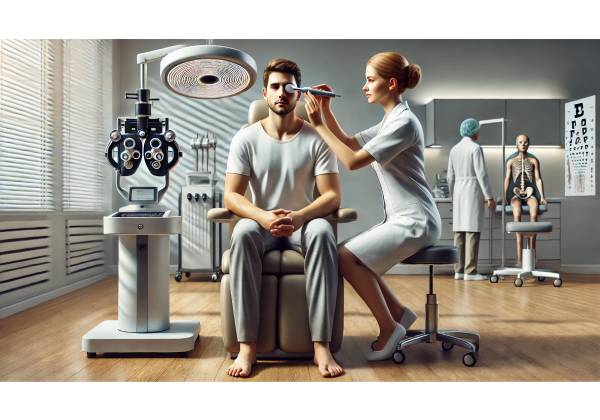
A Closer Look at Acoustic Wave Therapy for Dry Eye Disease
Acoustic wave therapy, sometimes known as shockwave therapy or low-intensity extracorporeal shockwave therapy (Li-ESWT), has gained attention across multiple medical fields due to its noninvasive and regenerative capabilities. While this form of therapy is more commonly associated with conditions like musculoskeletal injuries or erectile dysfunction, emerging studies now explore its potential to address chronic ophthalmologic issues—specifically, dry eye disease.
Dry eye disease, characterized by insufficient tear quality or quantity, can severely impact one’s comfort, vision, and overall quality of life. In the quest for more natural, less pharmacologically dependent solutions, acoustic wave therapy is being considered as a novel approach to stimulate the body’s intrinsic healing mechanisms within the eye’s delicate structures. Through the targeted application of low-intensity sound waves, practitioners aim to enhance local blood flow, improve glandular function, and restore the balance of the tear film.
This approach contrasts with more conventional methods that often focus on artificial tears, prescription eye drops, or surgical interventions. Instead, acoustic wave therapy attempts to harness the body’s own regenerative potential, potentially minimizing reliance on daily medications and invasive procedures. Early-stage clinical trials, pilot studies, and anecdotal patient reports have shown encouraging initial results, prompting further investigation into the technique’s underlying mechanisms, safety, and long-term viability.
Dry Eye Disease: What It Is and Why It Happens
Dry eye disease is a multifactorial condition affecting the tear film that coats the surface of the eye. A stable tear film is essential for clear vision, eye comfort, and overall ocular health. When the tear film is compromised—either through poor tear production, rapid evaporation, or an imbalance in tear composition—symptoms such as redness, irritation, burning, and blurred vision commonly arise.
Causes and Risk Factors:
A range of factors can contribute to dry eye disease. Common culprits include prolonged screen time, which reduces blink frequency; environmental conditions like dry or windy climates; the natural aging process; hormonal changes; and certain medications known to decrease tear production. Autoimmune conditions, including Sjögren’s syndrome, and eyelid disorders like meibomian gland dysfunction (MGD) also play a significant role. MGD, in particular, affects the glands that produce the oily layer of tears, leading to instability and rapid tear evaporation.
Anatomy of the Tear Film:
The tear film is composed of three layers:
- Lipid Layer: Produced by meibomian glands in the eyelids, this oily layer reduces evaporation.
- Aqueous Layer: Secreted by the lacrimal glands, this water-rich layer supplies essential nutrients and moisture.
- Mucin Layer: Derived from goblet cells in the conjunctiva, the mucin layer ensures the tear film spreads evenly across the cornea.
When any of these layers is disrupted, overall tear quality diminishes. The result is discomfort, inflammation, and, if left untreated, potential damage to the ocular surface.
Prevalence and Impact:
Dry eye disease is common, affecting millions of individuals worldwide. The severity ranges from mild irritation to chronic discomfort and substantial visual impairment. Patients often struggle with daily activities like reading, working at a computer, or driving at night. In some cases, persistent dryness can lead to serious complications, including corneal abrasions or ulcers, increasing the urgency for effective, sustainable treatments.
Current management strategies often revolve around symptom control. These include lubricating eye drops, artificial tears, warm compresses, and, in more severe cases, anti-inflammatory medications, punctal plugs, or intense pulsed light therapy. Each method has its limitations. Acoustic wave therapy, by contrast, aims to intervene at a foundational level—stimulating natural tear production and potentially offering longer-lasting relief.
How Acoustic Wave Therapy Boosts Natural Tear Production
Acoustic wave therapy applies low-intensity shockwaves or pressure waves to targeted tissues in the body. Although initially developed for breaking down kidney stones, more refined versions of the technology now use gentler energy levels intended to promote regenerative processes without damaging delicate tissues.
Biological Mechanisms at Play:
The exact mechanisms by which acoustic wave therapy may enhance tear production are still under investigation. However, several plausible explanations exist:
- Enhanced Blood Flow:
Applying low-intensity acoustic waves may cause mild mechanical stress on blood vessels within the eyelids and periorbital area. This stress can prompt vasodilation—the widening of blood vessels—thus improving circulation to meibomian glands, lacrimal glands, and related structures. Increased blood flow delivers more oxygen and nutrients, potentially reviving underperforming glands and supporting healthy tear production. - Stimulating Glandular Activity:
The meibomian and lacrimal glands are vital for creating the oily and aqueous layers of the tear film. Acoustic waves could positively influence the cells within these glands, encouraging them to secrete more balanced tear components. Improved gland function would better stabilize the tear film, reducing the rate of tear evaporation and improving overall eye surface health. - Microenvironmental Adjustments:
Chronic dry eye often involves subclinical inflammation or microscopic tissue damage. Studies of acoustic wave therapy in other fields suggest it may promote the release of growth factors, reduce localized inflammation, and support tissue remodeling. In the ocular region, these effects may translate to improved tear film integrity and more robust tear production over time. - Nerve Stimulation:
The ocular surface and glands that produce tears rely on a network of nerves. By gently stimulating these nerves, acoustic waves might improve the signaling pathways that coordinate tear production. More effective neural communication can lead to increased tear output or better tear quality.
Long-Term Regenerative Potential:
Unlike interventions that treat symptoms temporarily, acoustic wave therapy’s regenerative focus might enable longer-lasting improvements. If the therapy indeed triggers processes akin to tissue healing and regeneration, patients may find themselves relying less on daily artificial tears and other maintenance therapies. More research is needed to confirm these long-term outcomes, but initial findings show promise for a more sustained approach to dry eye management.
Implementing Acoustic Wave Treatment: Steps, Sessions, and Techniques
Considering the delicate structures around the eyes, the application of acoustic wave therapy must be performed with precision and care. Although clinical protocols are still evolving, certain foundational guidelines and steps help ensure a safe and effective treatment experience.
Initial Evaluation and Candidacy:
A thorough eye examination is a prerequisite for acoustic wave therapy. Patients are typically screened to confirm a dry eye diagnosis, often through clinical tests like the Schirmer’s test for tear production, tear breakup time (TBUT) assessments, and ocular surface staining. Advanced imaging techniques, such as meibography, can evaluate the structure of the meibomian glands. By establishing a clear baseline, clinicians can determine whether a patient is a suitable candidate. Patients with certain eyelid conditions, active infections, or severe ocular surface damage may require alternative approaches or adjunct therapies.
Treatment Protocols and Delivery Methods:
In a clinical setting, acoustic wave devices designed for ophthalmic use may employ specialized applicators that deliver gentle, focused waves to the eyelids and periorbital region. The practitioner usually follows a standardized protocol, adjusting settings based on the patient’s tolerance, gland function assessments, and clinical goals. Key elements of the treatment protocol may include:
- Localization:
The therapy is often delivered to the eyelids, particularly along the edges where meibomian glands reside. Precision ensures that the energy is directed at the target area without affecting surrounding delicate tissues. - Intensity and Frequency:
The device’s energy intensity, measured in millijoules or bar pressure, and frequency of the acoustic waves are carefully calibrated. Lower intensities may be used initially to gauge patient comfort and tissue response, followed by slight adjustments in subsequent sessions. - Session Duration and Schedule:
Each treatment session may last several minutes to around 15 minutes per eye, depending on the protocol. Patients might undergo a series of sessions spaced a few weeks apart. The exact number and frequency of sessions can vary, with some protocols involving four to six treatments over a couple of months. - Adjunct Measures:
While acoustic wave therapy may work on its own, pairing it with other supportive measures can enhance results. For example, warm compresses or eyelid hygiene techniques before or after treatment might help loosen meibomian gland blockages, improving the therapy’s overall effectiveness. Nutritional support, like omega-3 fatty acids, and artificial tears can also complement the regenerative effort until natural tear production stabilizes.
Post-Treatment Care:
Patients receiving acoustic wave therapy for dry eye should attend follow-up visits to track progress and monitor any potential side effects. Adjustments to the treatment protocol can be made based on improvement in tear production, reduction in dryness symptoms, and enhanced ocular surface health. Continuous communication between the patient and the healthcare provider ensures that therapy goals remain aligned with patient comfort and overall outcomes.
Evaluating the Safety and Efficacy of Acoustic Wave Therapy
Safety and effectiveness remain top priorities for any new or emerging therapy. While acoustic wave therapy is widely regarded as safe for certain medical applications, its role in eye care is still relatively new. Consequently, gathering safety and efficacy data is a critical step in validating this approach for dry eye disease.
Potential Side Effects:
When delivered properly, acoustic wave therapy is generally considered minimally invasive and comfortable. However, like any procedure, it may carry some risks. Potential side effects could include:
- Mild Redness or Irritation:
The eyelid skin or ocular surface may become slightly red or irritated immediately after treatment. This usually resolves within a few hours. - Temporary Discomfort:
Some patients might experience mild pressure or sensitivity during or right after the session, which typically subsides quickly. - Rare Complications:
Incorrect application or poorly calibrated devices could, in theory, cause tissue damage. This underscores the importance of seeking therapy from a trained and experienced eye care professional.
Efficacy Indicators and Outcome Measures:
Assessing effectiveness involves both objective and subjective measures. Clinicians may use tools like the Schirmer’s test to quantify tear production before and after therapy. Tear osmolarity testing, meibography, and tear film breakup time tests can offer insights into tear quality and gland function. Patients also report on their subjective symptoms, noting improvements in comfort, clarity of vision, and ability to perform daily tasks without eye strain.
Comparisons with Conventional Treatments:
Traditional treatments for dry eye disease often rely on artificial tears, prescription eye drops (such as cyclosporine or lifitegrast), punctal plugs, and lifestyle modifications. While these approaches can provide temporary relief, many patients remain interested in more durable solutions. Acoustic wave therapy, if proven effective through robust clinical trials, may emerge as a complementary or alternative option. Patients who fail to find relief with standard therapies or prefer minimally invasive interventions may find this appealing.
Regulatory Approvals and Guidelines:
Since acoustic wave therapy for dry eye is still an emerging application, regulatory bodies are closely monitoring clinical outcomes. Providers must adhere to safety guidelines, device standards, and ethical principles. Over time, as more data become available, clinical guidelines and best practices will likely be established, making it easier for eye care professionals to integrate this therapy into mainstream treatment plans.
Latest Research and Clinical Evidence Behind Acoustic Wave Interventions
Acoustic wave therapy’s role in dry eye disease remains in the investigative stage. While the concept is backed by principles observed in other applications—like improved microcirculation and tissue regeneration—formal research specific to dry eye is still developing.
Pilot Studies and Early Clinical Trials:
A handful of early-stage trials are exploring whether acoustic wave therapy improves tear production and gland function. Some studies evaluate patients with meibomian gland dysfunction, investigating whether gentle shockwaves can unclog glands and promote healthier oil secretion. Early results suggest modest improvements in tear film stability and patient-reported symptom relief. However, the sample sizes in these studies are often small, and the lack of long-term follow-up data means more research is necessary before drawing definitive conclusions.
Mechanistic Research and Animal Models:
To better understand how acoustic waves affect ocular tissues, researchers have turned to laboratory settings and animal models. By examining treated tissues under the microscope, they aim to identify cellular changes—such as increases in growth factor release or structural changes in glandular tissue—that might explain improved tear production. Understanding these mechanisms can guide refinements in therapy protocols, device settings, and patient selection criteria.
Collaborative Efforts and Data Sharing:
The advancement of this therapy depends on widespread collaboration. Ophthalmologists, optometrists, biomedical engineers, and researchers in regenerative medicine are sharing insights and pooling resources. Professional conferences, peer-reviewed journals, and clinical trial registries provide platforms for disseminating findings and building a robust scientific consensus.
Next Steps and Evolving Protocols:
As promising as the early data may be, acoustic wave therapy for dry eye has a long way to go before it becomes a standard treatment. Ongoing research endeavors focus on refining device design for ocular use, optimizing energy settings and treatment intervals, and comparing outcomes against established therapies. Longitudinal studies will track patients over months or years to confirm whether improvements are stable, reproducible, and cost-effective.
Cost Considerations and Making Acoustic Wave Therapy Accessible
While acoustic wave therapy has the potential to revolutionize how we address dry eye disease, it is crucial to consider cost and accessibility. As with many advanced treatments, initial expenses may be high, and availability may vary depending on geographic location and healthcare infrastructure.
Factors Influencing Cost:
Acoustic wave devices are specialized medical equipment that often require a significant investment by eye clinics or ophthalmology practices. Costs may encompass the purchase or leasing of devices, maintenance fees, staff training, and potential licensing arrangements. Additionally, each treatment session involves professional time, clinic overhead costs, and follow-up care. Together, these factors can raise the price of therapy sessions.
Insurance Coverage and Reimbursement:
Many new therapies face initial hurdles in securing insurance coverage. Without an established track record of efficacy supported by large-scale clinical trials, insurers may be reluctant to cover acoustic wave therapy for dry eye. Patients might have to pay out-of-pocket until evidence accumulates to justify reimbursement. Over time, if acoustic wave therapy proves to be both safe and effective, insurers may re-evaluate their policies and consider coverage.
Comparing Costs to Standard Therapies:
Traditional dry eye management often involves relatively affordable measures: lubricating drops, warm compresses, or nutrient supplements. Even prescription drops and punctal plugs may carry more predictable costs. Acoustic wave therapy, if priced significantly higher, may seem less accessible to some patients. However, if proven to provide longer-lasting relief, the upfront expense might be offset by reduced reliance on ongoing medications or repeated office visits.
Global Accessibility and Market Factors:
Healthcare resources and standards vary worldwide. While high-end medical centers in urban areas of wealthy nations may be early adopters, patients in resource-limited settings might find acoustic wave therapy harder to access. Expanding availability may require technology transfer, training local professionals, and exploring cost-reduction strategies. Additionally, competitive pressures as more manufacturers enter the market could drive down device and treatment costs.
Patient Education and Advocacy:
Patients and advocacy groups play a crucial role in shaping the future of treatments like acoustic wave therapy. By actively participating in clinical trials, sharing experiences, and voicing concerns, patients can guide healthcare providers, researchers, and insurers to focus on what matters most—improving health and well-being. Over time, patient-led advocacy may help lower barriers to entry, encourage insurers to consider coverage, and push for standardized guidelines that ensure everyone who could benefit has a pathway to access.
Disclaimer: This article is for informational purposes only and does not replace professional medical advice. Always consult a qualified healthcare provider regarding any questions or concerns related to medical conditions or treatments.


