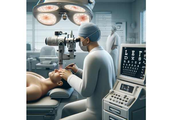
Epithelial Basement Membrane Dystrophy (EBMD), also known as map-dot-fingerprint dystrophy, is one of the most common corneal surface disorders, often leading to fluctuating vision, discomfort, and, in some cases, recurrent erosions. Understanding this condition’s nuances is vital for patients and eye care professionals alike. In this in-depth guide, we’ll explore every aspect of EBMD, from its causes and risk factors to today’s most effective conventional and surgical treatments, as well as the future of innovative care. Whether you’re seeking practical advice or clinical insight, this article provides authoritative yet accessible information to support your journey toward clearer, more comfortable vision.
Table of Contents
- Condition Background and Population Insights
- Evidence-Based Non-Surgical and Medication Options
- Operative Approaches and Interventional Advancements
- New Discoveries and Technological Breakthroughs
- Research Frontiers and the Future of Care
- Frequently Asked Questions
- Disclaimer
Condition Background and Population Insights
Epithelial Basement Membrane Dystrophy is a non-inflammatory disorder affecting the outermost layer of the cornea—the transparent front window of the eye. In EBMD, abnormalities develop in the basement membrane, the structure responsible for anchoring the corneal epithelium (surface cells) in place. These irregularities lead to characteristic map-like, dot-like, or fingerprint patterns seen under magnification.
Understanding EBMD’s Pathophysiology
- The corneal epithelium is kept smooth and clear by a healthy basement membrane. In EBMD, this membrane becomes thickened, wavy, or multi-layered, causing the epithelium to adhere poorly.
- As a result, tiny areas of the epithelium may become loose or lifted, which can cause visual symptoms and, at times, recurrent corneal erosions (painful breakdowns of the corneal surface).
Who Is Affected?
- EBMD is the most common corneal dystrophy, affecting about 2% of the general population, with higher prevalence in people over 40.
- Both men and women are equally susceptible, although symptoms often become more pronounced with age.
- Many individuals with EBMD remain asymptomatic, discovering the condition incidentally during routine eye exams.
Risk Factors
- Age: Incidence rises with advancing age.
- Family history: A genetic predisposition is suspected, though most cases are sporadic.
- Previous eye surgery or trauma: Procedures such as LASIK or cataract surgery can unmask or worsen EBMD.
- Dry eye disease: Poor ocular surface health can exacerbate symptoms.
Practical Takeaways
- Not every person with EBMD will experience symptoms; proactive eye care and regular exams are essential, especially before elective procedures like refractive surgery.
- If you’re experiencing frequent eye discomfort upon waking, blurry vision, or unexplained tearing, ask your eye doctor about EBMD.
Evidence-Based Non-Surgical and Medication Options
Many individuals with Epithelial Basement Membrane Dystrophy manage their condition successfully without surgery. Non-invasive therapies are especially effective for mild cases and for preventing recurrent corneal erosions.
Conservative and Pharmacological Interventions
- Lubricating Eye Drops and Ointments:
- Preservative-free artificial tears are the first-line therapy for EBMD. Used several times daily, they help smooth the ocular surface and reduce discomfort.
- At bedtime, thick lubricating ointments create a barrier, minimizing epithelial breakdown overnight.
- Hypertonic Saline Solutions and Ointments:
- Hypertonic saline (5%) drops or ointment draw excess fluid from the cornea, promoting adhesion of the epithelial cells to the underlying basement membrane.
- These are particularly helpful for patients experiencing recurrent erosions, especially upon waking.
- Anti-inflammatory Therapies:
- Short courses of mild topical corticosteroids may be prescribed for acute inflammation, though these are not a long-term solution due to potential side effects.
- Cyclosporine or lifitegrast eye drops are used in select cases to address underlying ocular surface inflammation.
- Bandage Contact Lenses:
- Special soft lenses may be worn short-term to protect the healing cornea, relieve pain, and promote regeneration after erosions.
- Strict hygiene and close follow-up are crucial to avoid infection.
Additional Supportive Measures
- Lid Hygiene: Gentle cleansing of the eyelid margins with diluted baby shampoo or commercial lid scrubs can reduce inflammation and bacterial load, supporting a healthier corneal surface.
- Lifestyle Adjustments:
- Avoid rubbing your eyes, which can disrupt the fragile epithelium.
- Use a humidifier in dry environments to prevent corneal dehydration.
- Wear sunglasses to shield eyes from wind, dust, and UV exposure.
Practical Self-Care Tips
- For those with morning discomfort, keep artificial tears and ointment on your nightstand for immediate use upon waking.
- Consider a consistent bedtime routine that includes eyelid hygiene and lubrication to lower the risk of overnight erosions.
Effectiveness and Limitations
- While most patients respond well to these therapies, some may experience persistent symptoms, especially if they have frequent erosions or blurred vision interfering with daily life.
- Early intervention helps minimize complications, so maintaining open communication with your eye care provider is essential.
Operative Approaches and Interventional Advancements
When conservative treatments are inadequate, or recurrent erosions significantly impact quality of life, surgical and interventional procedures are considered. Advances in corneal surgery have dramatically improved outcomes for EBMD patients.
Office-Based Procedures
- Debridement:
- The simplest and most common procedure, debridement involves gently removing the loose epithelium and irregular basement membrane, allowing healthy new tissue to regrow.
- It is performed under topical anesthesia in the office, with minimal discomfort.
- A bandage contact lens is typically applied post-procedure to protect the healing surface.
- Diamond Burr Polishing:
- After debridement, a special burr may be used to smooth the underlying corneal surface, reducing the risk of recurrence and improving visual quality.
- This procedure is safe, effective, and particularly beneficial for those with frequent erosions.
Advanced Surgical Interventions
- Phototherapeutic Keratectomy (PTK):
- This laser procedure removes the abnormal basement membrane and smooths the corneal surface at a microscopic level.
- PTK offers excellent outcomes for both vision and comfort, and is the gold standard for severe or refractory EBMD.
- Performed under local anesthesia, PTK requires careful postoperative care but boasts a low risk of complications.
- Anterior Stromal Puncture:
- Tiny punctures are made in the Bowman’s layer of the cornea with a fine needle, encouraging the epithelium to adhere more securely.
- This technique is used when PTK is not available or suitable.
Pre- and Post-Surgical Considerations
- Preoperative Evaluation: Detailed imaging and mapping of the cornea ensure accurate diagnosis and help tailor the procedure to each patient’s needs.
- Postoperative Care: Frequent lubrication, antibiotic drops, and sometimes mild steroids are essential for a smooth recovery. Follow all instructions carefully and attend scheduled checkups.
Potential Risks and Complications
- Infection, haze, or delayed healing are rare but possible; prompt reporting of worsening symptoms is crucial.
- Most surgical patients achieve significant, long-lasting improvement in comfort and vision.
Practical Advice for Recovery
- Rest your eyes and limit screen time during the initial healing period.
- Protect your eyes from injury and avoid swimming until cleared by your ophthalmologist.
New Discoveries and Technological Breakthroughs
The landscape of EBMD management continues to evolve, with research and technology driving better outcomes, safer procedures, and novel therapies.
Innovative Diagnostic Tools
- High-Resolution Anterior Segment Optical Coherence Tomography (AS-OCT):
- Provides cross-sectional imaging of the corneal layers, aiding in precise diagnosis and monitoring of EBMD.
- Allows for early detection and personalized treatment planning.
- In Vivo Confocal Microscopy:
- Enables real-time visualization of corneal microstructures, facilitating research into the disease process and identifying subtle abnormalities.
Advances in Laser and Surgical Technology
- Femtosecond Laser-Assisted Procedures:
- Allow for ultra-precise removal of abnormal tissue, minimizing damage to healthy structures and reducing recovery time.
- Customized PTK Algorithms:
- Emerging techniques use detailed corneal mapping to tailor laser treatments for each patient’s unique pattern of irregularity.
Cell-Based and Molecular Therapies
- Research into growth factors, stem cells, and biologic agents is paving the way for non-invasive regenerative therapies to restore a healthy basement membrane.
- Gene Editing:
- While not yet in clinical use, early research into gene therapy holds potential for correcting the underlying defects in hereditary forms of EBMD.
Artificial Intelligence in Diagnosis and Prognosis
- AI-powered software is being developed to analyze corneal images, identify early EBMD changes, and predict the risk of progression or recurrence after surgery.
Telemedicine and Digital Monitoring
- Remote assessment tools enable patients to monitor symptoms and healing after procedures, improving access to specialist care and reducing unnecessary clinic visits.
What Patients Should Know
- Ask your eye care provider about the availability of advanced diagnostic imaging if you’re planning surgery or experiencing ongoing symptoms.
- Keep up with new technologies, as many are rapidly becoming accessible in specialty eye centers.
Research Frontiers and the Future of Care
As our understanding of EBMD deepens, new frontiers in research are poised to revolutionize how the condition is managed and treated.
Current and Emerging Clinical Trials
- Next-Generation Lubricants:
- Studies are underway evaluating new artificial tear formulations designed to promote epithelial health and reduce inflammation more effectively.
- Novel Drug Delivery Systems:
- Investigational products include nanoemulsions, sustained-release inserts, and contact lenses embedded with slow-release medications, aimed at providing long-term relief.
- Biologics and Growth Factors:
- Early-phase trials are testing agents that stimulate healthy basement membrane regeneration, targeting the underlying cause of EBMD.
- Genetic and Stem Cell Therapies:
- Scientists are exploring ways to correct or replace faulty genes and cells responsible for dystrophy development.
Understanding the Disease at a Molecular Level
- Research is focusing on identifying the specific genetic mutations and molecular pathways that cause basement membrane abnormalities, potentially enabling pre-symptomatic diagnosis or even preventive care.
Personalized and Predictive Medicine
- The integration of genetic profiling, AI algorithms, and advanced imaging is paving the way for truly individualized treatment plans—matching the right therapy to the right patient at the right time.
Global Collaboration and Patient Engagement
- International research networks are accelerating progress by sharing data, pooling resources, and fostering patient participation in clinical trials.
What the Future Holds
- Within the next decade, patients can expect more effective, less invasive therapies; better prevention of recurrent erosions; and a growing role for digital health in managing chronic eye conditions like EBMD.
How Patients Can Contribute
- Consider joining clinical trials or patient registries if you are eligible.
- Stay informed about research developments and advocate for access to emerging treatments.
Frequently Asked Questions
What is the most effective treatment for epithelial basement membrane dystrophy?
The most effective treatment depends on severity. Mild cases respond to lubricating drops and ointments, while recurrent erosions may require office-based debridement or laser procedures like phototherapeutic keratectomy (PTK) for lasting relief and vision improvement.
Can EBMD go away on its own?
EBMD does not go away, but symptoms can fluctuate. Many people have long symptom-free periods, while others may require ongoing care to manage discomfort or recurrent erosions. Consistent use of artificial tears and regular checkups help maintain eye health.
Is epithelial basement membrane dystrophy hereditary?
EBMD can have a hereditary component, though most cases are sporadic. A family history of corneal dystrophies may increase risk, but genetics are not the only factor. If relatives have corneal problems, discuss this with your eye care provider.
Does EBMD affect vision permanently?
EBMD often causes fluctuating vision, but permanent vision loss is rare with proper care. Regular management and prompt treatment of erosions prevent scarring and long-term complications. Most patients maintain good vision with appropriate therapy.
Are there risks with EBMD surgery?
Surgical procedures for EBMD are generally safe and effective, with a low risk of complications such as infection, haze, or delayed healing. Following all post-operative instructions and attending follow-up visits helps ensure the best possible outcome.
Can I have LASIK if I have EBMD?
EBMD may increase the risk of complications from LASIK or other refractive surgeries. A thorough preoperative evaluation is essential. Your surgeon may recommend treating the EBMD first or choosing an alternative procedure to ensure safety and optimal results.
How can I prevent recurrent corneal erosions in EBMD?
Preventing recurrent erosions involves regular use of lubricating drops and ointments, avoiding eye rubbing, and following a strict eyelid hygiene routine. For frequent erosions, procedures like debridement or PTK may offer long-term relief.
Disclaimer
The information presented in this article is intended for educational purposes only and is not a substitute for professional medical advice, diagnosis, or treatment. Always consult your ophthalmologist or healthcare provider with questions about your symptoms or treatment options. Never ignore or delay seeking professional advice due to content from this guide.
If you found this guide helpful, please consider sharing it on Facebook, X (formerly Twitter), or your favorite social media platform. Your support enables us to continue producing reliable, high-quality health information for everyone. Follow us for more updates and thank you for being part of our community!


