Primary congenital glaucoma (PCG) is a rare but severe type of glaucoma that appears at birth or develops soon after. It is distinguished by abnormal development of the eye’s drainage system, specifically the trabecular meshwork and Schlemm’s canal, which results in elevated intraocular pressure (IOP). This elevated IOP can damage the optic nerve, potentially resulting in vision loss or blindness if not treated immediately.
PCG usually appears within the first year of life and is characterized by clinical symptoms such as enlarged eyes (buphthalmos), corneal clouding, excessive tearing (epiphora), and light sensitivity (photophobia). These symptoms occur as a result of increased pressure within the eye, causing the cornea to stretch and become hazy. A comprehensive eye examination is typically used to make a diagnosis, which includes measuring IOP, assessing corneal diameter, and evaluating the optic nerve head. Gonioscopy, which visualizes the anterior chamber angle, can also help confirm the diagnosis.
The condition is genetically heterogeneous, which means it can be caused by mutations in a variety of genes, with CYP1B1 being the most commonly linked. Understanding the genetic basis of PCG is critical for accurate diagnosis, genetic counseling, and possible future gene-targeted therapies.
Primary Congenital Glaucoma Management and Treatment
Primary congenital glaucoma management and treatment necessitate a multifaceted approach that includes surgical intervention, medical therapy, and ongoing monitoring in order to prevent vision loss and maintain ocular health.
Surgical Interventions
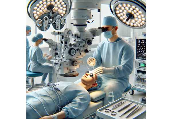
Surgery is the primary treatment for PCG because it directly addresses the structural abnormalities that cause high IOP. The main surgical options are:
- Goniotomy: This procedure involves making an opening in the trabecular meshwork to allow aqueous humor outflow. A goniotomy knife or laser are typically used, with a goniolens providing direct visualization.
- Trabeculotomy: Trabeculotomy, like goniotomy, involves incising the trabecular meshwork and Schlemm’s canal to improve fluid drainage. This procedure is frequently used when goniotomy is not feasible or has failed.
- Combined Trabeculotomy-Trabeculectomy: This method combines trabeculotomy and trabeculectomy, removing a small portion of the trabecular meshwork and surrounding sclera to create a new drainage pathway. It is used for more complex or refractory cases.
- Drainage Devices: Glaucoma drainage devices (shunts) may be implanted to provide an alternative pathway for aqueous humor outflow. These devices are especially useful in eyes with extensive scarring or after previous surgeries have failed.
Medical Therapy
While surgery is the primary treatment option for PCG, medical therapy can help manage IOP before and after surgery. Medications frequently used include:
- Beta-Blockers: These medications reduce aqueous humor production, which lowers IOP. Timolol is a commonly used beta-blocker for pediatric glaucoma.
- Carbonic Anhydrase Inhibitors: Both oral and topical forms of these medications reduce aqueous humor production. Acetazolamide and dorzolamide are examples of PCG medications.
- Prostaglandin Analogs: These medications stimulate uveoscleral outflow of aqueous humor. One such drug for older children is latanoprost.
- Alpha Agonists: These drugs decrease aqueous humor production while increasing uveoscleral outflow. Brimonidine is one example, though it should be used with caution due to potential side effects in infants and young children.
Post-operative Care and Monitoring
Postoperative care is critical for ensuring the success of surgical procedures and avoiding complications. This includes:
- Regular Follow-Up Visits: Frequent examinations are required to monitor IOP, evaluate surgical site function, and detect early signs of complications.
- Topical Medications: Anti-inflammatory and antibiotic eye drops are commonly prescribed after surgery to prevent infection and inflammation.
- Complication Monitoring: Potential complications, such as infection, scarring, or surgical procedure failure, must be identified and managed promptly.
Innovative Treatments for Primary Congenital Glaucoma
Recent advances in medical research and technology have significantly improved the treatment landscape for primary congenital glaucoma. These cutting-edge developments provide more effective, safer, and minimally invasive treatments for this difficult condition.
Minimal Invasive Glaucoma Surgery (MIGS)
Minimally invasive glaucoma surgery (MIGS) has transformed the treatment of many types of glaucoma, including PCG. These procedures aim to reduce IOP with less tissue disruption, fewer complications, and shorter recovery times than traditional surgeries. The key innovations in MIGS for PCG are:
- Microcatheter-Assisted Trabeculotomy: This technique circumnavigates Schlemm’s canal with a microcatheter, allowing for a more precise incision and greater trabecular meshwork opening. Studies have yielded promising results in terms of sustained IOP reduction.
- Gonioscopy-Assisted Transluminal Trabeculotomy (GATT): GATT entails threading a suture through Schlemm’s canal and then pulling it through to form a circular incision. This procedure improves aqueous outflow and has shown success in pediatric patients.
Laser-based Treatments
To improve precision and outcomes, glaucoma surgery now incorporates laser technology. Laser treatments for PCG include:
- Laser Goniotomy: This procedure uses a laser to create an opening in the trabecular meshwork. The precision of the laser reduces tissue damage and promotes faster healing.
- Cyclophotocoagulation: This laser procedure targets the ciliary body and reduces aqueous humor production. It is commonly used in refractory cases where conventional surgery has failed.
Advancements in Imaging and Diagnostic Tools
Accurate diagnosis and monitoring are essential for successful PCG management. Recent advances in imaging and diagnostic tools have increased the ability to detect and assess the severity of the condition.
- Anterior Segment Optical Coherence Tomography (AS-OCT): This non-invasive imaging technique produces high-resolution cross-sectional images of the anterior segment, which includes the trabecular meshwork and Schlemm’s Canal. AS-OCT aids in the diagnosis and preoperative planning of PCG.
- Ultrasound Biomicroscopy (UBM): UBM provides detailed visualization of anterior segment structures, allowing for precise assessment of anatomical abnormalities and surgical outcomes.
Gene and Molecular Therapies
Understanding the genetic basis of PCG has paved the way for future gene-targeted therapies. Research in this area is still in the early stages, but it shows promise for future treatment options:
- Gene Therapy: This method entails delivering functional copies of defective genes or silencing mutated genes to restore normal function. Gene therapy for PCG aims to address the underlying genetic causes, potentially leading to a long-term cure.
- CRISPR-Cas9 Gene Editing: CRISPR-Cas9 technology enables the precise modification of specific genes. Its application in correcting genetic mutations associated with PCG is under investigation.
Stem Cell Therapy
Stem cell therapy is another novel approach to treating PCG. Stem cells have the ability to regenerate damaged tissues and promote healing.
- Stem Cell-Derived Trabecular Meshwork Cells: Researchers are looking into the use of stem cells to create trabecular meshwork cells that can be implanted in the eye to restore normal aqueous outflow.
- Mesenchymal Stem Cells (MSCs): MSCs have immunomodulatory and regenerative properties that can help repair damaged ocular tissues and reduce inflammation.
Artificial Intelligence, Machine Learning
Artificial intelligence (AI) and machine learning are transforming ophthalmology, including the management of PCG. AI algorithms can analyze large datasets of imaging studies, clinical records, and genetic information to identify patterns and predict treatment outcomes:
- AI-Based Diagnostic Tools: AI algorithms can analyze imaging data to accurately diagnose PCG and determine the condition’s severity. These tools provide ophthalmologists with useful insights, allowing for more precise treatment planning.
- Predictive Modeling: Machine learning models can predict the efficacy of different treatment options based on individual patient characteristics. This aids in tailoring treatment plans for optimal results.
Telemedicine & Remote Monitoring
The COVID-19 pandemic has accelerated the adoption of telemedicine and remote monitoring technologies, opening up new opportunities for managing PCG.
- Virtual Consultations: Telemedicine platforms enable eye care professionals to provide virtual consultations, allowing patients to receive expert care from the comfort of their own homes. This approach is especially useful for routine follow-ups and initial assessments.
- Remote Monitoring Tools: Smartphone apps and wearable devices can track IOP and other relevant metrics in real time, providing valuable information to healthcare providers and allowing for timely interventions.
Future Prospects: Nanotechnology
Nanotechnology is a rapidly developing field with potential applications in the treatment of PCG. Nanoparticles can be engineered to deliver targeted therapies directly to the site of obstruction, increasing treatment efficacy while reducing side effects. Researchers are working to create nanoparticle-based drug delivery systems that can improve the outcomes of both medical and surgical treatments for PCG.


