Presbyopia, a common age-related condition, impairs the eye’s ability to focus on nearby objects. It is a natural part of the aging process and usually appears in people in their mid-40s to early 50s. The term “presbyopia” is derived from the Greek words “presbys,” meaning “old man,” and “ops,” meaning “eye,” indicating its prevalence in older adults. Presbyopia, unlike other refractive errors such as myopia (nearsightedness) and hyperopia (farsightedness), is caused by the gradual loss of flexibility in the lens rather than the shape of the eye itself.
Anatomy and Physiology of the Eye
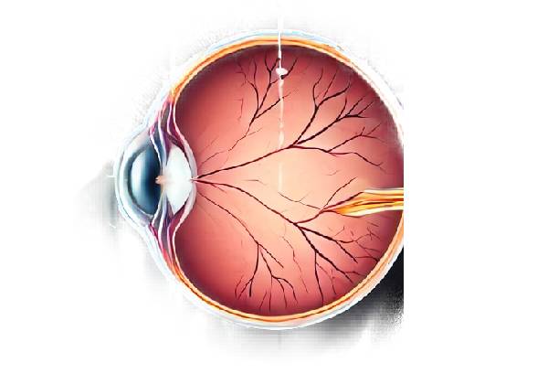
Understanding presbyopia requires a fundamental understanding of the eye’s anatomy and function. The eye is a complex organ with multiple structures that work together to provide clear vision. The cornea, lens, and ciliary muscles play critical roles in focusing light onto the retina.
- Cornea: The clear front part of the eye that refracts light entering the eye.
- Lens: A flexible, transparent structure located behind the iris that changes shape to direct light to the retina.
- Ciliary Muscles: These muscles surround the lens and change its shape by contracting and relaxing, allowing the eye to focus on objects of varying distances.
When the eye focuses on a nearby object, the ciliary muscles contract, making the lens thicker and more convex. This process, known as accommodation, enables the eye to sharply bend light rays and focus them on the retina, resulting in clear vision.
Pathophysiology of Presbyopia
Presbyopia develops as the lens’s flexibility decreases and the ciliary muscles weaken. Several factors contribute to the age-related change:
- Lens Hardening: As the lens ages, it becomes more rigid and less capable of changing shape. The accumulation of lens fibers and proteins causes this hardening, which reduces its elasticity.
- Ciliary Muscle Weakening: The ciliary muscles lose strength and the ability to contract effectively, making it more difficult for the lens to change shape.
- Lens Growth: The lens grows throughout life, resulting in an increase in thickness and size. This expansion restricts the space available for the lens to move and change shape.
As a result of these changes, the eye struggles to focus on nearby objects, making them appear blurry. The loss of near vision is the hallmark of presbyopia.
Symptoms of Presbyopia
Presbyopia develops gradually and is frequently discovered while reading small print or working on tasks that require close attention. Common symptoms include:
- Blurry Near Vision: Difficulty seeing objects up close, such as books, phones, or computer screens.
- Eye Strain: Eye discomfort or fatigue due to prolonged close-up work.
- Headache: Frequent headaches, particularly after activities that require concentration on nearby objects.
- Need for Brighter Light: A greater reliance on brighter lighting for reading or other close-up tasks.
- Holding Objects at Arm’s Length: The habit of holding reading materials at a greater distance in order to see them better.
Onset and Progress
Presbyopia typically begins around the age of 40 and progresses until about the age of 65. The rate of progression varies, but most people will notice a significant decline in near vision over the course of 10 to 20 years. The severity of symptoms can also vary depending on factors such as lighting, fatigue, and general eye health.
Risk Factors
While presbyopia is primarily age-related and affects almost everyone at some point, certain factors can influence its onset and progression.
- Age is the primary risk factor, as the lens hardens naturally and the ciliary muscles weaken with age.
- Genetics: Having a family history of presbyopia or other refractive errors increases the risk of developing the condition.
- Occupation: Jobs that require prolonged close proximity, such as reading, computer use, or fine detail work, can aggravate symptoms.
- Health Conditions: Diabetes and cardiovascular disease are two systemic conditions that can have an impact on eye health and contribute to presbyopia.
- Medications: Certain medications, such as antidepressants, antihistamines, and diuretics, can impair the eye’s focusing ability and hasten the development of presbyopia.
Impact on Daily Life
Presbyopia can have a significant impact on a person’s quality of life because it interferes with daily activities that require near vision. Reading, writing, using digital devices, and participating in hobbies like sewing or crafting can be difficult. The need for frequent breaks, additional lighting, or corrective lenses can all be inconvenient and frustrating. In severe cases, presbyopia can have an impact on job performance and productivity, especially in professions that require a lot of close-up work.
Identifying Presbyopia from Other Refractive Errors
It is critical to distinguish presbyopia from other refractive errors because the underlying causes and treatment options differ. Presbyopia, as opposed to myopia, hyperopia, and astigmatism, which are caused by irregularities in the shape of the eye, is associated with a loss of lens elasticity and ciliary muscle function. Individuals with preexisting refractive errors may still develop presbyopia, necessitating alternative or additional corrective measures.
Prevention and Delays
While presbyopia is an unavoidable part of aging, specific lifestyle choices and habits can help maintain overall eye health and potentially delay its onset:
- Regular Eye Exams: Routine eye exams can detect early signs of presbyopia and other eye conditions, allowing for timely treatment.
- Healthy Diet: A well-balanced diet rich in vitamins A, C, and E, as well as omega-3 fatty acids, can help maintain eye health.
- Protective Eyewear: Wearing UV-blocking sunglasses and protective goggles during activities that pose a risk to eye health can help prevent damage.
- Adequate Lighting: Having enough lighting while reading or working can help reduce eye strain and discomfort.
- Breaks from Screen Time: The 20-20-20 rule, which states that every 20 minutes, take a 20-second break to look at something 20 feet away. This can help reduce digital eye strain.
- Hydration: Keeping the eyes lubricated by staying hydrated and using artificial tears as needed can help relieve dryness and irritation.
Methods to Diagnose Presbyopia
A comprehensive eye examination is required to diagnose presbyopia and determine the severity of the condition. Several diagnostic methods are used to correctly diagnose presbyopia and distinguish it from other refractive errors and eye conditions.
Comprehensive Eye Examination
A thorough eye exam by an optometrist or ophthalmologist is the first step in diagnosing presbyopia. The examination typically includes:
- Patient History: The eye care professional will obtain a thorough medical and ocular history, including any symptoms the patient is experiencing, their duration, and any factors that aggravate or alleviate the symptoms. Also gathered is information about the patient’s occupation, lifestyle, and family history of eye conditions.
- Visual Acuity Test: This test determines the sharpness of the patient’s vision at different distances. Using a Snellen chart, the patient reads letters of decreasing size to determine their visual acuity. The test evaluates both distance and near vision, detecting any deficiencies in focusing ability.
- Refraction Assessment: The refraction test determines the patient’s precise prescription for corrective lenses. Using a phoropter, the eye care professional presents a series of lens options to the patient, who selects the lenses that provide the clearest vision. This procedure aids in the identification of any existing refractive errors, including myopia, hyperopia, and astigmatism, in addition to presbyopia.
- Near Vision Test: This test evaluates the patient’s ability to focus on close objects in order to diagnose presbyopia. The patient reads from a near vision chart at reading distance, and the eye care professional measures the smallest line of text that the patient can clearly read.
- Accommodation Testing: This test assesses the eye’s ability to shift focus from far away to near objects. The eye care professional may use a small, handheld device known as an accommodometer or perform subjective tests such as focusing on objects at various distances.
- Slit-Lamp Examination: A slit-lamp microscope is used to examine the eye’s structures at high magnification. This examination detects any abnormalities in the cornea, lens, or anterior segment that may contribute to vision problems.
Additional Diagnostic Tests
Additional tests may be required in some cases to rule out other conditions and confirm the diagnosis of presbyopia:
- Cycloplegic Refraction: This test uses eye drops to temporarily paralyze the ciliary muscles, which prevents accommodation. The eye care professional can accurately assess the patient’s prescription by measuring the refractive error without taking into account accommodation.
- Autorefraction: An autorefractor is an automated device that analyzes how light reflects off the retina to determine the eye’s refractive error. While autorefraction is not a replacement for a manual refraction assessment, it can provide a quick and objective measurement of the patient’s focusing ability.
- **Pupil *Examination*: The eye care professional looks at the size, shape, and response of the pupils to light and accommodation. Abnormal pupil function can indicate other ocular or neurological conditions that can impair vision.
Differential Diagnosis
It is critical to distinguish presbyopia from other refractive errors and eye conditions in order to ensure accurate diagnosis and treatment. The differential diagnosis can include:
- Myopia (Nearsightedness): Difficulty seeing distant objects clearly while near vision remains normal. Unlike presbyopia, myopia is caused by the eye’s shape, causing light to focus in front of the retina.
- Hyperopia (Farsightedness): Difficulty seeing nearby objects clearly, whereas distant vision is superior. Hyperopia occurs when the eye’s shape causes light to focus behind the retina.
- Astigmatism: Blurred vision at all distances caused by an irregularly shaped cornea or lens, which causes light to focus unevenly on the retina.
- Cataracts: Clouding of the eye’s natural lens, which causes blurred vision, glare, and difficulty seeing in low light conditions. Cataracts are typically associated with aging, but their pathology and treatment differ from that of presbyopia.
- Dry Eye Syndrome: Symptoms such as burning, itching, and blurred vision caused by insufficient or low-quality tears. Dry eye syndrome requires different management strategies than presbyopia.
Presbyopia Management
Managing presbyopia focuses on improving near vision and reducing symptoms in order to improve quality of life. Various corrective options and lifestyle changes can help manage presbyopia effectively.
Corrective Lenses
- Reading Glasses: Simple magnifying lenses used only for close-up tasks like reading or sewing. They are available over the counter in a variety of strengths or can be custom-made using a prescription.
- Bifocal Glasses: Glasses with lenses that have two distinct optical powers, typically the upper part for distance vision and the lower part for near vision. The line separating the two sections is visible.
- Trifocal Glasses are similar to bifocals but have three sections for distance, intermediate, and near vision. They are useful for activities that require clear vision at varying distances, such as computer use and reading.
- Progressive Lenses: Also known as no-line bifocals, these lenses offer a smooth transition between distance, intermediate, and near vision, with no visible lines. Many users prefer the natural visual experience that they provide.
Contact lenses
- Monovision Lenses: One eye has a lens for distance vision, while the other has a lens for near vision. The brain adapts to use each eye for its intended purpose, but some people may struggle to adjust.
- Bifocal and Multifocal Contact Lenses: These lenses have distinct zones for distance and near vision, similar to bifocal and progressive eyeglass lenses. They are available in both soft and rigid gas-permeable materials.
- Extended Depth of Focus (EDOF) Lenses: These lenses provide a continuous range of vision from distance to near with no distinct zones, resulting in a more consistent visual experience.
Surgical Options
- Refractive Surgery: Procedures such as LASIK, PRK, and SMILE can be used to achieve monovision by correcting one eye for distance and the other for near vision.
- Corneal Inlays: Tiny devices implanted into the cornea to improve close-up vision by increasing depth of focus. Examples include the Kamra inlay and the Raindrop Near Vision Inlay.
- Lens Implants: The replacement of the eye’s natural lens with a multifocal or accommodative intraocular lens (IOL) during or after cataract surgery. These IOLs have multiple focusing points, which improve near, intermediate, and distance vision.
Lifestyle Adjustments
- Adequate Lighting: Using proper lighting when reading or performing close-up tasks can reduce eye strain and improve visual clarity.
- Visual Breaks: Taking regular breaks from prolonged near work, such as the 20-20-20 rule, can help reduce eye fatigue.
- Ergonomic Adjustments: Place reading materials and computer screens at an appropriate distance and angle to reduce eye strain.
Regular Eye Exams
Regular eye exams are essential for monitoring vision changes and updating prescriptions as needed. Eye care professionals can also identify and treat any coexisting eye conditions that may impair vision.
Assistive devices
- Magnifiers: Handheld or stand magnifiers can be useful for detailed tasks like reading fine print or sewing.
- Electronic Reading Aids: E-readers and tablets with adjustable font sizes and brightness can make reading easier for people with presbyopia.
Trusted Resources and Support
Books
- “Presbyopia: Cause and Correction” by Ronald A. Schachar
- This book provides an in-depth look at the underlying causes of presbyopia and explores various correction methods, offering valuable insights for both patients and eye care professionals.
- “The Eye Book: A Complete Guide to Eye Disorders and Health” by Gary H. Cassel
- A comprehensive resource covering a wide range of eye conditions, including presbyopia, with practical advice on maintaining eye health and seeking appropriate treatment.
Organizations
- American Academy of Ophthalmology (AAO)
- Website: www.aao.org
- The AAO offers extensive resources on eye conditions, including presbyopia, with patient education materials, research updates, and professional guidelines.
- American Optometric Association (AOA)
- Website: www.aoa.org
- The AOA provides valuable information on eye care, vision health, and presbyopia, along with resources to find qualified optometrists and seek appropriate treatment.


