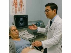
Aortic aneurysm is a life-threatening condition in which the aorta, the largest blood vessel in the body, becomes abnormally weakened and bulges outward. While some aortic aneurysms remain silent for years, others can grow rapidly and risk sudden rupture, leading to fatal internal bleeding. Early detection, understanding risk factors, and adopting preventive measures are vital in reducing complications. This comprehensive guide explores the origins, warning signs, advanced diagnostic techniques, and the latest treatments for aortic aneurysm to help patients and families make informed decisions about their vascular health.
Table of Contents
- Comprehensive Understanding of Aortic Aneurysm
- Underlying Causes, Contributing Factors, and Risk Profiles
- Recognizing Symptoms and Modern Diagnostic Strategies
- Current Approaches to Management and Treatment
- Frequently Asked Questions
Comprehensive Understanding of Aortic Aneurysm
An aortic aneurysm occurs when a segment of the aorta becomes weakened, causing it to balloon outward beyond its normal diameter. This condition is classified based on its location:
- Abdominal aortic aneurysm (AAA): The most common type, forming in the lower section of the aorta.
- Thoracic aortic aneurysm (TAA): Develops in the part of the aorta passing through the chest.
- Thoracoabdominal aneurysm: Spans both chest and abdominal segments.
- Aortic root and ascending aorta aneurysms: Affect the section closest to the heart, often linked to genetic disorders.
The aorta is critical for delivering oxygen-rich blood from the heart to the body. When its wall weakens, the risk of rupture or tearing (dissection) increases, posing an immediate, life-threatening emergency.
Key Characteristics:
- May remain asymptomatic until they reach a large size or rupture.
- Risk increases with the size and rate of aneurysm growth.
- Can affect anyone but is more common in older adults and people with certain risk factors.
Types of Aneurysms:
- True aneurysm: Involves all layers of the arterial wall.
- False (pseudo-) aneurysm: Involves only the outer layer or connective tissue.
Why Early Recognition Matters:
- Untreated aortic aneurysm can lead to catastrophic internal bleeding.
- Early intervention, especially with screening in at-risk populations, saves lives.
Practical Advice:
If you have a family history of aortic aneurysm, are over 65, or have risk factors such as smoking or hypertension, ask your doctor about screening.
Underlying Causes, Contributing Factors, and Risk Profiles
Understanding why aortic aneurysms develop is crucial for prevention and targeted management. Several factors, both genetic and environmental, contribute to weakening the aortic wall.
Primary Causes
- Atherosclerosis: Buildup of cholesterol and plaque weakens the arterial wall, making it prone to dilation.
- Hypertension: Chronic high blood pressure puts excessive stress on vessel walls.
- Connective tissue disorders: Genetic conditions such as Marfan syndrome, Ehlers-Danlos syndrome, and Loeys-Dietz syndrome can affect the strength and elasticity of the aorta.
- Infection: Rarely, infections (e.g., syphilis, mycotic aneurysms) can cause aortic wall damage.
- Inflammatory conditions: Vasculitis or inflammatory diseases can also predispose the aorta to weakening.
Risk Factors
- Age: Risk increases significantly after age 60.
- Gender: Males are at higher risk for abdominal aortic aneurysm.
- Family history: First-degree relatives with aneurysms dramatically raise risk.
- Smoking: The single most significant modifiable risk factor; increases the chance of aneurysm development and rupture.
- High cholesterol: Promotes atherosclerosis and arterial wall weakness.
- Chronic hypertension: Sustained high blood pressure accelerates aneurysm formation and growth.
- Obesity: Extra strain on the vascular system.
- Pre-existing vascular diseases: Peripheral artery disease, prior arterial aneurysms.
- Genetic syndromes: Connective tissue disorders, congenital bicuspid aortic valve.
Contributing Environmental and Lifestyle Factors
- Poor diet: Diets high in saturated fats and cholesterol.
- Sedentary lifestyle: Lack of physical activity increases vascular risk.
- Substance abuse: Cocaine or stimulant use may raise risk of vascular injury.
Special Populations at Risk
- Individuals with a known family history: Should undergo regular screening.
- Women: Though less common, women are more likely to die from ruptured aneurysm.
- People with history of trauma or chest injury: Sometimes implicated in thoracic aneurysm formation.
Practical Advice:
Quitting smoking and controlling blood pressure are the two most impactful steps you can take to reduce your risk of aortic aneurysm or slow its progression.
Recognizing Symptoms and Modern Diagnostic Strategies
Aortic aneurysms are known as “silent killers” because most remain asymptomatic until they reach a critical size or rupture. However, understanding potential warning signs and pursuing timely screening is vital for early intervention.
Typical Symptoms
Abdominal Aortic Aneurysm (AAA)
- Deep, constant pain in the abdomen, back, or side
- Pulsating feeling near the navel
- Unexpected pain or discomfort
- In advanced cases: symptoms of rupture (sudden severe pain, dizziness, shock, fainting)
Thoracic Aortic Aneurysm (TAA)
- Chest or upper back pain
- Hoarseness, cough, or shortness of breath (from pressure on nearby structures)
- Difficulty swallowing
- Sudden, severe chest or back pain if rupture or dissection occurs
Aneurysm Rupture or Dissection (Medical Emergency)
- Abrupt, tearing pain in chest, back, or abdomen
- Rapid heart rate and low blood pressure
- Loss of consciousness
- Signs of shock (pale, cold skin, clammy, confusion)
Physical Exam
- Palpable pulsatile mass in the abdomen (for AAA)
- Abdominal or chest bruits
- Evidence of diminished pulses in legs (advanced cases)
Screening and Early Detection
- Ultrasound: The gold standard for AAA screening, especially for men age 65-75 who have ever smoked.
- CT angiography: High-resolution images of the aorta, essential for surgical planning.
- MRI: Alternative imaging, especially useful for thoracic aneurysms and for patients who cannot undergo CT.
- X-ray: May reveal calcified outline of the aneurysm but is not definitive.
Diagnostic Tests
- Abdominal ultrasonography: Non-invasive, rapid, accurate.
- Contrast-enhanced CT scan: Provides detailed information on aneurysm size, location, and risk of rupture.
- Echocardiography: Transthoracic or transesophageal (TEE) for thoracic aneurysms.
- Magnetic resonance angiography (MRA): No radiation, excellent for serial monitoring.
- Blood tests: To assess for underlying inflammation, infection, or related vascular conditions.
Practical Advice:
If you are over 65, have smoked, or have a family history of aneurysm, ask your doctor about abdominal ultrasound screening. Early detection can be lifesaving.
Current Approaches to Management and Treatment
Treatment of aortic aneurysm depends on the size, growth rate, location, patient’s health, and risk for rupture. Management ranges from regular monitoring to advanced surgical procedures.
Medical Management
- Surveillance:
- Small aneurysms (AAA <5.5cm or TAA <5.0-5.5cm) are monitored with regular imaging (ultrasound, CT, or MRI) at intervals determined by aneurysm size.
- Aggressive blood pressure control (targeting <130/80 mmHg).
- Statins to control cholesterol and slow atherosclerosis.
- Beta-blockers or angiotensin receptor blockers, especially in Marfan syndrome or other connective tissue disorders.
- Lifestyle changes: smoking cessation, regular exercise, heart-healthy diet, weight management.
Interventional Procedures
Endovascular Aneurysm Repair (EVAR/TEVAR)
- Minimally invasive: A stent-graft is inserted through the femoral artery and positioned inside the aneurysm, reinforcing the vessel and preventing rupture.
- Most common for suitable AAA and some TAA patients.
- Advantages: Shorter recovery, less pain, lower short-term risk.
Open Surgical Repair
- Traditional method: Involves direct surgical exposure of the aorta, removal of the aneurysmal segment, and replacement with a synthetic graft.
- Preferred in younger patients, connective tissue disorders, or aneurysms with anatomy unsuitable for endovascular repair.
Hybrid and Novel Techniques
- Used for complex or thoracoabdominal aneurysms.
- May combine open and endovascular methods.
Emergency Management (Ruptured Aneurysm)
- Immediate emergency surgery is required for ruptured aneurysm.
- Survival depends on rapid diagnosis, transport, and intervention.
- Pre-hospital recognition is critical—call emergency services if a person with known aneurysm develops sudden, severe abdominal, chest, or back pain.
Post-Treatment and Long-Term Care
- Regular follow-up: Ongoing imaging to monitor for complications, new aneurysms, or graft issues.
- Medication: Continued blood pressure and cholesterol management.
- Lifestyle: Smoking cessation and heart-healthy habits remain essential.
- Support: Cardiac rehabilitation and patient support groups can help with recovery and ongoing wellness.
Prognosis
- Small aneurysms: May not require surgery, with proper monitoring and risk factor control.
- Large or rapidly growing aneurysms: High risk of rupture—require prompt repair.
- Post-surgical survival: Good if complications are avoided; risks include bleeding, infection, and graft-related problems.
Practical Advice:
Never ignore sudden severe chest, back, or abdominal pain—seek immediate care if you have an aortic aneurysm or significant risk factors.
Frequently Asked Questions
What is an aortic aneurysm?
An aortic aneurysm is a bulging or ballooning in the wall of the aorta, which can lead to life-threatening rupture or dissection if not monitored or treated appropriately.
What causes an aortic aneurysm?
The main causes are atherosclerosis (artery hardening), high blood pressure, genetic disorders like Marfan syndrome, and sometimes infection or trauma.
Who is most at risk for aortic aneurysm?
People over 60, men, those with a family history, smokers, and individuals with high blood pressure or high cholesterol have the highest risk.
What are the warning signs of an aortic aneurysm?
Most are silent until large or ruptured. Warning signs include deep abdominal or back pain, a pulsating abdominal mass, or sudden severe pain indicating possible rupture.
How is an aortic aneurysm diagnosed?
Diagnosis is usually made with ultrasound, CT scan, or MRI to visualize and measure the size and location of the aneurysm.
What are the treatment options for aortic aneurysm?
Small aneurysms are monitored, while larger or symptomatic ones require surgical repair—either minimally invasive endovascular stent-grafting or open surgery.
Can lifestyle changes help prevent aortic aneurysm?
Absolutely. Quitting smoking, maintaining a healthy blood pressure, cholesterol control, exercise, and a balanced diet are key to prevention and slow progression.
Disclaimer:
This article is for educational purposes only and should not be used as a substitute for professional medical advice, diagnosis, or treatment. If you have concerns about your vascular health or think you may have an aneurysm, consult your doctor immediately.
If you found this resource helpful, please consider sharing it on Facebook, X (formerly Twitter), or your favorite platform. Your support helps us continue to deliver reliable, practical health content—follow us on social media and stay informed!


