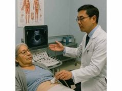
Aortic stenosis is a common and potentially serious heart valve disorder where the aortic valve narrows, making it harder for blood to flow from the heart into the aorta and onward to the rest of the body. Over time, this narrowing places extra strain on the heart and can lead to symptoms like chest pain, fatigue, and even heart failure if not addressed. Understanding the underlying causes, risk factors, warning signs, diagnosis, and the most effective treatment options is essential for anyone affected by this condition.
Table of Contents
- Detailed Overview of the Condition
- Leading Causes, Impacts, and Risk Factors
- Symptom Presentation and Diagnostic Steps
- Current Approaches to Management and Treatment
- Frequently Asked Questions
Detailed Overview of the Condition
Aortic stenosis develops when the aortic valve—the heart’s gateway to the body—becomes narrowed or stiffened, preventing it from opening fully. The aortic valve’s job is to regulate blood flow from the heart’s left ventricle into the aorta, the largest artery in the body. As the valve narrows, the heart must work harder to pump blood through the smaller opening. Over time, this extra effort can cause the left ventricle to thicken, weaken, and eventually fail to supply enough blood to the body’s organs and tissues.
The condition may be mild and stable for years or gradually worsen, eventually leading to symptoms such as chest pain (angina), breathlessness, fainting, or even sudden cardiac death. Aortic stenosis is especially common in older adults due to age-related calcification of the valve but can also be seen in younger individuals with congenital valve defects or a history of rheumatic fever.
With the advancement of diagnostic imaging and minimally invasive treatments, outcomes for aortic stenosis continue to improve. Early recognition, regular monitoring, and timely intervention are crucial to maintaining a healthy, active life.
Leading Causes, Impacts, and Risk Factors
Let’s explore what causes aortic stenosis, how it affects the heart, and who is most at risk.
Main Causes:
- Age-related calcification:
- Over time, calcium deposits build up on the aortic valve leaflets, making them stiff and less able to open fully. This is the most common cause, especially after age 65.
- Congenital bicuspid aortic valve:
- Some people are born with an aortic valve that has only two cusps (leaflets) instead of the usual three. This valve tends to wear out and narrow earlier in life.
- Rheumatic heart disease:
- After untreated strep throat or rheumatic fever, the immune system can damage the aortic valve, causing it to become scarred and thickened.
- Radiation therapy:
- Previous chest radiation for cancer can accelerate valve thickening and calcification.
- Rare inherited conditions:
- Some genetic disorders (such as Williams syndrome) can increase the risk in children or young adults.
Health Impacts and Complications:
- Left ventricular hypertrophy:
- The heart muscle thickens to push blood through the narrowed valve, which can eventually weaken the heart.
- Heart failure:
- As the heart struggles, fluid may build up in the lungs, legs, or abdomen.
- Arrhythmias:
- The risk of irregular heart rhythms increases, including atrial fibrillation.
- Angina (chest pain):
- Reduced blood flow to the heart muscle can cause pain, especially with exertion.
- Syncope (fainting):
- Inadequate blood flow to the brain during activity or sudden changes in position.
Major Risk Factors:
- Advancing age (most cases after 60)
- Male sex
- High blood pressure
- High cholesterol
- Diabetes
- Smoking
- Chronic kidney disease
- Family history of heart valve disease
- History of rheumatic fever or congenital heart conditions
Actionable Tips to Reduce Risk:
- Control blood pressure and cholesterol with healthy eating and medication if needed.
- Avoid smoking and limit alcohol.
- Treat strep throat promptly to prevent rheumatic fever.
- Get regular heart checkups if you have risk factors or a family history.
Recognizing your risk profile and making positive lifestyle choices can delay or prevent complications from aortic stenosis.
Symptom Presentation and Diagnostic Steps
Aortic stenosis often progresses silently for years before any symptoms appear. When symptoms do develop, they can signal that the condition has become severe and may require urgent attention.
Common Symptoms:
- Shortness of breath:
- Especially during physical activity or when lying flat.
- Chest pain or pressure (angina):
- May worsen with exertion or emotional stress.
- Fatigue or weakness:
- A sense of “slowing down” or tiring more easily.
- Dizziness or fainting (syncope):
- Especially during exercise, due to reduced blood flow to the brain.
- Heart palpitations:
- Sensation of a racing or irregular heartbeat.
- Swollen ankles or feet:
- Signs of fluid buildup and possible heart failure.
Subtle Signs:
- Some people may only notice decreased ability to exercise or increased breathlessness when climbing stairs.
Physical Exam Findings:
- Heart murmur:
- A harsh, “crescendo-decrescendo” sound heard best at the right upper chest.
- Weak or delayed pulse:
- The pulse may feel faint or slow to rise.
- Enlarged or thickened left ventricle:
- Detected on imaging.
Diagnostic Tests:
- Echocardiogram:
- The gold standard for diagnosis. Visualizes valve structure, movement, and measures severity.
- Electrocardiogram (ECG):
- Checks for heart muscle thickening or abnormal rhythms.
- Chest X-ray:
- May show heart enlargement or fluid in the lungs.
- Cardiac MRI/CT scan:
- Provides detailed images for surgical planning or unclear cases.
- Exercise stress test:
- Evaluates heart response during physical activity, especially in borderline cases.
- Cardiac catheterization:
- Sometimes needed to confirm diagnosis and measure pressures if other tests are inconclusive.
When to See a Doctor:
- Any unexplained shortness of breath, chest pain, fainting, or declining exercise ability should prompt medical attention, especially if you have known heart valve disease.
Practical Monitoring Advice:
- Keep a log of your symptoms and share updates at every appointment.
- Schedule regular echocardiograms as directed by your cardiologist.
- Do not ignore new or worsening symptoms—early action can be lifesaving.
Current Approaches to Management and Treatment
Managing aortic stenosis requires a tailored, stage-based approach. Treatment depends on the severity of narrowing, presence of symptoms, and overall heart function.
Medical Management:
- Observation and monitoring:
- People with mild or moderate stenosis may need only regular checkups and echocardiograms.
- Medication:
- No drugs can “cure” aortic stenosis, but medications can help manage symptoms and blood pressure.
- Diuretics may relieve fluid buildup; beta-blockers or ACE inhibitors may help control blood pressure.
- Treat other risk factors like high cholesterol and diabetes.
- Lifestyle adjustments:
- Stay physically active with doctor approval, but avoid extreme exertion.
- Eat a heart-healthy diet rich in fruits, vegetables, whole grains, and lean proteins.
- Maintain a healthy weight and avoid smoking.
Interventional and Surgical Treatments:
When symptoms develop or tests show severe narrowing, procedures to relieve the obstruction are usually needed.
- Aortic valve replacement:
- The mainstay of treatment for severe aortic stenosis.
- Surgical aortic valve replacement (SAVR): Open-heart surgery to remove the diseased valve and replace it with a mechanical or tissue valve.
- Transcatheter aortic valve replacement (TAVR): A minimally invasive option for those at higher surgical risk; a new valve is delivered via a catheter, usually through an artery in the groin.
- Balloon valvuloplasty:
- Involves stretching the narrowed valve using a balloon catheter. Usually reserved for young patients, pregnant women, or those who cannot undergo surgery; effects may be temporary.
- Other procedures:
- Some may need additional surgery to repair nearby heart structures or treat associated conditions.
After Valve Replacement:
- Lifelong follow-up:
- Regular checkups and imaging to monitor valve function and heart health.
- Anticoagulation:
- Mechanical valves require lifelong blood thinners; tissue valves may not.
- Dental hygiene:
- Good oral health and preventive antibiotics before dental procedures help prevent endocarditis.
- Physical activity:
- Gradually increase as tolerated, with guidance from your healthcare team.
Coping and Living Well:
- Connect with support groups or counselors if you feel anxious or overwhelmed.
- Engage your family and friends in your care—they can help notice early changes and support recovery.
Prognosis:
With timely intervention, most people enjoy significant improvements in symptoms and quality of life. Untreated severe aortic stenosis, however, is life-threatening.
Frequently Asked Questions
What is aortic stenosis and how serious is it?
Aortic stenosis is a narrowing of the aortic valve opening, restricting blood flow from the heart. It can be mild but often progresses to cause chest pain, fainting, or heart failure if not treated.
What causes aortic stenosis?
The main causes are age-related calcification, congenital bicuspid aortic valve, and rheumatic heart disease. Less commonly, prior chest radiation or certain inherited conditions play a role.
What are the warning signs of aortic stenosis?
Typical symptoms are breathlessness, chest pain (angina), fatigue, fainting, and swollen ankles or feet. These signs may appear gradually as the condition worsens.
How is aortic stenosis diagnosed?
Aortic stenosis is diagnosed primarily with echocardiogram imaging, supported by ECG, chest X-ray, and sometimes cardiac MRI or catheterization for detailed assessment.
When is valve replacement needed for aortic stenosis?
Valve replacement is advised when severe narrowing causes symptoms or impairs heart function. Options include surgical or minimally invasive (TAVR) procedures.
Can aortic stenosis be managed without surgery?
Mild cases can be managed with regular monitoring, lifestyle changes, and symptom management. Surgery is required for severe or symptomatic cases.
What is the outlook after valve replacement?
Most people feel better and live longer after valve replacement, especially with regular follow-up and heart-healthy habits.
Disclaimer
This article is for educational purposes only and is not a substitute for professional medical advice, diagnosis, or treatment. Always consult your healthcare provider with any health concerns or questions about heart valve disease.
If you found this article helpful, please share it on Facebook, X (formerly Twitter), or your favorite platform, and follow us for more health content. Your support helps us keep producing high-quality, accessible information for all.


