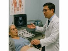
Aortic valve prolapse is a rare but impactful heart valve disorder where one or more leaflets of the aortic valve sag backward (prolapse) into the left ventricle during the heart’s pumping cycle. While it can be silent for years, it sometimes leads to aortic regurgitation—allowing blood to leak backward into the heart—which can cause symptoms ranging from subtle fatigue to severe heart failure if not addressed. This comprehensive guide covers the underlying causes, major risk factors, classic symptoms, diagnosis, and the full spectrum of treatment options, ensuring you are informed and empowered to take charge of your cardiovascular health.
Table of Contents
- Detailed Overview of the Condition
- Main Causes, Consequences, and Risk Factors
- Identifying Clinical Signs and Modern Diagnostic Tools
- Comprehensive Approaches to Management and Treatment
- Frequently Asked Questions
Detailed Overview of the Condition
Aortic valve prolapse is a structural abnormality involving the aortic valve, which sits between the heart’s left ventricle and the aorta—the body’s main artery. In prolapse, one or more of the valve’s leaflets (flaps) bend or bow back into the ventricle during the heartbeat, rather than closing neatly. This deformation can prevent the valve from sealing properly, which may cause aortic regurgitation—a condition where blood leaks backward with each heartbeat.
The degree of prolapse varies from mild to severe. Some people live with the condition for years with no symptoms, while others may develop complications such as heart failure, arrhythmias, or endocarditis (infection of the valve). Prolapse may be congenital (present from birth), acquired from disease, or the result of trauma. It is far less common than mitral valve prolapse but warrants careful monitoring due to its potential impact on heart function.
Aortic valve prolapse is most often identified during imaging for unrelated symptoms or during routine heart exams. Prompt recognition and expert management can help prevent serious complications and maintain heart health.
Main Causes, Consequences, and Risk Factors
Let’s explore what leads to aortic valve prolapse, who’s at greatest risk, and what potential complications may arise.
Primary Causes:
- Congenital (present at birth):
- Bicuspid aortic valve: The most common congenital abnormality, with only two valve leaflets instead of three, leading to abnormal motion and prolapse.
- Connective tissue disorders: Conditions like Marfan syndrome or Ehlers-Danlos syndrome, which weaken the supporting tissues, may predispose to prolapse.
- Acquired (developed later in life):
- Aortic root dilation: Expansion of the base of the aorta stretches the valve’s attachments, causing the leaflets to sag.
- Trauma: Direct injury to the chest (car accidents, blunt trauma) may damage valve structures, resulting in prolapse.
- Infective endocarditis: Infection can erode valve tissue, causing one or more leaflets to prolapse.
- Rheumatic heart disease: Scar tissue from past infection may distort valve anatomy.
- Degenerative changes: Age-related wear, calcium buildup, or degeneration can destabilize the valve.
Major Consequences:
- Aortic regurgitation (insufficiency):
- The most significant outcome, where the valve fails to close tightly, allowing blood to leak back into the left ventricle.
- Left ventricular enlargement and dysfunction:
- The heart works harder to compensate for the leak, which over time may cause it to stretch and weaken.
- Arrhythmias:
- Structural changes may trigger abnormal heart rhythms.
- Endocarditis:
- Irregular valve surfaces are more prone to infection.
- Heart failure:
- Persistent volume overload leads to heart failure if not managed.
Key Risk Factors:
- Congenital valve abnormalities (especially bicuspid valve)
- Connective tissue disorders
- Aortic aneurysm or root dilation
- History of chest trauma
- Previous episodes of endocarditis or rheumatic fever
- Advancing age
- Family history of valve disease
- High blood pressure
- Certain genetic mutations (rare)
Practical Risk Reduction Strategies:
- Control blood pressure: Keep it in a healthy range.
- Protect against infections: Practice excellent dental hygiene and seek immediate care for infections.
- Heart-healthy lifestyle: Avoid smoking, eat a balanced diet, and stay active as advised by your physician.
- Monitor high-risk individuals: Family members of those with congenital or inherited disorders may need screening.
By understanding these causes and risk factors, you’re better equipped to prevent or address complications associated with aortic valve prolapse.
Identifying Clinical Signs and Modern Diagnostic Tools
Aortic valve prolapse often develops quietly, but vigilance is essential—especially for those at risk. Early recognition, guided by symptoms and the right diagnostic approach, can make all the difference.
Classic Symptoms:
- Many are asymptomatic at first, but as regurgitation progresses, the following may develop:
- Fatigue or weakness
- Shortness of breath, especially with exertion or when lying flat
- Palpitations (awareness of heartbeats or irregular rhythm)
- Chest pain or pressure
- Dizziness or fainting
- Swelling in the legs or abdomen (a sign of heart failure)
- Reduced exercise tolerance
- Unexplained cough or nocturnal breathing difficulty
Acute Symptoms (Emergency):
- Sudden severe shortness of breath
- Chest pain and rapid heart rate
- Signs of shock (pale, clammy, confused)
Seek emergency care if these occur, as they may indicate acute valve failure.
Physical Exam Findings:
- Heart murmur: A “blowing” or “diastolic” murmur heard by stethoscope due to regurgitant blood flow.
- Water hammer pulse or bounding pulse.
- Wide pulse pressure (large difference between systolic and diastolic blood pressure).
- Visible pulsations in the neck or chest (rare).
State-of-the-Art Diagnostic Methods:
- Echocardiogram (heart ultrasound):
- The gold standard for visualizing valve structure, movement, and function. Doppler imaging detects the degree of regurgitation.
- Transesophageal echocardiogram (TEE):
- Provides clearer images of the valve, especially when standard echo is inconclusive.
- Electrocardiogram (ECG):
- Detects arrhythmias, chamber enlargement, or strain.
- Chest X-ray:
- Assesses heart size and detects fluid in the lungs.
- Cardiac MRI or CT scan:
- Advanced imaging for detailed anatomy, useful for surgical planning.
- Cardiac catheterization:
- Measures pressures, evaluates severity, and checks for other heart disease if intervention is planned.
Monitoring Advice for Patients:
- If you have a known valve disorder, keep regular follow-up appointments—even if you feel well.
- Track your symptoms—write down any changes and bring your notes to medical visits.
- Ask your provider about the frequency of echocardiograms.
When to Seek Testing:
- New or worsening breathlessness, palpitations, chest pain, fainting, or swelling should prompt timely evaluation, especially in those with risk factors.
Comprehensive Approaches to Management and Treatment
The management of aortic valve prolapse is tailored to the severity of valve dysfunction, symptoms, and your individual health profile. Let’s explore both medical and surgical strategies.
Medical (Nonsurgical) Management:
- Observation:
- Mild prolapse or regurgitation, especially if you are symptom-free, may require only regular monitoring and lifestyle changes.
- Medications:
- Diuretics to reduce fluid overload and control symptoms.
- Beta-blockers or ACE inhibitors to support heart function and control blood pressure.
- Vasodilators in selected patients to reduce regurgitant flow.
- Antiarrhythmics if irregular heart rhythms develop.
- Antibiotics for high-risk individuals before dental or invasive procedures to prevent endocarditis.
- Lifestyle Modifications:
- Heart-healthy diet low in salt and saturated fats.
- Moderate, regular exercise as tolerated and approved by your doctor.
- Maintain a healthy weight.
- Avoid tobacco and excessive alcohol.
- Meticulous dental hygiene.
Interventional and Surgical Treatments:
When significant regurgitation, symptoms, or ventricular dysfunction develop, intervention is usually indicated:
- Aortic valve repair:
- Preferred in children, younger adults, or when anatomy allows; preserves your natural valve and may avoid long-term anticoagulation.
- Aortic valve replacement (AVR):
- Surgical AVR (SAVR): Open-heart surgery replaces the diseased valve with a mechanical or tissue (bioprosthetic) valve.
- Transcatheter aortic valve replacement (TAVR): Less invasive; used mainly in high-risk or elderly patients.
- Aortic root or ascending aorta repair:
- If dilation or aneurysm is present, these structures may be repaired or replaced along with the valve.
Post-Procedure and Long-Term Management:
- Anticoagulation:
- Required lifelong for mechanical valves (e.g., warfarin); not for most tissue valves.
- Regular follow-up:
- Lifelong echocardiograms, checkups, and lab tests to monitor heart and valve function.
- Dental care:
- Good oral hygiene and antibiotic prophylaxis as recommended.
- Physical activity:
- Gradual return; cardiac rehabilitation may be helpful after surgery.
- Education:
- Know your valve type, operation history, and carry this information with you.
Living Well with Aortic Valve Prolapse:
- Take charge with consistent appointments and medication adherence.
- Seek emotional support through groups or counseling if you feel anxious.
- Educate your family on the symptoms of worsening valve disease or heart failure.
When to Call Your Doctor:
- Sudden or severe shortness of breath, chest pain, fainting, or rapid swelling require immediate medical attention.
Frequently Asked Questions
What is aortic valve prolapse and how is it different from other valve disorders?
Aortic valve prolapse occurs when a valve leaflet bends back into the ventricle, often causing regurgitation. It is rarer than mitral valve prolapse and can lead to aortic insufficiency.
What are the main causes of aortic valve prolapse?
The most common causes include congenital bicuspid valve, connective tissue disorders, aortic root dilation, infection, trauma, and age-related degeneration.
What symptoms should prompt medical evaluation?
Shortness of breath, palpitations, chest pain, fainting, or swelling should always prompt medical attention, especially for those at risk or with a known valve disorder.
How is aortic valve prolapse diagnosed?
Diagnosis is made by echocardiogram, often supported by ECG, chest X-ray, MRI, or cardiac catheterization for severe cases or surgical planning.
What are the treatment options for aortic valve prolapse?
Mild cases may be monitored, while significant regurgitation or symptoms usually require valve repair or replacement (surgical or transcatheter).
Can I live a normal life with aortic valve prolapse?
With regular monitoring, healthy habits, and timely treatment if needed, most people can lead a full, active life.
How can I reduce my risk of complications?
Control blood pressure, practice good dental hygiene, avoid smoking, and keep all follow-up appointments.
Disclaimer
This article is for educational purposes only and should not replace professional medical advice, diagnosis, or treatment. Always consult your healthcare provider with questions or concerns about your heart health.
If you found this guide valuable, please share it on Facebook, X (formerly Twitter), or your favorite social media platform, and follow us for more expert, up-to-date health content. Your support helps us continue providing trusted resources for everyone.


