Introduction
The most common malignant eyelid tumor is basal cell carcinoma (BCC), which accounts for roughly 90% of all eyelid cancers. This condition primarily affects older adults and is strongly linked to prolonged exposure to ultraviolet radiation from the sun. BCC is a slow-growing skin cancer that develops from basal cells in the epidermis and often appears as a pearly or waxy bump on the eyelid. While BCC rarely metastasizes, it can cause significant local tissue destruction if not treated, resulting in functional and cosmetic deformities. Early detection and treatment are critical for preventing complications.
Understanding The Condition
Basal cell carcinoma (BCC) of the eyelid is a non-melanoma skin cancer caused by basal cells found in the epidermis’s lowest layer. BCC is known for its slow growth and low metastatic potential, but it can cause significant local damage. Understanding the pathogenesis, risk factors, clinical characteristics, and progression of BCC is critical for clinicians and researchers seeking to improve patient outcomes.
Pathogenesis
BCC develops as a result of genetic mutations caused primarily by UV radiation, which causes DNA damage in the epidermis’ basal cells. BCC is frequently associated with the tumor suppressor gene PTCH1 and the oncogene SMO, both of which are part of the Hedgehog signaling pathway. Mutations in these genes cause uncontrolled cell proliferation and tumor formation. Other risk factors for BCC include exposure to ionizing radiation, arsenic, and certain genetic conditions such as Gorlin-Goltz syndrome.
Risk Factors
Several risk factors raise the possibility of developing BCC of the eyelid:
- UV Radiation: The most important risk factor is chronic sun exposure, especially for people with fair skin, light-colored eyes, and a history of sunburns.
- Age: BCC primarily affects older adults, usually those over 50, as a result of cumulative UV exposure over time.
- Gender: Men are more likely than women to be diagnosed with BCC, which could be attributed to increased occupational and recreational sun exposure.
- Genetic Predisposition: People with genetic conditions like xeroderma pigmentosum or Gorlin-Goltz syndrome are at a higher risk.
- Immunosuppression: Patients with weakened immune systems, such as organ transplant recipients or those with HIV/AIDS, are more likely to develop BCC.
- History of Skin Cancer: A personal or family history of skin cancer increases the likelihood of developing BCC.
Clinical Features
BCC of the eyelid frequently presents with distinct clinical features that aid in its diagnosis:
- Nodular BCC: This is the most common type, which appears as a small, pearly nodule with telangiectasias (small visible blood vessels) on its surface. It may ulcerate and create a central crater, also known as a “rodent ulcer.”
- Superficial BCC: This type is less common on the eyelid and appears as a red, scaly patch that can be confused with eczema or psoriasis.
- Morpheaform (Sclerosing) BCC: This variant is distinguished by a scar-like, white, and firm lesion with ill-defined borders, making it more difficult to detect and manage.
- Pigmented BCC: This type, which appears as a brown or black lesion, is similar to melanoma but distinguished by pearly borders and telangiectasias.
Progress and Complications
BCC grows slowly and rarely metastasizes, but it can cause extensive local tissue damage. If left untreated, BCC can invade deeper tissues such as muscle, bone, and the orbit, resulting in severe functional impairment and disfigurement. Perineural invasion, or cancer spreading along nerves, can cause pain, numbness, and muscle weakness. In severe cases, BCC can cause eye loss or necessitate extensive reconstructive surgery.
Effects on Quality of Life
The functional and cosmetic effects of BCC on the eyelid can be significant. Tumor growth and subsequent surgical excision can impair eyelid function, resulting in conditions such as lagophthalmos (inability to fully close the eyelids), ectropion (outward turning of the eyelid), and ptosis (drooping of the upper eyelid). These conditions can cause discomfort, dryness, corneal exposure, and secondary infections, reducing the patient’s quality of life. Furthermore, both the tumor and its treatment can cause a cosmetic deformity, which can have an impact on a patient’s self-esteem and psychological health.
Genetic and Molecular Insights
Advances in genetic and molecular research have shed light on the pathogenesis of BCC. The Hedgehog signaling pathway is critical in basal cell carcinogenesis. Mutations in the PTCH1 or SMO genes cause this pathway to be activated continuously, promoting cellular proliferation and tumour growth. Targeting the Hedgehog pathway with inhibitors such as vismodegib has shown promise in treating advanced BCC, emphasizing the importance of molecular insights in developing new therapies.
Epidemiology
BCC is the most common type of eyelid cancer, accounting for roughly 90% of all cases. The incidence varies globally, with higher rates found in areas with high sun exposure. Over 2 million people in the United States suffer from BCC each year, with eyelid BCC accounting for a sizable proportion of the total. The lower eyelid and medial canthus are the most commonly affected areas, most likely due to increased UV exposure.
Reducing Risk of Eyelid Basal Cell Carcinoma
- Use Sunscreen: To protect against harmful UV rays, apply broad-spectrum sunscreen with SPF 30 or higher to all exposed skin, including the eyelids.
- Wear Protective Eyewear: Use sunglasses that block 100% of UVA and UVB rays to protect your eyes and skin from direct sun exposure.
- Avoid Peak Sun Hours: Limit outdoor activities between 10 a.m. and 4 p.m., when UV radiation is most intense.
- Wear Hats: A wide-brimmed hat can help protect your face and eyelids from direct UV exposure.
- Seek Shade: Stay in the shade whenever possible, especially during peak sunlight hours, to reduce UV exposure.
- Regular Skin Examinations: Perform self-examinations on a regular basis and see a dermatologist for annual skin checks to catch any suspicious lesions early.
- Avoid Tanning Beds: Avoid using tanning beds because they emit harmful UV radiation, increasing the risk of skin cancer.
- Be Aware of Photosensitizing Medications: Some medications can make you more sensitive to UV radiation. Consult your healthcare provider about potential risks and precautions.
- Maintain a Healthy Lifestyle: Eating a well-balanced diet rich in antioxidants, exercising regularly, and quitting smoking can all help to improve skin health and lower cancer risk.
- Educate Yourself and Others: Increase awareness about the importance of sun protection and early detection of skin cancer, which can help prevent BCC and other skin cancers.
Diagnostic methods
Basal cell carcinoma (BCC) of the eyelid is diagnosed using a combination of clinical evaluation, imaging, and histopathological analysis. Early detection is critical for effective management and preventing complications.
Clinical Examination
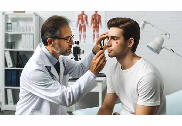
The first step in diagnosing BCC of the eyelid is a comprehensive clinical examination by an ophthalmologist or dermatologist. The examination is centered on the lesion’s appearance, size, and location. Pearly nodules, ulceration, telangiectasias, and potential involvement of the eyelid margin are all important features. A detailed patient history, including sun exposure and family history of skin cancer, helps to assess risk factors.
Dermoscopy
Dermoscopy, a non-invasive diagnostic tool, improves visibility of skin lesions. This method magnifies the lesion with a dermatoscope, revealing features like arborizing blood vessels, blue-gray ovoid nests, and ulceration. Dermoscopy can help distinguish BCC from other eyelid tumors and benign lesions, increasing diagnostic accuracy.
Biopsy
A biopsy is required to make a definitive diagnosis of BCC. Different biopsy techniques, such as shave, punch, or excisional biopsy, are used depending on the size and location of the lesion. The tissue sample is sent for histopathological examination, which confirms the presence of basaloid cells, peripheral palisading, and stromal retraction artifacts as BCC.
Imaging Techniques
Imaging modalities such as high-frequency ultrasound and optical coherence tomography (OCT) produce detailed images of the lesion’s depth and extent. These methods are especially useful for determining tumor invasion into deeper structures, which is essential for surgical planning. High-frequency ultrasound provides real-time imaging, whereas OCT provides high-resolution cross-sectional images, allowing for more precise tumor localization.
Mohs Micrographic Surgery(MMS)
Mohs micrographic surgery is both diagnostic and therapeutic. The tumor is removed sequentially, followed by an immediate microscopic examination of the margins. This technique allows for complete tumor excision while preserving as much healthy tissue as possible. MMS is especially useful for BCCs in cosmetically and functionally important areas, such as the eyelid.
Advanced Diagnostic Tools
Emerging diagnostic tools, such as reflectance confocal microscopy (RCM) and Raman spectroscopy, provide non-invasive in vivo imaging of skin lesions. RCM provides cellular-level resolution, allowing for the visualization of BCC-specific features without the need for a biopsy. Raman spectroscopy examines the molecular composition of the lesion, identifying malignant and benign tissues based on their distinct spectral signatures.
Genetic Testing
While not commonly used for initial diagnosis, genetic testing can provide useful information in patients with recurrent or multiple BCCs, particularly those with genetic syndromes such as Gorlin-Goltz syndrome. Mutations in genes like PTCH1 can confirm a predisposition to BCC and help guide personalized treatment plans.
Treatment Management
The primary goal of treating basal cell carcinoma (BCC) of the eyelid is to completely remove the tumor while maintaining eyelid function and appearance. Treatment options vary according to the tumor’s size, location, and depth of invasion.
Surgical Excision
The standard treatment for eyelid BCC is surgical excision with clear margins. The tumor is removed along with a margin of healthy tissue to ensure complete excision. This method is extremely effective, but it may necessitate reconstructive surgery to repair the eyelid and restore its functionality and appearance.
Mohs Micrographic Surgery(MMS)
Mohs micrographic surgery is the preferred method for treating BCCs in sensitive areas such as the eyelid. MMS is the systematic removal and microscopic examination of tumor layers until clear margins are achieved. This technique maximizes tissue conservation while reducing recurrence rates, making it ideal for eyelid BCC.
Cryotherapy
Cryotherapy destroys cancerous cells by exposing them to extreme cold, usually liquid nitrogen. This treatment is less invasive and ideal for superficial BCCs. However, its application to the eyelid is limited due to the risk of damaging adjacent healthy tissue and causing scarring.
Topical Treatments
Topical treatments for superficial BCCs include imiquimod and 5-fluorouracil (5-FU) creams. Imiquimod activates the immune system to attack cancer cells, whereas 5-FU prevents cell proliferation. These treatments are non-invasive, but less effective for deeper or nodular BCCs and require longer application times.
Radiation Therapy
Radiation therapy is an option for patients who are not good candidates for surgery. It entails sending high-energy radiation at the tumor to kill cancer cells. Radiation therapy can be used as a primary treatment or as an add-on to surgery, particularly in cases of incomplete excision or recurrence. However, it can cause long-term skin changes and is usually reserved for elderly patients or those with large, inoperable tumors.
Innovative and Emerging Therapies
Hedgehog Pathway Inhibitors.
Vismodegib and sonidegib are oral medications that block the Hedgehog signaling pathway, which is frequently abnormally activated in BCC. These drugs are especially effective for advanced or metastatic BCC that cannot be surgically removed. Clinical trials have yielded promising results, including significant tumor shrinkage and manageable side effects.
Photodynamic therapy (PDT)
Photodynamic therapy (PDT) involves applying a photosensitizing agent to the skin, which is then absorbed by cancer cells. The area is then exposed to a specific wavelength of light, which activates the agent and kills cancer cells. PDT is effective for superficial BCCs and has the benefit of being minimally invasive while producing excellent cosmetic results.
Laser Therapy.
Laser therapy uses focused light to destroy cancerous tissue. Carbon dioxide (CO2) lasers are commonly used in this application. Laser therapy is appropriate for small, superficial BCCs and can be performed under local anesthesia. This method is precise and leaves little scarring, but it is less effective for deeper tumors.
Immunotherapy.
BCC immunotherapy research is ongoing, with the goal of harnessing the body’s immune system to target and destroy cancer cells. Immune checkpoint inhibitors, similar to those used in melanoma, are being tested for their efficacy against advanced BCC.
Trusted Resources
Books
- “Skin Cancer: Recognition and Management” by Robert A. Schwartz
- “Cutaneous Malignancy of the Head and Neck: A Multidisciplinary Approach” by Randal Weber and Terry Day
- “Eyelid Tumors: Clinical Diagnosis and Surgical Treatment” by Jayne S. Weiss
Online Resources
- Skin Cancer Foundation: https://www.skincancer.org
- American Academy of Dermatology: https://www.aad.org
- National Cancer Institute: https://www.cancer.gov
- British Association of Dermatologists: https://www.bad.org.uk


