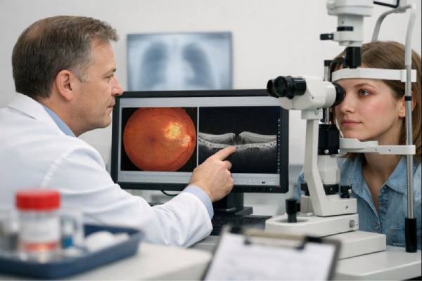
The bacterium Mycobacterium tuberculosis causes ocular tuberculosis, a type of extrapulmonary tuberculosis. Tuberculosis (TB) primarily affects the lungs, but it can spread to other parts of the body, including the eyes. Ocular tuberculosis can manifest in a variety of ways, affecting different parts of the eye, and can result in significant visual impairment if not diagnosed and treated promptly.
Understanding Tuberculosis’s Ocular Manifestations
Tuberculosis is an infectious disease that has been afflicting humans for centuries. It spreads primarily through airborne droplets produced when an infected person coughs or sneezes. While the lungs are the primary site of infection, Mycobacterium tuberculosis can spread throughout the bloodstream to other organs, including the eyes. When an infection enters the eye, it can cause inflammation and damage to several ocular structures.
Ocular tuberculosis can be primary (infection begins in the eye) or secondary (infection spreads from another site, such as the lungs). Secondary ocular tuberculosis is more common and can occur in people who have active pulmonary tuberculosis or have a history of TB.
Pathophysiology of Ocular Tuberculosis
The pathophysiology of ocular tuberculosis is complex and varies depending on the part of the eye involved. The bacteria can infect nearly every part of the eye, including the conjunctiva, cornea, sclera, uvea, retina, optic nerve, and orbit. Ocular tuberculosis can present with a wide range of clinical symptoms, making diagnosis difficult.
- Uveitis: Uveitis is a common ocular tuberculosis manifestation that causes inflammation of the uveal tract, which includes the iris, ciliary body, and choroid. Tuberculous uveitis can manifest as anterior, intermediate, posterior, or panuveitis, depending on which part of the uveal tract is involved. Granulomatous inflammation is typically characterized by the formation of granulomas, which are small immune-cell nodules. These granulomas can disrupt the uveal tract’s normal function, resulting in complications such as synechiae (iris-lens adhesions), cataracts, and glaucoma.
- Choroiditis: Choroiditis, also known as choroid inflammation, is another common ocular tuberculosis manifestation. The choroid is the vascular layer of the eye that brings blood to the retina. Tuberculous choroiditis usually manifests as multiple, yellowish lesions in the choroid that can merge to form larger patches. If left untreated, these lesions can cause scarring and atrophy of the choroid and retina, eventually leading to permanent vision loss.
- Scleritis: Scleritis, or inflammation of the sclera, the white outer layer of the eye, can also occur with ocular tuberculosis. Tuberculous scleritis is usually chronic and painful, with redness and swelling of the sclera. The inflammation can spread to the adjacent cornea, resulting in sclerokeratitis, corneal ulcers, and perforation.
- Optic Neuritis: Optic neuritis, or inflammation of the optic nerve, is a rare but serious manifestation of ocular tuberculosis. The optic nerve transmits visual information from the retina to the brain, and inflammation of this nerve can cause sudden vision loss, pain with eye movement, and a reduction in color vision. Tuberculous optic neuritis is difficult to diagnose because it can mimic other types of optic neuropathy.
- Conjunctivitis and Keratitis: Tuberculosis can also affect the conjunctiva and cornea, though these symptoms are uncommon. Conjunctival tuberculosis can manifest as chronic conjunctivitis with redness, irritation, and discharge. In some cases, conjunctival granulomas may be visible. Tuberculous keratitis, or corneal tuberculosis, is uncommon but can result in chronic corneal ulcers that are resistant to standard treatments.
- Orbital Tuberculosis: In rare cases, tuberculosis can affect the orbit, a bony cavity that houses the eye. Orbital tuberculosis can present as a mass lesion that causes proptosis (eye bulging), restricted eye movement, and pain. Orbital TB is frequently associated with sinus involvement and may necessitate imaging studies to distinguish it from other orbital diseases.
Clinical Features of Ocular Tuberculosis
The clinical presentation of ocular tuberculosis varies greatly, depending on the part of the eye involved and the severity of the infection. Symptoms can range from mild discomfort to severe visual impairment, and in some cases, ocular tuberculosis may be asymptomatic, detected only during an eye examination.
- Visual Disturbances: Patients with ocular tuberculosis may experience a variety of visual disturbances, such as blurry vision, floaters, or vision loss. The severity and location of the infection determine the extent of vision loss. Choroiditis and optic neuritis, for example, can result in significant vision loss if left untreated.
- Eye Pain: Pain is a common symptom of scleritis and optic neuritis associated with ocular tuberculosis. Deep, aching pain that worsens with eye movement. Uveitis can cause pain, redness, and photophobia.
- Redness and Swelling: Redness and swelling of the eye can occur in a variety of ocular tuberculosis, particularly scleritis and conjunctivitis. The inflammation may be limited to one area of the eye or affect the entire eye.
- Floaters: Floaters, or small spots that move across the field of vision, are common in cases of uveitis or choroiditis. The presence of inflammatory cells or debris in the vitreous humor, the gel-like substance that fills the inside of the eye, causes floaters.
- Photophobia: Another common symptom of ocular tuberculosis is light sensitivity, or photophobia, which occurs in cases of anterior uveitis. The inflammation of the uveal tract increases the eye’s sensitivity to light, causing discomfort in bright environments.
- Discharge and Tearing: Patients suffering from conjunctivitis or keratitis may experience discharge, tearing, and a grittiness or foreign body sensation in their eyes. These symptoms are frequently mistaken for more common forms of conjunctivitis, delaying the diagnosis of tuberculosis.
Epidemiology and Risk Factors
Ocular tuberculosis is a relatively uncommon form of tuberculosis, but its true prevalence is likely underreported due to the difficulty of diagnosing the condition. The prevalence of ocular tuberculosis varies geographically, with higher rates seen in areas where pulmonary tuberculosis is endemic, such as Sub-Saharan Africa, Southeast Asia, and parts of Latin America.
Several risk factors may increase the likelihood of developing ocular tuberculosis:
- Immunosuppression: People with weakened immune systems, such as those with HIV/AIDS, diabetes, or who are receiving immunosuppressive therapy, are more likely to develop extrapulmonary tuberculosis, including ocular tuberculosis. Immunosuppression allows the bacteria to spread more easily beyond the lungs, raising the possibility of ocular involvement.
- Previous TB Infection: Even if successfully treated, people with a history of tuberculosis are more likely to develop ocular tuberculosis. The bacteria can remain dormant in the body for years before reactivating when the immune system is weak.
- Close Contact with TB Patients: Living or working close to people who have active tuberculosis increases the risk of exposure and infection. This is especially true in crowded or poorly ventilated areas, where bacteria can easily spread through the air.
- Geographic Location: People residing in regions with
Clinical Evaluation (Continued)
- Patient History: The diagnostic process starts with a thorough patient history, which includes any history of tuberculosis, contact with people who have TB, and the presence of systemic symptoms like chronic cough, weight loss, fever, or night sweats. The clinician will also ask about the onset, duration, and type of ocular symptoms, such as pain, redness, visual disturbances, and photophobia. Understanding the patient’s overall health, immune status, and risk factors is critical to guiding the diagnostic process.
- Ocular Examination: A thorough ocular examination is required to diagnose ocular tuberculosis. This includes a slit-lamp examination of the eye’s anterior segment (cornea, conjunctiva, iris, and lens) and a fundoscopic examination of the posterior segment (retina, choroid, and optic nerve). Granulomatous inflammation, choroidal lesions, scleritis, or optic neuritis can all point to ocular tuberculosis, particularly in a patient with known tuberculosis risk factors.
Lab Tests
- Tuberculin Skin Test (TST): The tuberculin skin test, also known as the Mantoux test, is a popular screening method for tuberculosis infection. A small amount of purified protein derivative (PPD) is injected intradermally, and the injection site is checked 48 to 72 hours later for induration (a raised, hard area). A positive result indicates prior exposure to Mycobacterium tuberculosis, but it does not distinguish between latent and active tuberculosis or determine ocular involvement. A positive TST, however, when combined with ocular symptoms, can help to confirm the diagnosis of ocular tuberculosis.
- Interferon-Gamma Release Assays (IGRAs): IGRAs, such as the QuantiFERON-TB Gold test, detect the immune system’s release of interferon-gamma in response to tuberculosis antigens. A positive IGRA, like the TST, indicates tuberculosis infection but does not confirm active disease. IGRAs are especially useful in patients who have received the Bacille Calmette-Guérin (BCG) vaccine because they are unaffected by previous BCG vaccination.
- Polymerase Chain Reaction (PCR): PCR testing can identify Mycobacterium tuberculosis DNA in ocular fluid samples such as aqueous humor or vitreous fluid. PCR is highly specific and can confirm the presence of bacteria, providing strong evidence for diagnosing ocular tuberculosis. The small number of bacteria in the eye may limit PCR sensitivity, and a negative result does not rule out the disease.
- Culture and Staining: Although less common due to the difficulty in obtaining samples and the slow growth of Mycobacterium tuberculosis, cultures of ocular fluids can be definitive in diagnosing TB. Acid-fast bacilli (AFB) staining can also be used to detect mycobacteria in tissue samples or fluids, but its sensitivity is limited.
- Chest X-Ray: Because ocular tuberculosis is frequently secondary to pulmonary tuberculosis, a chest X-ray is a valuable diagnostic tool. It can detect active or previous pulmonary tuberculosis infection, such as cavitary lesions, nodules, or fibrosis in the lungs. The presence of radiographic findings consistent with tuberculosis, along with ocular symptoms, can aid in the diagnosis.
Imaging Studies
- Optical Coherence Tomography (OCT) is a non-invasive imaging technique that generates detailed cross-sectional images of the retina and choroid. OCT can detect choroidal thickening, subretinal fluid, and other changes in ocular tuberculosis that indicate choroiditis or retinal involvement. OCT is especially useful for tracking treatment outcomes and detecting complications like retinal detachment.
- Fluorescein Angiography (FA) is a procedure that involves injecting a fluorescent dye into the bloodstream and then imaging the retinal and choroidal vasculature. In ocular tuberculosis, FA can reveal choroiditis symptoms such as early hyperfluorescence (indicating active inflammation) and late staining or leakage. It can also help detect retinal vasculitis and neovascularization, both of which are possible complications of ocular tuberculosis.
- B-Scan Ultrasonography: B-scan ultrasonography is useful for evaluating the posterior segment of the eye when direct visualization is difficult due to media opacities, such as vitreous hemorrhage or dense cataracts. B-scan can detect choroidal masses, retinal detachment, and other structural changes associated with ocular tuberculosis (TB).
- Magnetic Resonance Imaging (MRI): An MRI of the orbit may be recommended in cases of orbital tuberculosis or when there is a suspicion of optic nerve involvement. MRI provides detailed images of the orbit, optic nerve, and surrounding tissues, which aid in distinguishing tuberculosis from other causes of orbital inflammation or masses.
Treating Tuberculosis of the Eye
Managing tuberculosis of the eye necessitates a multidisciplinary approach that includes both anti-tubercular therapy (ATT) to treat the underlying infection and additional treatments to control ocular inflammation and prevent complications. The primary goals of treatment are to eliminate the Mycobacterium tuberculosis infection, reduce inflammation, preserve vision, and prevent recurrences. The severity of the ocular involvement, the patient’s overall health, and the presence of any associated systemic tuberculosis all influence treatment decisions.
Anti-Tubercular Treatment (ATT)
Anti-tubercular therapy is the cornerstone of ocular tuberculosis management, and it typically consists of a combination of four first-line drugs: isoniazid, rifampicin, pyrazinamide, and ethambutol. These medications are taken for a minimum of 6 to 12 months, depending on the severity of the infection and the response to treatment.
- Isoniazid: Isoniazid is a bactericidal agent that prevents the synthesis of mycolic acids, which are critical components of the mycobacterial cell wall. It is typically administered for the duration of the treatment course and is an essential component of the ATT regimen.
- Rifampicin: Rifampicin is another important first-line antibiotic that inhibits RNA synthesis in mycobacteria, effectively killing them. It is known for its ability to penetrate tissues, including ocular tissues, making it especially effective in treating ocular tuberculosis.
- Pyrazinamide: Pyrazinamide is introduced into the regimen during the first intensive phase of treatment. It works by impairing mycobacteria’s ability to maintain an acidic environment, which is required for their survival in host tissues.
- Ethambutol: The primary purpose of ethambutol is to prevent drug resistance. It inhibits cell wall synthesis by preventing the production of arabinogalactan, a key component of the mycobacterial cell wall. Ethambutol’s role in ocular tuberculosis is noteworthy because of its ability to cause optic neuritis, a condition that necessitates close monitoring during treatment.
The treatment typically begins with a two-month intensive phase that includes all four drugs, followed by a four- to ten-month continuation phase with isoniazid and rifampicin. Monitoring for side effects is critical, especially given the risk of ethambutol-induced optic neuropathy and rifampicin-induced liver toxicity. Patients taking ATT for ocular tuberculosis should have regular eye exams and liver function tests.
Corticosteroid Treatment
In addition to ATT, corticosteroids are frequently used to manage the inflammatory response associated with ocular tuberculosis. Corticosteroids help to reduce tissue damage caused by inflammation and can be administered in a variety of forms.
- Topical Corticosteroids: Prednisolone eye drops are a common treatment for anterior uveitis and scleritis. They reduce inflammation in the eye, limiting systemic side effects. However, long-term use of topical corticosteroids necessitates monitoring for complications such as high intraocular pressure and cataract formation.
- Periocular Corticosteroid Injections: For more severe inflammation, periocular corticosteroid injections (such as triamcinolone acetonide) may be used. These injections deliver the medication directly to the affected area, resulting in a higher concentration and longer-lasting effect. Periocular injections are especially effective in cases of intermediate or posterior uveitis.
- Systemic Corticosteroids: Systemic steroids, such as oral prednisone, may be necessary in cases of severe ocular inflammation or if multiple parts of the eye are affected. Systemic steroids are frequently tapered gradually to reduce the risk of adverse effects such as weight gain, osteoporosis, and immunosuppression. In some cases, systemic corticosteroids may be given in addition to periocular injections.
Immunosuppressive Therapy
Immunosuppressive agents may be considered for patients with refractory ocular tuberculosis or who cannot tolerate corticosteroid treatment. Methotrexate, azathioprine, and cyclosporine can be used to treat inflammation and prevent recurrence. These agents work by suppressing the immune system, which reduces the inflammatory response that causes tissue damage in ocular tuberculosis. However, immunosuppressive therapy increases the risk of infection and necessitates close monitoring.
Surgical Management
Surgical intervention may be required in certain cases of ocular tuberculosis, especially when complications threaten vision:
- Vitrectomy: Patients with vitreous hemorrhage, chronic vitritis, or retinal detachment caused by ocular tuberculosis may benefit from a vitrectomy. This procedure removes the vitreous gel from the eye, allowing for better visualization and treatment of the retina and choroid.
- Cataract Surgery: Patients with ocular tuberculosis who develop cataracts as a result of chronic inflammation or corticosteroid use may need cataract surgery to restore their vision. The timing of surgery is critical and must be carefully considered to avoid exacerbating inflammation.
- Glaucoma Surgery: If secondary glaucoma develops as a result of chronic uveitis or corticosteroid use, surgical intervention, such as trabeculectomy or implantation of a glaucoma drainage device, may be required to control intraocular pressure and prevent optic nerve damage.
Follow-Up and Monitoring
Regular follow-up is required in the management of ocular tuberculosis to monitor treatment response, detect potential side effects, and identify signs of recurrence. To ensure comprehensive care, patients should be evaluated by an ophthalmologist as well as an infectious disease specialist. Imaging studies, such as OCT and FA, may be repeated on a regular basis to assess the condition of the retina and choroid, while visual field testing and intraocular pressure measurements are used to monitor for complications such as glaucoma or optic neuropathy.
Trusted Resources and Support
Books
- “Ocular Tuberculosis: A Clinical Guide to Diagnosis and Management” by Atul Kumar: This book provides a comprehensive overview of the diagnosis, management, and treatment of ocular tuberculosis, with detailed case studies and clinical insights.
- “Tuberculosis and Nontuberculous Mycobacterial Infections” by David Schlossberg: This resource offers a thorough exploration of tuberculosis, including its ocular manifestations, with emphasis on diagnostic challenges and treatment strategies.
Organizations
- World Health Organization (WHO): WHO offers extensive resources on tuberculosis, including guidelines on the management of TB and its extrapulmonary manifestations, such as ocular TB.
- American Academy of Ophthalmology (AAO): The AAO provides educational resources and clinical guidelines for ophthalmologists managing patients with ocular tuberculosis, including updates on the latest research and treatment protocols.


