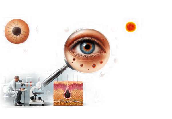
What is Melanoma of the Eyelid?
Melanoma of the eyelid is a rare but potentially fatal cancer that arises from melanocytes, the pigment-producing cells in the skin. This cancer accounts for only a small percentage of all skin cancers, but it poses significant risks due to its proximity to the eye and the possibility of local invasion and distant metastases. Early detection and accurate diagnosis are critical for successful treatment and a good prognosis. The condition usually manifests as a pigmented lesion on the eyelid that varies in size, shape, and color over time.
Detailed Investigation of Melanoma of the Eyelid
Melanoma of the eyelid, while uncommon, is a serious condition in ocular oncology due to its aggressive nature and the difficulties it presents in diagnosis and treatment. Understanding this cancer entails investigating its etiology, risk factors, clinical presentation, histopathology, and possible complications.
Etiology and Pathogenesis
Melanoma develops from melanocytes, which produce melanin, the pigment that gives color to the skin, hair, and eyes. Melanoma develops as a result of malignant transformation of these cells caused by a variety of genetic and environmental factors. Melanomagenesis is characterized by key genetic mutations in the BRAF, NRAS, and KIT genes, all of which play roles in cell growth and proliferation.
Risk Factors
Several risk factors contribute to the development of eyelid melanoma:
- UV Radiation: The most significant risk factor is prolonged exposure to ultraviolet (UV) radiation from the sun. UV radiation can damage skin cells’ DNA, resulting in cancer-causing mutations.
- Fair Skin: People with fair skin, light-colored eyes, and blonde or red hair are more vulnerable due to lower levels of melanin, which provides some protection against UV radiation.
- Sunburn History: Severe or blistering sunburns, particularly during childhood, are associated with an increased risk of melanoma.
- Family History: A family history of melanoma raises the risk, indicating a genetic predisposition to the disease.
- Atypical Moles: The presence of atypical or dysplastic nevi, or unusual-looking moles, can raise the risk of melanoma.
- Age: The risk of melanoma rises with age, but it can occur at any age.
Clinical Presentation
Melanoma of the eyelid can manifest in a variety of ways, making early detection challenging. Common clinical characteristics include:
- Pigmented Lesion: The most common presentation is a pigmented lesion on the eyelid that can be brown, black, or multicolored. These lesions can be flat or raised, and their appearance may change with time.
- Asymmetry: Asymmetrical lesions, with one half differing in shape or color from the other, raise concerns about melanoma.
- Border Irregularity: Melanomas frequently have irregular, notched, or scalloped borders, whereas benign moles have smooth, even borders.
- Color Variation: A lesion with multiple colors or an uneven distribution of color is more likely to be cancerous.
- Diameter: Lesions larger than 6 millimeters in diameter are more likely to be melanomas, but smaller lesions can also be malignant.
- Evolving: Any change in a lesion’s size, shape, color, or symptoms (such as itching or bleeding) is a red flag.
Histopathology
A histopathological examination of melanoma reveals several distinct features:
- Cellular Atypia: Melanoma cells have pronounced cellular atypia, with large, irregular nuclei and prominent nucleoli.
- Mitotic Activity: Melanoma cells exhibit increased mitotic activity, which indicates rapid cell division.
- Pagetoid Spread: Melanoma is characterized by melanocytes spreading upward into the epidermis in a pagetoid pattern.
- Invasion: Melanoma can penetrate deeper into the dermis and beyond, and the depth of invasion (Breslow thickness) is an important prognostic indicator.
- Ulceration: The presence of ulceration around the melanoma is associated with a poor prognosis.
Complications
Melanoma of the eyelid can cause a number of complications if not detected and treated promptly:
- Local Invasion: The tumor can invade nearby structures, including the orbit, causing significant morbidity.
- Metastasis: Melanoma has a high risk of metastasis. It has the potential to spread to both regional lymph nodes and distant organs such as the liver, lungs, brain, and bones.
- Functional Impairment: Eyelid involvement can cause functional impairment, affecting eyelid closure and ocular surface health, and may result in exposure keratopathy and vision loss.
Staging
Melanoma staging involves assessing the tumor’s thickness (Breslow thickness), ulceration status, and the presence of metastasis.
- Breslow Thickness: This is the depth of the tumor from the top layer of skin to the deepest point of invasion. Thick tumors have a poor prognosis.
- Clark Level: This refers to the anatomical level of invasion; higher levels indicate deeper invasion.
- Sentinel Lymph Node Biopsy: This procedure determines whether the cancer has spread to the regional lymph nodes.
- Imaging Studies: Advanced imaging, such as CT or PET scans, can detect distant metastasis.
Prognosis
The prognosis of eyelid melanoma is determined by a variety of factors, including tumor thickness, ulceration, and the presence of metastases. Early-stage melanomas have a better prognosis with proper treatment, whereas advanced melanomas with regional or distant spread have a worse prognosis.
Diagnostic methods
Melanoma of the eyelid is diagnosed using a combination of clinical examination, imaging studies, and histopathology. Early and accurate diagnosis is critical for effective treatment and better patient outcomes.
Clinical Examination
Diagnosing eyelid melanoma begins with a thorough clinical examination by an ophthalmologist or dermatologist. The key components of the examination are:
- Visual Inspection: Examine the eyelid and surrounding skin for pigmented lesions, asymmetry, border irregularities, color variation, diameter, and changing characteristics.
- Dermatoscopy: Also known as dermoscopy, this non-invasive technique uses a specialized magnifying instrument to provide a close-up view of skin lesions. Dermatoscopy can help distinguish between benign and malignant lesions by examining specific patterns and structures.
- Palpation: Feeling the lesion and surrounding tissues to determine firmness, fixation to underlying structures, and lymph node involvement.
Biopsy and histopathological analysis
Melanoma diagnosis requires a biopsy and histopathological examination. Several biopsy techniques are available:
- Excisional Biopsy: The preferred method for suspected melanoma, this procedure involves completely removing the lesion while leaving a margin of normal tissue. It provides a complete thickness sample for accurate histopathological analysis.
- Incisional Biopsy: If the lesion is too large to remove completely, an incisional biopsy may be performed to collect a sample of the lesion for diagnostic purposes.
- Punch Biopsy: A circular blade is used to collect a core sample of the lesion, which includes both the epidermis and the dermis.
Histopathological analysis of the biopsy sample entails examining the tissue under a microscope to identify melanoma-specific features such as cellular atypia, mitotic activity, pagetorial spread, and invasion depth.
Imaging Studies
Imaging studies play an important role in staging melanoma and determining the extent of local invasion and distant metastasis.
- Ultrasound: High-frequency ultrasound can assess the depth of the lesion and its involvement in underlying structures.
- CT and MRI: These imaging modalities produce detailed cross-sectional images of the eyelid, orbit, and regional lymph nodes, which aid in determining the extent of the tumor and detecting metastases.
- PET Scan: Positron emission tomography (PET) scans can detect areas of increased metabolic activity, which may indicate metastasis to other organs.
Sentinel Lymph Node Biopsy
A sentinel lymph node biopsy is a surgical procedure that determines whether melanoma has spread to the regional lymph nodes. It entails injecting a tracer near the tumor site and determining the first lymph node (sentinel node) to which cancer cells will most likely spread. The sentinel node is then removed and checked for the presence of melanoma cells.
Genetic and Molecular Testing
Advances in genetic and molecular testing have improved the accuracy and prognosis of melanoma. Tests for specific genetic mutations, such as BRAF, NRAS, and KIT, can provide insight into the tumor’s behavior and potential response to targeted therapies.
Melanoma of the eyelid Treatment
Treatment for eyelid melanoma requires a multidisciplinary approach, with ophthalmologists, dermatologists, oncologists, and plastic surgeons frequently working together. The primary goals are to completely remove the tumor, prevent recurrence, and maintain the function and appearance of the eyelid.
Surgical Treatments
- Excisional Surgery: The primary treatment option for eyelid melanoma is surgical excision with clear margins. This entails removing the tumor along with a margin of healthy tissue to ensure that all malignant cells are removed. The size of the tumor, its location, and histopathological findings all influence the extent of the excision.
- Mohs Micrographic Surgery: This technique is especially useful for eyelid melanoma because it preserves healthy tissue while completely removing the tumor. During Mohs surgery, thin layers of tissue are removed and examined under a microscope until no cancer cells are found.
- Reconstructive Surgery: Depending on the amount of tissue removed, reconstructive surgery may be required to restore the eyelid’s function and appearance. The techniques range from simple primary closure to complex flap or graft procedures, depending on the size and location of the defect.
Non-surgical Treatments
- Radiation Therapy: Radiation therapy may be considered if surgical excision is not possible or if surgery results in positive margins. It can also be used as a supplement to surgery in more advanced cases.
- Topical Treatments: Topical treatments such as imiquimod or 5-fluorouracil can be considered for in situ melanomas or very superficial lesions. These treatments are less commonly used, but they can be effective in certain situations.
Systematic Treatments
- Immunotherapy: Immune checkpoint inhibitors like pembrolizumab (Keytruda) and nivolumab (Opdivo) have transformed the treatment of advanced melanoma. These drugs enhance the immune system’s ability to recognize and attack melanoma cells.
- Targeted Therapy: For melanomas with specific genetic mutations, such as BRAF, targeted therapies like vemurafenib (Zelboraf) and dabrafenib (Tafinlar) can be extremely effective. These drugs specifically target and inhibit the activity of the mutated proteins that promote tumor growth.
- Chemotherapy: Traditional chemotherapy is less commonly used for melanoma due to the introduction of more effective targeted and immunotherapies. However, it may still be an option in some advanced cases.
Innovative and Emerging Therapies
- Gene Therapy: Researchers are looking into gene therapy approaches that could directly target melanoma-causing genetic mutations. These therapies aim to correct or silence the faulty genes that cause tumor growth.
- Oncolytic Virus Therapy: This novel approach uses genetically modified viruses to selectively infect and kill cancer cells. Talimogene laherparepvec (T-VEC) is one such approved melanoma treatment.
- Adoptive Cell Transfer: This therapy involves harvesting and expanding a patient’s own immune cells before reintroducing them into the body to attack melanoma cells. CAR-T cell therapy is being studied for its potential in treating melanoma.
Effective Methods to Improve and Avoid Melanoma of the Eyelid
- Regular Skin Checks: Examine your skin on a regular basis, including your eyelids, for new or changing lesions. Early detection is critical to successful treatment.
- Use Sun Protection: To protect your skin from UV radiation, wear a broad-spectrum sunscreen with an SPF of at least 30, wide-brimmed hats, and UV-protective sunglasses.
- Avoid Tanning Beds: Avoid using tanning beds, as they emit harmful UV radiation and increase the risk of melanoma.
- Stay in the Shade: Seek shade during peak sun hours (10 a.m. to 4 p.m.) to avoid direct sun exposure.
- Wear Protective Clothing: When spending extended periods of time outdoors, cover your skin with long-sleeved shirts, pants, and protective eyewear.
- Monitor Moles and Lesions: Keep an eye on moles and skin lesions to see if they change size, shape, color, or texture. Report any changes to a healthcare professional right away.
- Maintain a Healthy Lifestyle: A well-balanced diet high in antioxidants, regular exercise, and quitting smoking can all help you maintain good skin and boost your immune system.
- Educate Yourself and Others: Be aware of the risks and symptoms of melanoma, and inform your family and friends about preventive measures.
- Make Regular Dermatology Appointments: Regular check-ups with a dermatologist can aid in the early detection and treatment of melanoma and other skin conditions.
Trusted Resources
Books
- “Cutaneous Melanoma: Etiology and Therapy” by William H. Ward and John M. Farma
- “Skin Cancer: Recognition and Management” by Robert A. Schwartz
- “Melanoma: Prevention, Detection, and Treatment” by Catherine M. Poole


