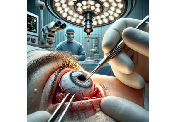
A penetrating eye injury is a severe ocular condition in which a foreign object pierces the eye and damages its internal structures. This type of injury can affect the cornea, sclera, lens, retina, and even the optic nerve, causing severe vision impairment or loss. Accidents involving sharp objects, high-speed projectiles, or blunt trauma resulting in an eye laceration or perforation are common causes of these injuries.
The presentation of penetrating eye injuries varies greatly depending on the severity and location of the damage. Severe pain, decreased vision, bleeding, and the sensation of a foreign body are all common symptoms. In some cases, there may be visible entry and exit wounds on the eye’s surface, as well as a misshapen or collapsed globe. Immediate medical attention is required to determine the severity of the injury, prevent infection, and limit permanent damage.
A comprehensive eye examination, including slit-lamp biomicroscopy, ocular ultrasound, and imaging studies such as computed tomography (CT) scans, is usually used to determine the extent of the injury. Early and accurate diagnosis is critical for developing an effective treatment strategy and improving the likelihood of visual recovery.
Traditional Penetrating Eye Injury Treatments
Management and treatment of penetrating eye injuries necessitate a systematic and multidisciplinary approach. The initial treatment focuses on stabilizing the patient, managing pain, preventing infection, and determining the severity of the injury. Here are the standard treatment methods widely used:
Initial Stabilization and Assessment: The first step after presentation is to stabilize the patient and conduct a thorough evaluation of the injury. This includes a thorough investigation of the incident, visual acuity testing, and an examination for signs of an open globe injury. Protective measures, such as shielding the eyes and avoiding pressure on the globe, are essential.
Antibiotic Therapy: Early administration of systemic and topical antibiotics is critical to preventing infection, which can exacerbate the injury and lead to endophthalmitis—a severe inflammation of the internal eye structures. Broad-spectrum antibiotics are usually chosen first, with adjustments based on culture results.
Tetanus Prophylaxis: Due to the risk of contamination in penetrating injuries, tetanus prophylaxis is given if the patient’s immunization status is not up to date.
Surgical Intervention: Surgical repair is frequently required to address the injury’s structural damage. The goals of surgery are to close the wound, restore the globe’s integrity, and remove any foreign bodies. The most common surgical procedures are:
- Primary Wound Repair: Suturing the corneal or scleral laceration to restore eye integrity.
- Vitrectomy: This procedure involves removing any vitreous hemorrhage, foreign bodies, or retinal detachment caused by the injury. It helps to clear the visual axis and avoid further complications.
- Lens Surgery: If the lens is damaged, cataract surgery or lens extraction may be required, followed by the implantation of an intraocular lens (IOL), as needed.
Management of Complications: Penetrating eye injuries can cause retinal detachment, glaucoma, or sympathetic ophthalmia (an inflammatory response in the uninjured eye). Continuous monitoring and management of these complications are critical for a successful recovery.
Innovative Penetrating Eye Injury Therapies
Recent advances in medical technology and surgical techniques have greatly improved the treatment outcomes for penetrating eye injuries. These advancements are revolutionizing the treatment of such injuries, providing new hope for a better visual prognosis and faster recovery. Below, we will go over some of the most innovative treatments in detail.
Advanced Imaging Techniques
Advanced imaging technology has transformed the diagnosis and treatment of penetrating eye injuries. High-resolution imaging modalities enable detailed visualization of ocular structures, facilitating precise diagnosis and treatment planning.
Optical Coherence Tomography (OCT): OCT is a non-invasive imaging technique for obtaining high-resolution cross-sectional images of the retina and other ocular structures. It enables clinicians to evaluate the severity of retinal and choroidal damage, plan surgical interventions, and track postoperative healing. OCT angiography, a type of OCT, can visualize blood flow within the retina and choroid, aiding in the detection and management of complications such as neovascularization.
Ultra-high frequency ultrasound (UHFU): UHFU generates detailed images of the eye’s anterior and posterior segments using sound waves. It is especially useful when the cornea is opaque and other imaging techniques fail to visualize it. UHFU can help detect and locate foreign bodies, assess intraocular hemorrhage, and guide surgical planning.
Bioengineered Tissues and Implants
The development of bioengineered tissues and advanced implants has created new opportunities for treating penetrating eye injuries. These innovations aim to improve ocular structure and function.
Amniotic Membrane Transplantation The amniotic membrane, which originates from the placenta’s innermost layer, has anti-inflammatory, anti-scarring, and healing properties. It is useful for covering corneal and scleral defects, promoting epithelialization, and reducing inflammation. Amniotic membrane transplantation has demonstrated promising results in improving wound healing and lowering postoperative complications.
Artificial Corneas (Keratoprostheses): When the cornea is severely damaged and traditional corneal grafts are not viable, keratoprostheses provide an alternative. These synthetic corneas can replace damaged corneal tissue, restore vision, and shield the eye from further damage. Biomaterials and surgical techniques have advanced, increasing keratoprostheses’ success rates and biocompatibility.
Nanotechnology and Drug Delivery Systems
Nanotechnology has developed novel drug delivery systems that improve the efficacy of treatments for penetrating eye injuries. These systems ensure the targeted and sustained release of therapeutic agents, which improves outcomes and reduces side effects.
Nanoparticle-Based Drug Delivery: Nanoparticles can be engineered to transport medications directly to the site of injury. This targeted delivery maximizes drug concentrations in the affected area while minimizing systemic exposure. Nanoparticles, for example, can be loaded with anti-inflammatory or antimicrobial agents to more effectively treat infection and inflammation.
Delivery of Drugs via Hydrogen Hydrogels are three-dimensional polymer networks that can contain and release drugs in a controlled manner. Injectable hydrogels can be used to treat injuries by delivering therapeutic agents over time and promoting tissue healing. They can also deliver growth factors that promote cell regeneration and repair.
Stem Cell Therapy
Stem cell therapy is a rapidly developing field with enormous potential for treating penetrating eye injuries. Stem cells can differentiate into a variety of cell types, allowing for tissue repair and regeneration.
Limbal Stem Cell Transplant: Limbal stem cells, found at the cornea and sclera’s border, are essential for corneal transparency and healing. In cases of injury-induced limbal stem cell deficiency, transplanting healthy limbal stem cells can restore the ocular surface and promote corneal regeneration. This technique has shown potential for improving visual outcomes and reducing scarring.
Retinal Stem Cell Therapy: Researchers are studying retinal stem cells in order to regenerate damaged retinal tissue and restore vision. While still in experimental stages, retinal stem cell therapy has the potential to treat retinal detachment and other complications of penetrating eye injuries. Early studies showed that stem cells can integrate into the retina and improve visual function.
Genetic Therapy
Gene therapy is the delivery of genetic material to cells with the goal of treating or preventing disease. Gene therapy for penetrating eye injuries aims to promote tissue repair, reduce inflammation, and prevent complications.
Anti-Inflammatory Gene Therapy: Delivering genes encoding anti-inflammatory proteins can modulate the immune response and reduce inflammation in the injured eye. This approach has the potential to reduce tissue damage while promoting healing.
Gene Therapy for Neuroprotection: The goal of neuroprotective gene therapy is to prevent optic nerve damage and maintain vision. This therapy, which introduces genes that encode neuroprotective factors, can improve the survival and function of retinal ganglion cells, which are essential for vision.
Robotic Surgery
Robotic surgery is an advanced technique that improves precision and control during surgical procedures. Robotic systems can help perform delicate and complex surgeries with greater precision, lowering the risk of complications.
Microsurgical Robots: Microsurgical robots help ophthalmic surgeons perform complex procedures. These robots have superior dexterity and stability, allowing for precise manipulation of delicate tissues. Robotic surgery can improve outcomes in complex penetrating eye injuries, such as retinal detachment or intraocular foreign bodies.


