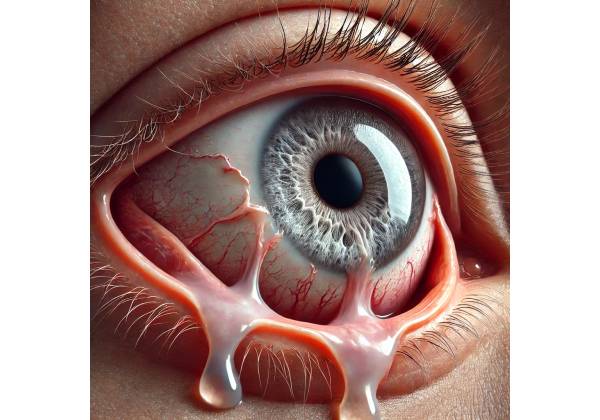
Punctal stenosis is defined as the narrowing or complete occlusion of the lacrimal puncta, which are tiny openings on the inner corners of the upper and lower eyelids that drain tears into the tear ducts. This narrowing can cause a variety of symptoms, including chronic tearing (epiphora), eye irritation, and recurring eye infections. Punctal stenosis can have a significant impact on quality of life because it interferes with normal tear drainage, causing tears to overflow onto the face.
Anatomy and Function of the Lacrimal System
Understanding punctal stenosis requires knowledge of the lacrimal system’s basic anatomy and function. The lacrimal system produces, distributes, and drains tears. It consists of several components:
- Lacrimal Glands: These glands, located in the upper outer region of each eye, create the aqueous layer of the tear film.
- Lacrimal Puncta: Small openings in the inner corners of the upper and lower eyelids that allow tears to enter the drainage system.
- Canaliculi are small channels that transport tears from the puncta to the lacrimal sac.
- Lacrimal Sac: A reservoir located in the medial canthus to collect tears from the canaliculi.
- Nasolacrimal Duct: This duct transports tears from the lacrimal sac to the nasal cavity, where they eventually dissolve.
Etiology and Risk Factors
Punctal stenosis may be congenital or acquired. The condition’s etiology is highly variable, and several risk factors can contribute to its development:
- Congenital Punctal Stenosis: Some people are born with punctal stenosis due to developmental issues. Congenital cases are uncommon and frequently associated with other ocular or systemic issues.
- Acquired Punctal Stenosis: This is the most common form and can result from a variety of factors, including:
- Chronic Inflammation: Conditions such as chronic blepharitis, conjunctivitis, or meibomian gland dysfunction can cause punctal inflammation and scarring, resulting in stenosis.
- Infections: Recurrent eye infections, particularly those involving the eyelid margins, can cause punctal narrowing.
- Trauma: Injuries to the eyelids or surrounding structures can mechanically damage the puncta, resulting in stenosis.
- Surgical Procedures: Certain eyelid surgeries or radiation treatments may inadvertently damage the puncta, causing stenosis.
- Medications: Prolonged use of certain medications, such as topical glaucoma medications or chemotherapeutic agents, can result in local toxicity and punctal stenosis.
- Age-Related Changes: As people age, the tissues of their eyelids and puncta lose elasticity and become more susceptible to narrowing.
Pathophysiology
The pathophysiology of punctal stenosis involves the progressive narrowing of the punctal openings, which prevents tears from draining normally from the ocular surface into the lacrimal system. This obstruction can be partial or complete, affecting one or both eyes. Inflammation, fibrosis, or punctal tissue scarring are common causes of narrowing.
Inflammation and Fibrosis
Chronic inflammation, such as in blepharitis or conjunctivitis, can cause thickening of the punctal epithelium and surrounding tissues. This inflammatory response can lead to fibrosis, which is the replacement of normal tissue with scar tissue, resulting in the punctal opening narrowing or closing completely.
Structural Changes
Age-related changes, trauma, and surgical interventions can all affect the puncta’s structural integrity. These changes can cause the punctal opening to become smaller, irregular, or completely blocked, preventing tears from entering the lacrimal drainage system.
Clinical Presentation
Patients with punctal stenosis usually have symptoms related to impaired tear drainage. The severity and type of symptoms can differ depending on the extent of punctal narrowing and whether the condition affects one or both eyes.
Common Symptoms
- Epiphora (Excessive Tearing): The most common symptom of punctal stenosis is chronic tearing, which occurs when tears overflow onto the cheeks rather than draining properly through the lacrimal system. This can be inconvenient and socially awkward for patients.
- Eye Irritation: Patients may experience grittiness, burning, or foreign body sensations in their eyes as a result of tears pooling on the ocular surface.
- Recurrent Infections: Poor tear drainage can cause recurring conjunctivitis or dacryocystitis (lacrimal sac infection).
- Redness and Swelling: Inflammation of the eyelid margins or conjunctiva may accompany punctal stenosis, resulting in redness and swelling around the eyes.
Effects on Quality of Life
The symptoms of punctal stenosis can have a significant impact on a patient’s quality of life. Chronic tearing can disrupt daily activities like reading, driving, and working. The constant wiping of tears can irritate and chafe the skin around the eyes. The condition’s visibility and discomfort may also have an impact on social interactions.
Differential Diagnosis
Several other conditions can present with similar symptoms to punctal stenosis, so differential diagnosis is important.
- Nasolacrimal Duct Obstruction: This condition is characterized by a blockage of the nasolacrimal duct, which can cause similar symptoms such as tearing and recurring infections. Diagnostic tests like dacryocystography or dacryoscintigraphy can help distinguish between punctal stenosis and nasolacrimal duct obstruction.
- Blepharitis: Chronic inflammation of the eyelid margins can result in irritation and tears. Blepharitis, on the other hand, is more commonly associated with diffuse inflammation of the eyelids than with localized punctal narrowing.
- Conjunctivitis: Infections or allergic reactions to the conjunctiva can cause tearing, redness, and irritation. A thorough examination and patient history can aid in distinguishing conjunctivitis from punctal stenosis.
- Dry Eye Syndrome: While dry eye syndrome is primarily associated with insufficient tear production, it can occasionally cause reflex tearing, resulting in symptoms similar to punctal stenosis. Schirmer’s test, also known as tear film breakdown time (TBUT), can aid in the diagnosis of dry eye syndrome.
Complications
Punctal stenosis, if left untreated, can cause several complications:
- Chronic Conjunctivitis: Consistent tearing can create a moist environment for bacterial growth, resulting in recurrent conjunctivitis.
- Dacryocystitis: A blockage in tear drainage can cause an infection of the lacrimal sac, resulting in pain, swelling, and redness in the inner corner of your eye.
- Skin Irritation: Constant wiping of tears can irritate, chafe, and infect the skin around the eyes.
- Visual Disturbance: Excessive tearing can impair vision, especially during activities requiring clear and focused vision.
Prognosis
The prognosis for patients with punctal stenosis varies according to the underlying cause and the effectiveness of treatment. Early detection and effective treatment can significantly improve symptoms and quality of life. When necessary, surgical interventions can provide long-term symptom relief while also restoring normal tear drainage.
Punctal Stenosis Diagnostic Approaches
A comprehensive clinical evaluation, including a patient history, physical examination, and specialized tests to assess the extent of punctal narrowing and its impact on tear drainage, is required to diagnose punctal stenosis.
Clinical Examination
- Patient History: A complete patient history is required to determine symptoms, onset, duration, and any underlying conditions or risk factors. Clinicians should inquire about chronic eye irritation, recurring infections, and any prior eye surgeries or trauma.
- Visual Acuity Testing: Measuring visual acuity can help determine whether punctal stenosis is impairing the patient’s vision. While punctal stenosis does not usually significantly impair vision, associated conditions such as conjunctivitis or dacryocystitis can.
- External Examination: Examine the eyelids, puncta, and surrounding skin for signs of inflammation, swelling, or scarring. The presence of discharge, redness, or skin irritation may indicate underlying infections or chronic irritation.
- Slit-Lamp Examination: A slit-lamp microscope magnifies the anterior segment of the eye, allowing for a thorough examination of the puncta, conjunctiva, and cornea. The examiner can evaluate the size and shape of the punctal openings, looking for signs of narrowing, fibrosis, or obstruction.
Specialized Tests
- Punctal Dilation and Irrigation: This test consists of dilating the punctal openings with a small instrument and flushing the tear drainage system with saline. The procedure helps to determine whether the puncta are patent (open) or obstructed. If saline flows freely through the system and exits via the nose, the puncta and nasolacrimal duct are most likely patent. Resistance to flow or failure to irrigate may indicate punctal stenosis or distal obstruction.
- Tear Drainage Tests: There are several tests that can evaluate tear drainage function.
- Fluorescein Dye Disappearance Test (FDDT): The examiner administers a drop of fluorescein dye into the conjunctival sac and observes how long it takes for the dye to disappear from the ocular surface. Prolonged dye retention indicates impaired tear drainage.
- Jones Test: There are two versions of the Jones test: primary and secondary. The primary test involves instilling fluorescein dye into the eye and placing a cotton-tipped applicator in the nose to detect the presence of dye after a set period of time. The secondary test entails irrigating the tear duct system and then injecting dye to see if it passes through, which aids in localizing the site of obstruction.
- Lacrimal Scintigraphy: This nuclear medicine test involves injecting a radioactive tracer into the tear film and using a gamma camera to monitor its movement through the lacrimal system. This test aids in the identification of specific sites of obstruction within the lacrimal drainage pathway.
- Dacryocystography is a radiographic procedure that involves injecting a contrast dye into the tear ducts and taking X-rays to see the lacrimal drainage system. This test produces detailed images of the anatomy, including any blockages.
Punctal Stenosis Treatments
Managing punctal stenosis entails addressing the underlying causes, alleviating symptoms, and restoring normal tear drainage. Treatment options range from conservative measures to surgical interventions, depending on the severity of the condition and the patient’s specific needs.
Conservative Management
- Lubricating Eye Drops: Artificial tears can help with dryness and irritation by keeping the ocular surface moist. These drops are especially beneficial for mild cases of punctal stenosis, where the primary concern is discomfort rather than significant tear overflow.
- Warm Compresses: Placing warm compresses on the eyelids can help reduce inflammation and open partially blocked puncta. This simple home remedy can help with symptoms of irritation and mild obstruction.
- Topical Anti-inflammatory Medications: If chronic inflammation is a contributing factor, topical corticosteroids or nonsteroidal anti-inflammatory drugs (NSAIDs) can be used to reduce inflammation and improve punctal patency.
Surgical Management
Surgical intervention is frequently required for moderate to severe punctal stenosis or when conservative measures fail to provide relief. There are several surgical options available.
- Punctal Dilation: This procedure entails using progressively larger dilators to widen the punctal openings. Punctal dilation is usually done in the office under local anesthesia and can provide short- or long-term relief, depending on the underlying cause of the stenosis.
- Punctoplasty: Punctoplasty is a surgical procedure that involves incision and widening of the punctal openings to restore normal tear drainage. There are several techniques available, including:
- Three-Snip Procedure: This entails making three small incisions at the punctal opening to increase its size and facilitate drainage.
- Micro-punctoplasty: A more precise technique that uses micro-instruments to create a larger punctal opening while causing minimal trauma to the surrounding tissues.
- Canalicular Bypass Surgery: When punctal dilation and punctoplasty are ineffective, a more invasive procedure may be required. Canalicular bypass surgery involves inserting a small stent or tube through the obstructed punctum, allowing tears to drain directly into the canaliculi.
- Laser-Assisted Procedures: Laser punctoplasty is a minimally invasive technique that employs laser energy to create or expand punctal openings. This method is effective in cases of mild to moderate stenosis and has the advantage of less bleeding and faster recovery.
Adjunctive therapies
- Silicone Punctal Plugs: Temporary or permanent silicone plugs can be inserted into the puncta to keep them open and allow tears to drain properly. This method is frequently used as a supplement to punctal dilation or punctoplasty to prevent re-stenosis.
- Mitomycin C Application: Mitomycin C is an anti-fibrotic agent that can be used on the surgical site during punctoplasty to reduce scarring and prevent recurrence. It is especially useful in patients who are at high risk of fibrosis and re-stenosis.
Post-operative Care and Follow-Up
- Postoperative Medications: Typically, patients are given antibiotic and anti-inflammatory eye drops to prevent infection and inflammation following surgery. Cold compresses may also be recommended to help reduce swelling and discomfort.
- Follow-Up Visits: Regular follow-up visits are required to monitor the healing process, evaluate the procedure’s success, and address any complications. Adjustments or additional procedures may be required in some cases to achieve the best results.
- Patient Education: It is critical to educate patients about proper eye hygiene, the importance of follow-up care, and the signs of complications in order for them to recover successfully.
Managing Complications
- Infection: Postoperative infections are a risk factor. Prompt antibiotic treatment is required to avoid further complications and ensure proper healing.
- Re-stenosis: Recurrent narrowing of the puncta is possible, especially if the underlying causes are not addressed. Regular follow-up and possibly repeated dilation or other procedures may be required.
- Scarring: Excessive scarring can impair the success of surgical procedures. Antifibrotic agents, such as Mitomycin C, can help reduce scarring and improve outcomes.
Trusted Resources and Support
Books
- “Lacrimal System Surgery” by John V. Linberg: This comprehensive book covers various aspects of lacrimal system surgery, including the diagnosis and treatment of punctal stenosis. It provides detailed surgical techniques and postoperative care guidelines.
- “Oculoplastic Surgery Atlas: Eyelid and Lacrimal Disorders” by Geoffrey J. Gladstone and Evan H. Black: This atlas offers a visual guide to oculoplastic surgical procedures, including those for punctal stenosis. It is a valuable resource for both clinicians and patients.
Organizations
- American Academy of Ophthalmology (AAO): The AAO provides extensive resources, guidelines, and continuing education for ophthalmologists and patients dealing with punctal stenosis and other ocular conditions. AAO Website
- American Society of Ophthalmic Plastic and Reconstructive Surgery (ASOPRS): ASOPRS offers valuable resources and information on lacrimal system disorders, including punctal stenosis. Their website provides access to patient education materials and professional guidelines. ASOPRS Website
- National Eye Institute (NEI): Part of the National Institutes of Health, the NEI conducts and supports research on eye diseases and provides comprehensive educational resources on various ocular conditions, including punctal stenosis. NEI Website


