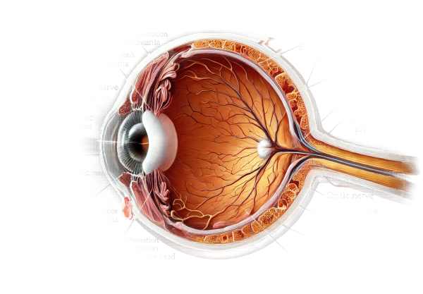
What is the Optic Nerve Pit?
Optic nerve pits are a rare congenital anomaly of the optic disc that cause small, crater-like depressions in the optic nerve heads. These pits can cause serous retinal detachment and macular changes, resulting in visual impairment. The condition is frequently discovered during routine eye exams and can remain asymptomatic unless complications arise. Understanding the optic nerve pit is critical for early diagnosis and treatment to avoid vision loss.
Comprehensive Analysis of Optic Nerve Pit
Anatomy and Pathophysiology
The optic nerve transmits visual information from the retina to the brain. In optic nerve pit, a defect in the development of the optic disc causes a small depression or pit on the surface of the optic nerve head. These pits are typically located temporally, but they can occur in other parts of the optic disc.
Causes and Risk Factors
Optic nerve pits are congenital, meaning they are present from birth. The exact cause is unknown, but it is thought to be due to incomplete closure of the embryonic fissure during fetal development. There are no known risk factors for the development of optic nerve pits, and the condition manifests sporadically with no clear hereditary pattern.
Clinical Presentation
Optic nerve pits are frequently asymptomatic and discovered coincidentally during routine eye exams. However, when symptoms appear, they can include:
- Visual disturbances:
- Patients may experience visual problems such as blurred vision, metamorphopsia (distorted vision), or scotomas (blind spots).
- Serious retinal detachment:
- Fluid leakage from the optic pit can result in serous retinal detachment, a condition in which fluid accumulates beneath the retina, causing it to detach from the surrounding tissue. If not treated promptly, this can lead to severe vision loss.
- Maculopathy:
- Fluid leakage can cause changes in the macula, the central part of the retina responsible for fine detail vision. This can result in central vision loss and difficulty performing tasks that require fine visual acuity, such as reading or recognizing faces.
Pathophysiology
The primary concern about optic nerve pits is their ability to cause serous retinal detachment. The pit allows fluid from the vitreous cavity or cerebrospinal fluid to enter the subretinal space, resulting in detachment. The exact mechanism of fluid leakage and accumulation is unknown, but it is believed that the pit serves as a communication pathway between the vitreous cavity and the subretinal space.
Complications
Optic nerve pits can cause a number of complications, the most common of which is serous retinal detachment.
- Permanent vision loss:
- If not treated promptly, retinal detachment can cause permanent vision loss due to retinal cell damage.
- Macular Changes:
- Chronic fluid leakage can cause macular changes such as cystoid macular edema or macular hole formation, which further impairs vision.
- Recurrence:
- Even after successful treatment, retinal detachment can reoccur, necessitating ongoing monitoring and possibly re-treatment.
Prognosis
The prognosis for people with optic nerve pits varies according to the presence and severity of complications. Many patients can keep their vision intact with prompt diagnosis and treatment. However, ongoing monitoring is required to quickly detect and address any recurrences or new complications.
Methods to Diagnose Optic Nerve Pit
To accurately identify the presence of an optic nerve pit and assess any associated complications such as serous retinal detachment, a combination of clinical examination and advanced imaging techniques is required. Here are the main diagnostic methods used:
Clinical Examination
- Patient history:
- A thorough patient history is required to diagnose optic nerve pit. The ophthalmologist will investigate the onset, duration, and progression of any visual symptoms, such as blurred vision, metamorphopsia (distorted vision), or scotomas. Furthermore, any family history of ocular conditions could be relevant.
- Ophthalmological Examination:
- Visual Acuity Testing: This simple yet important test measures vision clarity to determine the severity of visual impairment. It is typically carried out using a Snellen chart or other age-appropriate visual acuity tests.
- Fundoscopy: An ophthalmologist can examine the optic disc directly using an ophthalmoscope or a slit-lamp biomicroscope equipped with a fundus lens. An optic nerve pit is a small, grayish, crater-like depression on the optic disc. This exam may also reveal any associated macular changes or retinal detachment.
- Pupil reactions:
- Assessing the pupil’s response to light can aid in determining any associated optic nerve dysfunction. An afferent pupillary defect (APD) could indicate significant optic nerve involvement.
Imaging Studies
- Optical Coherence Tomography (OCT):
- OCT is a non-invasive imaging technique for obtaining high-resolution cross-sectional images of the retina and optic nerve head. It is especially useful for examining the optic pit, detecting subretinal fluid, and evaluating macular changes. OCT can help track the progression of retinal detachment and treatment efficacy over time.
- Fluorescein Angiogram:
- This diagnostic procedure entails injecting a fluorescent dye into the bloodstream and taking a series of retinal photographs to visualize retinal circulation. Fluorescein angiography can help identify leakage points in the optic pit as well as areas of retinal detachment or macular edema. It is particularly useful for distinguishing optic nerve pit-associated serous retinal detachment from other types of retinal detachment.
- B-Scan Ultrasonography:
- When fundus visualization is difficult due to media opacities (e.g., dense cataract or vitreous hemorrhage), B-scan ultrasonography can reveal important information about the optic nerve head and the presence of retinal detachment. This imaging technique employs high-frequency sound waves to produce detailed images of the eye’s internal structures.
Additional Diagnostic Tests
- Visual Field Test:
- Automated perimetry or other visual field tests can assist in quantifying visual field defects, such as scotomas or peripheral vision loss, which may be associated with optic nerve pits. These tests provide a functional evaluation of the visual impact of the condition.
- Electrophysiological Test:
- In some cases, electrophysiological tests such as visual evoked potentials (VEP) may be used to evaluate the functional integrity of the optic nerve pathways. These tests assess the electrical responses of the visual cortex to visual stimuli and can help determine the severity of optic nerve dysfunction.
Optic Nerve Pit: Available Treatment Methods
Standard Treatment Options
The primary goal of treating optic nerve pits is to manage complications such as serous retinal detachment, as the pits themselves rarely require intervention unless they cause visual problems.
- Observation:
- In asymptomatic cases or those with minimal visual impact, regular monitoring may suffice. Routine ophthalmic examinations and imaging studies such as OCT can detect any changes in the condition over time.
- Laser photocoagulation:
- In this procedure, a laser is used to create small burns around the optic pit. The goal is to create a barrier that prevents fluid from seeping into the subretinal space, lowering the risk of serous retinal detachments. Laser photocoagulation can help stabilize the condition and prevent further visual deterioration.
- Vitrectomy:
- In cases of serous retinal detachment, pars plana vitrectomy may be required. This surgical procedure consists of removing the vitreous gel from the eye and possibly injecting gas or oil to reattach the retina. This procedure helps to reduce fluid accumulation and reattach the retina, which improves or stabilizes vision.
- Gas Tamponade:
- During a vitrectomy, a gas bubble may be injected into the eye to help flatten the detached retina against the back wall. The patient must maintain a specific head position to keep the bubble in place and allow the retina to properly reattach.
- Macular Buckling:*
- This less common surgical technique involves wrapping a buckle around the eye to indent the wall and relieve traction on the retina. This procedure can help reattach the retina and is usually considered when other methods fail.
Innovative and Emerging Therapies
- Anti-VEGF injections:
- While not widely used for optic nerve pits, intravitreal injections of anti-VEGF (vascular endothelial growth factor) agents, which are commonly used in conditions such as age-related macular degeneration, are being investigated for their ability to reduce retinal edema and fluid accumulation.
- Stem Cell Treatment:
- Research into stem cell therapy for retinal diseases is still ongoing. Stem cell therapies seek to regenerate damaged retinal cells and restore vision. While still in the experimental stage, this approach may provide new hope for patients suffering from optic nerve pit complications.
- Genetic Therapy:
- Gene therapy is the process of modifying or correcting defective genes that cause ocular conditions. Although not yet used to treat optic nerve pits, advances in gene therapy may eventually lead to targeted treatments for this and other congenital eye conditions.
Supportive Care
- Visual rehabilitation:
- Patients with severe vision loss may benefit from visual rehabilitation services. This includes working with low vision aids like magnifiers or specialized software to improve functional vision and quality of life.
- Psychological support:
- Dealing with vision loss can be difficult. Psychological support and counseling can assist patients and families in managing the emotional and mental health aspects of living with a chronic ocular condition.
Regular follow-up with an ophthalmologist is required to monitor the condition, manage complications, and adjust treatment plans as necessary.
Effective Ways to Improve and Prevent Optic Nerve Pit
- Regular Eye Examination:
- Schedule regular eye exams to check for changes in the optic nerve and detect early signs of complications. Early detection enables timely intervention and improved management.
- Awareness of symptoms:
- Be aware of any changes in vision, such as blurring, distortions, or the appearance of blind spots. Report these symptoms to an eye care professional right away.
- A Healthy Lifestyle:
- Eat a balanced diet high in antioxidants and nutrients that promote eye health. Regular exercise and avoiding smoking can also help with overall ocular health.
- Managing Systemic Conditions:
- Manage systemic conditions such as hypertension and diabetes, which can exacerbate vision problems. Proper treatment of these conditions can help reduce the risk of complications from optic nerve pits.
- Protect the eyes from trauma:
- Wear protective eyewear when participating in activities that may cause eye injury. Preventing trauma can lower the likelihood of exacerbating an existing optic nerve pit or causing further ocular damage.
- Stay informed:
- Learn more about optic nerve pits and their potential complications. Understanding the condition allows you to take proactive measures to manage your eye health effectively.
- Treatment Plans:
- Follow the treatment plans and follow-up schedules recommended by your ophthalmologist. Consistent monitoring and adherence to treatment can halt the progression of complications.
- Using Low Vision Aids:
- Use low vision aids prescribed by a specialist to improve remaining vision and daily functioning.
- Engage in Support Networks:
- Participate in support groups or online communities for people with optic nerve pits or similar conditions. Sharing experiences and strategies can offer both emotional support and practical advice.
Trusted Resources
Books
- “Clinical Ophthalmology: A Systematic Approach” by Jack J. Kanski
- “Vitreoretinal Surgery: Strategies and Tactics” by Thomas A. Deutsch
- “Retina” by Stephen J. Ryan


