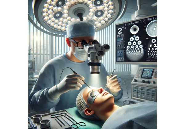
Definition of Lens Subluxation
Subluxation of the lens, also known as lens dislocation, is an ocular condition in which the natural lens of the eye is displaced from its original position. This can happen due to weakened or broken zonules, which are the fibers that hold the lens in place. The condition can significantly impair vision, resulting in blurred vision, double vision, and, in severe cases, complete vision loss. Depending on the degree of displacement of the lens, subluxation can be partial or complete.
Lens subluxation can result from a variety of causes. It can be congenital and linked to genetic conditions such as Marfan syndrome, Ehlers-Danlos syndrome, and homocystinuria, which affect connective tissue. Trauma to the eye, such as blunt force injuries or penetrating wounds, can also cause lens subluxation. Additionally, advanced cataracts or high myopia (nearsightedness) can predispose people to this condition.
A comprehensive eye examination, including a slit-lamp examination, is usually required to diagnose lens subluxation. Ultrasound biomicroscopy or optical coherence tomography (OCT) can also be used to determine the extent of displacement and any resulting damage to other ocular structures. Early detection and appropriate management are critical to avoiding complications like glaucoma, retinal detachment, and further vision loss.
Lens Subluxation: Standard Management and Treatment
The management of lens subluxation entails a combination of non-surgical and surgical approaches, each tailored to the severity of the condition and its underlying cause. The primary goals are to stabilize the lens, improve visual acuity, and avoid additional complications.
Non-surgical Management
Conservative management may be appropriate in mild cases of lens subluxation with minimal displacement and no significant impact on visual acuity. This includes regular monitoring by an ophthalmologist to detect changes in lens position or the onset of complications. Patients may be prescribed corrective lenses or contact lenses to improve their vision.
Medications
If subluxation is linked to inflammation or secondary glaucoma, medications may be prescribed to treat both conditions. Anti-inflammatory eye drops can reduce inflammation, and intraocular pressure-lowering medications can help control glaucoma. However, these medications do not address the displacement of the lens.
Surgical Management
Surgical intervention is frequently required for moderate to severe cases of lens subluxation, especially when vision is significantly impaired or complications develop. Several surgical techniques are used depending on the individual patient’s needs and the degree of lens displacement.
Lens Extraction and IOL Implantation
A common surgical approach is to remove the dislocated natural lens and replace it with an artificial intraocular lens (IOL). Lensectomy, also known as lens extraction, is a type of cataract surgery that uses a small incision. If the capsular bag is intact, the IOL is implanted there, but if the capsular support is insufficient, it is implanted in the ciliary sulcus instead.
Scleral fixation of the IOL
In cases where capsular support is insufficient, scleral fixation of the IOL may be required. Suturing the IOL to the sclera (the white part of the eye) ensures stable positioning. This technique is more complex and necessitates precise surgical skills to avoid complications like retinal detachment and infection.
Capsular Tension Rings (CTR)
Capsular tension rings (CTRs) are devices that stabilize the capsular bag during zonular weakness. They are inserted into the capsular bag to provide structural support, preventing further lens displacement and making it easier to place an IOL. CTRs can be especially beneficial for patients with underlying connective tissue disorders.
Pars Plana Vitrectomy
For severe dislocations in which the lens has moved into the vitreous cavity, pars plana vitrectomy (PPV) may be required. This procedure entails removing the vitreous gel and the dislocated lens, followed by implanting an IOL. PPV is frequently combined with other techniques, such as scleral fixation or the use of capsular tension rings, to ensure the best results.
Regular Follow-up and Monitoring
Post-surgical follow-up is critical for monitoring the stability of the IOL, assessing visual acuity, and detecting complications early. Patients may require additional treatments, such as laser therapy for capsular opacification, or changes to their medication regimen to control intraocular pressure.
Innovative Subluxation Lens Treatment
Recent advances in medical research and technology have resulted in significant improvements in the treatment of lens subluxation. These cutting-edge approaches seek to improve the efficacy of traditional therapies, introduce new treatment options, and improve patient outcomes. The following are some of the most promising developments in the management and treatment of lens subluxation.
Femtosecond Laser-Assisted Surgery
Femtosecond laser technology has revolutionized cataract surgery and is now being used to treat lens subluxations. This laser is capable of making precise, bladeless incisions and fragmenting the lens, allowing for its removal with minimal trauma to surrounding tissues. Femtosecond laser-assisted surgery improves the accuracy and safety of lens extraction and IOL implantation, especially in cases involving weak zonules or complex anatomy. The use of femtosecond lasers reduces the risk of complications while improving visual outcomes by ensuring more precise placement of the IOL.
Customized Intraocular Lenses
Advances in the design and customization of intraocular lenses (IOLs) have opened up new treatment options for patients with lens subluxation. Toric IOLs, which correct astigmatism, and multifocal IOLs, which provide both near and distance vision, can be customized to meet the unique visual requirements of each patient. The development of adjustable IOLs, which can be fine-tuned postoperatively using light-based technology, opens the door to even greater precision in achieving optimal visual outcomes. Customized IOLs improve visual acuity and quality of life for patients undergoing lens replacement surgery.
Suture-Free Scleral Fixation Techniques
Surgical techniques have evolved to include sutureless scleral fixation methods for IOL implantation. These techniques use specialized devices, such as anchors or clips, to secure the IOL to the sclera without the use of sutures. Sutureless scleral fixation shortens surgical time, reduces the risk of suture-related complications, and provides consistent long-term results. These advancements are especially beneficial to patients who have complex cases of lens subluxation or require reoperation.
Gene and Molecular Therapies
Research into the genetic and molecular basis of connective tissue disorders has led to new treatment options for lens subluxation. Gene therapy, which involves replacing or repairing defective genes with healthy ones, has the potential to correct the underlying genetic mutations that cause conditions such as Marfan syndrome or Ehlers-Danlos syndrome. Molecular therapies targeting specific pathways involved in the maintenance of zonular fibers and lens stability are also under investigation. These novel approaches seek to address the underlying cause of lens subluxation and prevent further progression.
Minimal Invasive Lens Stabilization Devices
The development of minimally invasive devices for lens stabilization has opened up new avenues for managing lens subluxation. Devices like the Cionni ring and the Ahmed segment are designed to be inserted into the eye through small incisions, supporting the capsular bag and preventing further lens displacement. These devices, when combined with other surgical techniques, can improve IOL stability and visual outcomes. Minimally invasive lens stabilization devices are a less traumatic alternative to traditional surgical methods, reducing recovery time.
Robotic Assisted Surgery
Robotic-assisted surgery is a growing field that improves precision and control for complex ophthalmic procedures such as lens subluxation. Robotic systems improve surgeons’ dexterity and accuracy, allowing them to perform delicate maneuvers with more confidence. This technology is especially useful for intricate surgeries that require scleral fixation or the placement of capsular tension rings. Robotic surgery improves surgical outcomes while lowering the risk of complications, paving the way for more advanced and effective lens subluxation treatments.
Bioengineered Zonular Repair
Advances in tissue engineering and regenerative medicine have prompted the investigation of bioengineered solutions for zonular repair. Researchers are looking into how bioengineered scaffolds and growth factors can promote zonular fiber regeneration and restore lens stability. These novel approaches aim to provide a long-term solution for patients suffering from lens subluxation due to zonular weakness or damage. Bioengineered zonular repair has the potential to transform the treatment of lens subluxation by addressing underlying structural issues and preventing recurrence.
Artificial Intelligence, Machine Learning
Artificial intelligence (AI) and machine learning (ML) are becoming more widely used in ophthalmology, providing new tools for diagnosing and treating lens subluxation. AI algorithms can use large datasets of clinical and imaging data to identify patterns and predict disease progression. Machine learning models can help with treatment planning by assessing factors such as subluxation severity, patient compliance, and response to previous therapies before recommending the best treatment strategies for individual patients. AI and ML improve the precision and personalization of lens subluxation management, which leads to better patient outcomes.


