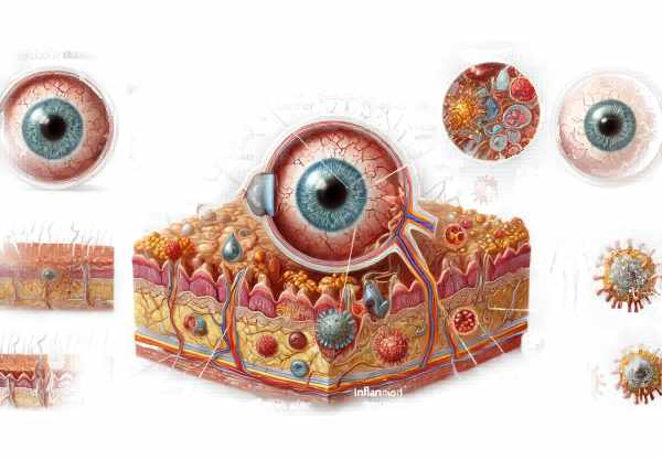
Introduction to Ebola Virus Disease Ocular Symptoms
Ebola Virus Disease (EVD) is a severe, often fatal illness caused by the Ebola virus, which belongs to the Filoviridae family. While EVD primarily affects the immune system, resulting in severe hemorrhagic fever, it also has significant ocular symptoms that can last even after recovery. Ocular complications include uveitis, optic neuritis, and other vision-threatening conditions. Understanding these manifestations is critical for managing and supporting Ebola survivors’ recovery, as they can cause long-term vision impairment and have a significant impact on quality of life.
Ebola Virus Disease Ocular Manifestations: Insights
Pathophysiology Of Ocular Involvement
The Ebola virus can infect a variety of body tissues, including the eyes. Ocular complications result from the virus’s ability to cross the blood-ocular barrier, causing inflammation and direct viral damage to ocular tissues. The immune response triggered by the infection can exacerbate the inflammation, causing additional damage.
Common Ocular Manifestations
Uveitis
Uveitis is the most commonly reported ocular complication among Ebola survivors. It causes inflammation of the uvea, the eye’s middle layer that includes the iris, ciliary body, and choroid.
- Anterior Uveitis: Inflammation primarily affects the front part of the eye (iris and ciliary body), resulting in symptoms like redness, pain, photophobia, and blurred vision. Topical corticosteroids are commonly used to treat anterior uveitis and reduce inflammation.
- Intermediate Uveitis: This type affects both the vitreous and peripheral retina. Symptoms include floaters and blurred vision. Depending on the severity, treatment may include both topical and systemic approaches.
- Posterior Uveitis: Affects the choroid and retina, resulting in more severe visual disturbances such as floaters, scotomas, and significant vision loss. It is treated with systemic corticosteroids and immunosuppressive medications.
- Panuveitis: The most severe form, characterized by inflammation of all layers of the uvea, can cause widespread symptoms and vision loss.
Optic Neuritis
Optic neuritis, or inflammation of the optic nerve, can result from either direct viral invasion or a secondary immune response. The symptoms include painful eye movements and vision loss. To avoid permanent damage, optic neuritis must be treated promptly with systemic corticosteroids.
Conjunctivitis
Conjunctivitis, or inflammation of the conjunctiva, can develop during the acute stage of Ebola infection. The symptoms include redness, tearing, and discomfort. This condition is usually treated with supportive care, but it can complicate the clinical picture.
Retinal Lesions
Ebola survivors may develop retinal lesions, which can be detected with a fundoscopic examination. If not treated properly, these lesions can cause scarring and permanent vision loss.
Long-term Ocular Complications.
Even after recovering from the acute phase of Ebola virus disease, survivors may develop long-term ocular complications. Chronic uveitis, cataract formation as a result of prolonged inflammation and steroid use, and glaucoma caused by high intraocular pressure are all possibilities.
Mechanisms for Ocular Manifestations
The precise mechanisms by which the Ebola virus causes ocular manifestations are not fully understood, but they are thought to involve several factors:
- Direct Viral Invasion: The virus can enter the ocular tissues, causing cell damage and inflammation.
- Immune-Mediated Damage: The immune system’s reaction to the virus can cause inflammation and damage to ocular structures.
- Blood-Ocular Barrier Disruption: When the blood-ocular barrier is compromised, inflammatory cells and mediators can infiltrate ocular tissues.
- Vascular Compromise: Vascular involvement can cause ischemia and hemorrhage in the eye, resulting in a variety of complications.
Effects on Quality of Life
The ocular manifestations of the Ebola virus can have a significant impact on survivors’ quality of life. Vision loss and chronic eye pain can impair daily activities, mobility, and independence. The fear of permanent vision loss, as well as the social stigma associated with EVD, can cause psychological effects such as depression and anxiety.
Epidemiology
The prevalence of ocular complications in Ebola survivors varies, but studies have shown that a sizable proportion of survivors develop some form of ocular involvement. A study conducted in West Africa during the 2014-2016 Ebola outbreak discovered that approximately 20% of survivors developed uveitis. Risk factors for developing ocular complications include the severity of the initial infection, delays in receiving adequate medical care, and underlying health problems.
Prevention Tips
- Early Detection and Treatment: Ebola survivors should have regular eye examinations to detect ocular complications early. Prompt treatment of conditions such as uveitis and optic neuritis is critical to avoiding permanent damage.
- Vaccination: Promoting the use of the Ebola vaccine in high-risk areas can reduce the spread of Ebola virus disease and, as a result, its ocular complications.
- Education and Awareness: Informing healthcare providers and Ebola survivors about the potential ocular manifestations of the disease can lead to earlier detection and treatment. Awareness campaigns should include information about symptoms and the importance of regular eye exams.
- Infection Control Measures: Strict infection control measures in Ebola-affected areas can help to slow the spread of the virus. This includes using personal protective equipment (PPE) properly, maintaining hand hygiene, and isolating infected individuals.
- Supportive Care During Acute Infection: Offering comprehensive supportive care during the acute phase of Ebola virus disease can help to reduce the severity of systemic and ocular complications. This includes preventing dehydration, maintaining electrolyte balance, and looking for secondary infections.
- Ongoing Research: Continued research into the pathogenesis, treatment, and prevention of Ebola virus disease and its ocular manifestations is critical. Research can lead to novel therapeutic approaches and improved management strategies.
- Comprehensive Survivor Care Programs: Creating and implementing comprehensive care programs for Ebola survivors, including ophthalmologic evaluation and follow-up, can aid in the management of long-term complications. These programs should be accessible and affordable.
- Nutritional Support: Providing adequate nutritional support to Ebola survivors can help boost their immune systems and improve overall health, potentially lowering the risk of complications.
- Regular Complication Monitoring: Long-term monitoring of Ebola survivors for ocular and other systemic complications can result in timely intervention and better management outcomes. This includes regular check-ups and access to specialised care.
- Mental Health Support: Giving Ebola survivors psychological support can help them deal with the disease’s emotional impact and complications. Mental health services should be integrated into survivor support programs.
Diagnostic methods
The ocular manifestations of Ebola Virus Disease (EVD) are diagnosed using a combination of standard ophthalmic examinations, advanced imaging techniques, and laboratory tests. The goal is to identify the specific ocular complications, determine the extent of damage, and devise effective treatment strategies.
- Comprehensive Eye Examination: This preliminary evaluation is critical for detecting visible signs of ocular inflammation or infection. An ophthalmologist conducts a detailed evaluation, which includes:
- Visual Acuity Test: Determines how well the patient can see at different distances.
- Slit-Lamp Examination: This procedure allows the ophthalmologist to examine the front part of the eye (cornea, iris, and lens) for signs of inflammation or other abnormalities.
- Fundus Examination: Using an ophthalmoscope, inspect the back of the eye (retina, optic disc, and blood vessels) for signs of uveitis, retinal lesions, or hemorrhages.
- Tonometry: This test measures intraocular pressure (IOP) inside the eye and is useful for detecting glaucoma, which can develop as a result of uveitis.
Advanced Imaging Techniques
- Optical Coherence Tomography (OCT) is a non-invasive imaging test that generates detailed cross-sectional images of the retina. It aids in detecting retinal edema, vitreous opacities, and structural changes in the retina and choroid associated with Ebola-related uveitis.
- Fluorescein Angiography: This technique involves injecting a fluorescent dye into the bloodstream and photographing the dye as it travels through the retinal blood vessels. It aids in detecting leakage, ischemia, and neovascularization in the retina.
- Indocyanine Green Angiography (ICGA): Like fluorescein angiography, ICGA uses indocyanine green dye to visualize choroidal circulation. It is particularly effective at detecting choroidal inflammation and vascular changes.
- Ultrasound B-Scan: This imaging technique is used when there is significant vitreous hemorrhage or other opacities that block the view of the retina. It aids in evaluating the posterior segment and detecting retinal detachments or other structural anomalies.
Lab Tests
- PCR (Polymerase Chain Reaction): Aqueous or vitreous fluid can be tested for Ebola virus RNA to confirm active or recent infection in the eye.
- Serological Tests: These tests detect antibodies to the Ebola virus in the blood, indicating previous infection and the body’s immune response.
- Inflammatory Markers: Blood tests for inflammatory markers like ESR (erythrocyte sedimentation rate) and CRP (C-reactive protein) can help determine the level of systemic inflammation, which may be related to ocular inflammation.
Comprehensive Diagnostic Approach
Combining these diagnostic methods enables a thorough assessment of Ebola virus disease’s ocular manifestations. Early and accurate diagnosis is critical for successful management and prevention of long-term complications. Regular follow-up exams are also essential for tracking disease progression and response to treatment.
Treatment
The treatment of Ebola Virus Disease ocular manifestations entails reducing inflammation, avoiding complications, and maintaining vision. A multidisciplinary approach, involving ophthalmologists and infectious disease specialists, is frequently required.
Standard Treatment Options
- Corticosteroids: Corticosteroids can be applied topically, periocularly, or systemically to reduce inflammation in uveitis and optic neuritis. The choice of administration is determined by the severity and location of the inflammation.
- Topical Steroids: Prednisolone acetate drops are a common treatment for anterior uveitis.
- Periocular Steroids: Injections around the eye can be used in severe cases.
- Systemic Steroids: Corticosteroids can be administered orally or intravenously to treat severe posterior uveitis or optic neuritis.
- Anti-VEGF Therapy: Intravitreal injections of anti-VEGF (vascular endothelial growth factor) agents, such as bevacizumab, can treat neovascularization and macular edema caused by uveitis.
- Antibiotics and Antivirals: If there is a concurrent bacterial or viral infection, appropriate antibiotics or antiviral medications are used to control the infection.
Innovative and Emerging Therapies
- Biologic Agents: Newer biologic agents that target specific inflammatory pathways are being investigated to treat severe uveitis. These include TNF-alpha and interleukin inhibitors.
- Immunomodulatory Therapy: Immunosuppressive agents such as methotrexate, azathioprine, and mycophenolate mofetil may be used to treat chronic or refractory uveitis in order to reduce corticosteroid use and control inflammation.
- Gene Therapy: Although still in experimental stages, gene therapy has the potential to treat viral infections and associated complications by delivering therapeutic genes to target cells.
- Stem Cell Therapy: Researchers are looking into stem cell therapy to regenerate damaged retinal tissue and restore vision. This emerging field may offer new hope to patients suffering from severe ocular damage caused by the Ebola virus.
Supportive Treatments
- Regular Monitoring: Regular follow-up visits with an ophthalmologist are required to monitor treatment response and adjust therapy as necessary.
- Visual Rehabilitation: For patients with severe vision loss, visual rehabilitation and the use of low-vision aids can help improve their quality of life.
- Mental Health Support: Psychological support and counseling are essential for patients dealing with the emotional impact of vision loss and the fear of recurrence.
- Nutritional Support: Proper nutrition promotes overall health and may enhance the body’s ability to respond to treatment.
Trusted Resources
Books
- “Ebola: The Natural and Human History of a Deadly Virus” by David Quammen
- “Ebola: Clinical Patterns, Public Health Concerns” by Adnan I. Qureshi
- “Ocular Inflammatory Disease and Uveitis: Fundamentals and Clinical Practice” by Peizeng Yang, Vishali Gupta


