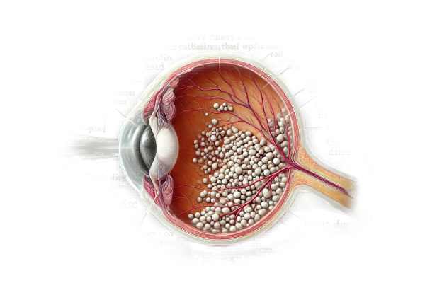
What is Optic Disc Drusen?
Optic disc drusen are abnormal calcified deposits that form within the optic nerve head. These deposits can cause a variety of visual problems and are frequently discovered incidentally during routine eye exams. Optic disc drusen are typically benign, but they can occasionally cause visual field defects and other complications. Understanding optic disc drusen, including their causes, symptoms, and diagnostic methods, is critical for successful monitoring and treatment.
Detailed Insight into Optic Disc Drusen
Anatomy and Pathophysiology
The optic disc, also known as the optic nerve head, is where the retinal nerve fibers converge to form the optic nerve, which transmits visual information from the eye to the brain. Optic disc drusen are deposits of extracellular calcium that accumulate within the optic nerve head. These deposits calcify over time and are detectable using a variety of imaging techniques.
The exact pathophysiology of optic disc drusen is unknown, but it is thought that these deposits are caused by axoplasmic stasis and subsequent degeneration of retinal ganglion cells. This process produces drusen, which can compress optic nerve fibers and cause visual field defects.
Epidemiology
Optic disc drusen are fairly common, with an estimated prevalence of 0.3% to 2% in the general population. They can affect people of all ages, but young adults are the most commonly diagnosed. There is no clear gender preference, and the condition may be unilateral or bilateral.
Causes and Risk Factors
The precise cause of optic disc drusen is unknown, but several factors are believed to contribute to their development:
- Genetic predisposition: There is evidence that there is a hereditary component, as optic disc drusen can occur in families. Mutations in genes involved in optic nerve development and maintenance could play a role.
- Congenital Abnormalities: Some people are born with structural abnormalities of the optic disc that make them prone to the formation of drusen.
- Vascular Factors: It is believed that impaired blood flow and ischemia within the optic nerve head contribute to the development of drusen.
Symptoms
Optic disc drusen are frequently asymptomatic and can be discovered coincidentally during routine eye exams. However, when symptoms appear, they can include:
- Visual Field Defects: These are the most common symptoms, which can vary from mild to severe. Peripheral vision loss is common, with central vision usually spared.
- Transient Visual Obscurations: Some people may experience brief episodes of vision loss or dimming, especially during posture changes.
- Photopsia: The presence of flashing lights or flickering vision may indicate optic disc drusen.
- Blurred Vision: In rare cases, optic disc drusen can cause a general decrease in visual acuity.
Complications
While optic disc drusen are generally benign, they can cause a number of complications, including:
- Nonarteritic anterior ischemic optic neuropathy (NAION): This condition causes sudden vision loss due to reduced blood flow to the optic nerve. Individuals with optic disc drusen are at a higher risk of NAION.
- Retinal Vascular Occlusions: The presence of drusen can cause compression of retinal blood vessels, increasing the risk of vascular occlusion and retinal damage.
- Choroidal Neovascularization:** Abnormal blood vessel growth beneath the retina may occur in conjunction with optic disc drusen, potentially resulting in vision-threatening complications.
Prognosis
Individuals with optic disc drusen have a good prognosis, particularly if they do not have any significant visual symptoms. Regular monitoring and management of any associated complications are critical for maintaining vision and quality of life.
Diagnostic Techniques for Optic Disc Drusen
To confirm the presence of drusen and assess their impact on the optic nerve and visual function, clinicians use a combination of clinical evaluation, advanced imaging techniques, and differential diagnosis.
Clinical Evaluation
- ophthalmoscopy: This is the primary tool for the initial diagnosis. During an ophthalmoscopic examination, the ophthalmologist looks for yellowish, glistening deposits that indicate drusen. Buried drusen may not be visible, necessitating additional imaging.
- Visual Field Testing: Automated perimetry tests can detect visual field defects caused by optic disc drusen. Peripheral vision loss is common, and these tests can assess the severity and pattern of vision loss.
Advanced Imaging Techniques
- B-Scan Ultrasonography: B-scan ultrasonography is a non-invasive imaging method that uses high-frequency sound waves to produce detailed images of the optic nerve head. It is especially effective for detecting buried drusen that are not visible under ophthalmoscopy. The characteristic highly reflective ultrasound signals confirm the presence of calcified drusen.
- Optical Coherence Tomography (OCT): OCT can generate high-resolution cross-sectional images of the retina and optic nerve head. It aids in visualizing the structure of the optic nerve, detecting drusen, and evaluating any associated retinal changes. Enhanced depth imaging OCT can be especially effective for detecting buried drusen.
- Fluorescein Angiogram: This imaging technique involves injecting a fluorescent dye into the bloodstream to reveal retinal and choroidal circulation. It aids in separating optic disc drusen from other conditions such as optic disc edema. In optic disc drusen, the dye may cause hyperfluorescent spots.
Optical Disc Drusen Treatment
Treatment of Symptoms and Complications
Optic disc drusen are usually asymptomatic and do not always require treatment. However, when symptoms or complications appear, management focuses on monitoring and addressing them in order to preserve vision and prevent further damage.
- Regular ophthalmologic examinations are necessary for patients with optic disc drusen to monitor progression and detect complications early. This includes visual field testing and OCT imaging to detect any changes in the optic nerve or retinal structure.
- Managing Visual Field Defects: Regular perimetry tests can monitor optic disc drusen-related visual field defects. In some cases, low vision aids and vision therapy may be recommended to assist patients in maximizing their remaining vision and adapting to visual field changes.
Treatment for Complications
- Nonarteritic anterior ischemic optic neuropathy (NAION): Patients with optic disc drusen are at a higher risk for developing NAION. Controlling vascular risk factors like hypertension, diabetes, and hyperlipidemia is part of the management process. To reduce the risk of future vascular events, doctors may prescribe aspirin or other antiplatelet agents.
- Retinal Vascular Occlusions: Prompt treatment is necessary for retinal vascular occlusions caused by optic disc drusen to avoid vision loss. This could include intravitreal injections of anti-VEGF (vascular endothelial growth factor) agents to reduce macular edema and improve visual outcomes.
- Choroidal Neovascularization: – Anti-VEGF therapy can treat abnormal blood vessels under the retina. Regular monitoring with OCT and fluorescein angiography is essential for detecting and treating this complication early.
Innovative and Emerging Therapies
- Neuroprotective Agents: – Neuroprotective drugs aim to protect retinal ganglion cells and optic nerve fibers from damage. These agents may slow the progression of visual field loss in patients with optic disc drusen.
- Advances in gene therapy provide hope for treating hereditary conditions like optic disc drusen. While still in the experimental stage, gene therapy may one day provide a way to correct underlying genetic mutations and prevent the formation of drusens.
- Stem Cell Therapy: – Stem cell therapy is being investigated as a treatment option for various ocular conditions, including optic nerve damage. This method attempts to regenerate damaged optic nerve cells and restore some level of vision.
Lifestyle and Supportive Measures
- Maintaining a healthy lifestyle, such as eating a balanced diet with antioxidants, exercising regularly, and quitting smoking, can improve eye health and lower the risk of vascular complications.
- Patient Education: – Educating patients on optic disc drusen, potential symptoms, and the need for regular eye exams can aid in early detection and management of complications.
Effective Ways to Improve and Avoid Optic Discs Drusen
- Schedule regular eye exams with an ophthalmologist to check for optic disc drusen and detect changes early.
- Control Vascular Risk Factors: – Manage hypertension, diabetes, and hyperlipidemia with medication, diet, and lifestyle changes to prevent complications like NAION.
- Maintain a healthy diet high in antioxidants, vitamins, and minerals for optimal eye health. Foods like leafy greens, nuts, and fish are healthy.
- Avoid Smoking: Smoking and exposure to secondhand smoke can worsen vascular issues and accelerate optic nerve damage.
- Wear protective eyewear during activities that may cause eye injury to prevent worsening the condition.
- Stay Hydrated: – Proper hydration promotes vascular health and eye function.
- Patient Education: – Understand optic disc drusen, its symptoms, and the need for regular monitoring to manage the condition effectively.
- Seek immediate medical attention if experiencing symptoms like sudden vision loss, flashing lights, or significant vision changes, as these may indicate complications.
- Use Low Vision Aids: – Use low vision aids and vision therapy to improve remaining vision and adapt to visual field defects.
Trusted Resources
Books
- “Clinical Neuro-Ophthalmology: A Practical Guide” by Ambar Chakravarty
- “Optic Nerve Disorders: Diagnosis and Management” by Jane W. Chan
- “Clinical Ophthalmology: A Systematic Approach” by Jack J. Kanski and Brad Bowling


