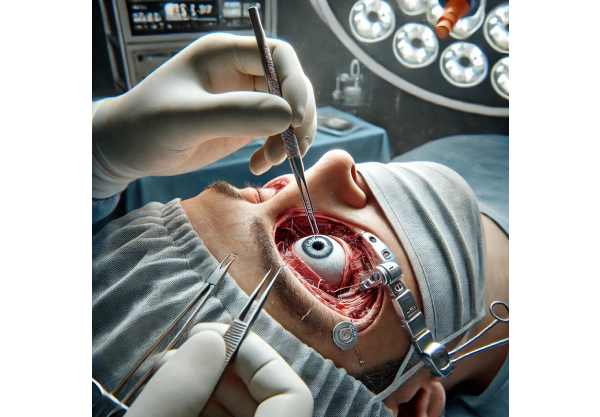
Orbital fractures are breaks or cracks in the bones surrounding the eye, known as the orbit. These fractures are most commonly caused by blunt trauma to the face, such as sports injuries, falls, car accidents, or physical assaults. The orbit is made up of several thin bones that form the eye socket, and fractures can affect one or more of them, resulting in a variety of symptoms and complications.
An orbital fracture can cause pain around the eye, swelling, bruising, double vision, and difficulty moving the eye. In severe cases, there may be a visible change in the position of the eyeball (enophthalmos), vision loss, or nerve damage. The diagnosis is typically confirmed through a combination of physical examination and imaging studies, such as X-rays or computed tomography (CT) scans, which provide detailed images of the orbital bones and any associated injuries.
Prompt diagnosis and treatment are critical to avoiding long-term complications like persistent double vision, cosmetic deformities, or impaired vision. Understanding the nature and extent of orbital fractures is critical for successful treatment and patient outcomes.
Orbital Fractures: Management and Treatment
Orbital fractures require a multidisciplinary approach that includes ophthalmologists, maxillofacial surgeons, and, in some cases, neurosurgeons. The primary goals are to alleviate symptoms, restore normal function, and avoid complications. Standard treatments for orbital fractures include:
- Observation and Conservative Management: If the fracture is minor and does not impair the eye’s function or position, a conservative approach may be used. This includes using ice to reduce swelling, taking pain medications, and giving the bones time to heal naturally. Patients are closely monitored to ensure that no new symptoms arise and that healing occurs as expected.
- Medical Therapy: Antibiotics may be prescribed to prevent infection, especially if there is an open wound associated with the fracture. Decongestants can be used to relieve nasal congestion and avoid sinus complications. Steroids may be administered to reduce inflammation and swelling around the eyes.
- Surgical Intervention: Surgical repair is frequently required for more severe fractures, particularly when there is significant displacement of bone fragments, entrapment of eye muscles, or a risk of vision impairment. The surgical approach is based on the fracture type and severity:
- Orbital Floor Fractures: Repair entails repositioning fractured bone fragments and utilizing implants or bone grafts to restore the orbital floor’s integrity and support the eye.
- Medial Wall Fractures: These fractures are repaired by repositioning bone fragments and reconstructing the orbital wall with materials like titanium mesh or absorbable plates.
- Complex Orbital Fractures: When there are multiple fractures or significant disruptions in the orbital structure, a combination of techniques and materials may be used to reconstruct the orbit and ensure proper alignment and function of the eye.
- Postoperative Care and Rehabilitation: After surgical repair, patients need careful postoperative care to prevent complications and promote healing. This includes regular follow-up visits, imaging studies to evaluate the surgical outcome, and rehabilitation to address any remaining problems with eye movement or vision.
- Visual Rehabilitation: Patients who have persistent double vision or other visual disturbances following an orbital fracture can benefit from visual rehabilitation services to improve their visual function. This could include prism glasses, vision therapy, or surgical procedures to correct alignment issues.
Cutting-Edge Innovations in Orbital Fractures Treatment
Recent advances in medical research and technology have resulted in novel approaches that provide new hope for patients with orbital fractures. These cutting-edge innovations include advanced imaging techniques, new surgical methods, regenerative medicine, and integrative care models. Each of these innovations offers distinct advantages and has the potential to improve orbital fracture management.
Advanced Imaging Techniques
Imaging advancements have greatly improved the diagnosis and monitoring of orbital fractures. High-resolution imaging modalities enable detailed visualization of the orbital bones and surrounding structures, resulting in early and accurate diagnosis.
Three-Dimensional (3D) Imaging: Techniques like CT scans and MRI provide comprehensive views of the orbital anatomy. These images can be reconstructed into 3D models, allowing surgeons to plan and simulate surgeries with greater accuracy. 3D imaging aids in the precise location and extent of fractures, guiding surgical planning and improving outcomes.
Intraoperative Imaging: Systems such as intraoperative CT and cone-beam CT provide real-time visualization of the surgical field during procedures. This technology improves the accuracy of surgical interventions, ensuring that fractures are correctly aligned and repaired. Intraoperative imaging lowers the risk of postoperative complications while improving overall surgical outcomes.
Novel Surgical Methods
Innovative surgical techniques have been developed to improve the outcomes for patients with orbital fractures. These methods aim to improve the precision, safety, and efficacy of surgical interventions.
Endoscopic Orbital Surgery: Endoscopic techniques access and repair orbital fractures through small incisions and specialized instruments. Endoscopic surgery has several advantages, including less scarring, faster recovery, and a lower risk of complications. This minimally invasive method is especially effective for repairing medial wall and floor fractures.
Computer-Assisted Surgery: Computer-assisted surgical systems use advanced software to plan and carry out precise surgical interventions. These systems can guide surgeons through procedures, ensuring proper implant placement and reconstruction of the orbital anatomy. Computer-assisted surgery improves fracture repair accuracy and overall surgical outcomes.
Custom implants and 3D printing: Custom-made implants, created with 3D printing technology, are tailored to the patient’s specific anatomy. These implants provide optimal support and fit, lowering the risk of complications while improving aesthetic results. 3D printing enables the creation of complex implant designs that are not possible using traditional manufacturing methods.
Regenerative Medicine
Regenerative medicine provides novel approaches to repairing and restoring damaged orbital tissues, opening up new opportunities for patients with orbital fractures.
Stem Cell Therapy: Stem cells are used to regenerate damaged bones and soft tissues in the orbit. Recent advances in stem cell technology have allowed for the creation of induced pluripotent stem cells (iPSCs), which can be generated from the patient’s own cells, lowering the likelihood of immune rejection. Researchers are investigating the potential of iPSCs in regenerating orbital tissues and improving outcomes in patients with orbital fractures.
Biomaterials and Tissue Engineering: Techniques are being developed to create scaffolds that aid in tissue regeneration. These scaffolds can be implanted into the orbit to promote the growth of new bone and soft tissues, thereby accelerating healing and restoring the orbit is structural integrity. Biomaterial advancements, such as biocompatible polymers and hydrogels, are accelerating the development of novel orbital reconstruction solutions.
Integrative and Complementary Approaches
Integrative approaches combine traditional medical treatments with complementary therapies to provide comprehensive care for patients with orbital fractures.
Nutritional Support: Proper nutrition is critical for promoting fracture recovery and assisting with the healing process. Nutritional interventions, such as the use of specific vitamins and minerals that promote bone health, can supplement surgical and medical treatments while improving patient outcomes.
Physical Therapy and Rehabilitation: Physical therapy and rehabilitation are critical for restoring function and mobility following an orbital fracture. Specialized exercises and therapies can assist patients in regaining strength, improving eye movement, and reducing pain and stiffness. Physical therapy programs are tailored to each patient’s specific needs, resulting in optimal recovery and functional outcomes.
Pain Management: Effective pain management is a critical component of treating patients with orbital fractures. Complementary therapies, such as acupuncture and mindfulness meditation, can help relieve pain and reduce the need for opioids. Integrative pain management approaches offer holistic relief and promote overall health.
Personalized Medicine
Personalized medicine tailors treatment plans to each patient’s unique characteristics, including genetic profile, lifestyle, and specific injury manifestations.
Genomic Testing: Advances in genomic testing have enabled the identification of genetic factors that may influence a patient’s healing process and response to treatment. Understanding these genetic factors can help guide personalized treatment strategies, ensuring that patients receive the most effective therapies based on their individual genetic makeup.
Lifestyle and Environmental Modifications: Personalized medicine emphasizes the importance of lifestyle and environmental factors in managing orbital fractures. Patients can benefit from personalized recommendations for nutrition, exercise, and environmental changes that promote healing and prevent future injuries.
Artificial Intelligence, Machine Learning
The use of artificial intelligence (AI) and machine learning (ML) in healthcare has the potential to revolutionize orbital fracture management.
AI-Powered Diagnostics: Artificial intelligence algorithms can analyze large datasets of imaging and clinical data to identify patterns and predict disease progression. AI-powered diagnostics can improve the accuracy and efficiency of detecting orbital fractures, allowing for earlier intervention and personalized treatment strategies.
Predictive Modeling: Machine learning models can forecast the likelihood of complications and guide treatment decisions based on individual patient data. Predictive modeling assists clinicians in developing proactive management plans, which improves long-term outcomes for patients with orbital fractures.


