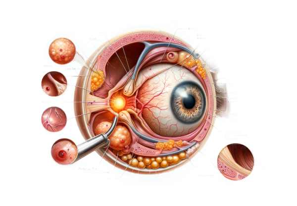
What is Orbital Rhabdomyosarcoma?
Orbital rhabdomyosarcoma is a malignant tumor that develops from skeletal muscle cells in the orbit, the bony cavity that houses the eyeball. It is the most common primary orbital malignancy in children, usually presenting between the ages of five and seven. This aggressive tumor can cause sudden symptoms like eye swelling, proptosis (eye bulging), and impaired vision. Early diagnosis and treatment are critical for avoiding serious complications and improving outcomes.
In-Depth Look at Orbital Rhabdomyosarcoma
Orbital rhabdomyosarcoma is a subtype of rhabdomyosarcoma, a cancer of the primitive muscle cells. The rapid growth of this malignancy can have a significant impact on the orbit, which contains vital structures such as the eye, optic nerve, and muscles controlling eye movements.
Epidemiology
Orbital rhabdomyosarcoma is uncommon, accounting for only 4% of all rhabdomyosarcomas and about 10% of pediatric soft tissue sarcomas. It primarily affects children, with the highest incidence between the ages of 5 and 7. There is a slight male majority. Although uncommon in adults, it can occur at any age.
Pathophysiology
Rhabdomyosarcoma develops from mesenchymal cells destined to become skeletal muscle. In orbital rhabdomyosarcoma, these cells become malignant and proliferate uncontrollably within the orbit. The tumor can develop from any of the orbit’s striated muscles, but the most common are the superior rectus and levator palpebrae superioris.
Rhabdomyosarcoma’s aggressive nature allows it to rapidly invade surrounding tissues such as the optic nerve, extraocular muscles, and even the bony walls of the orbit. Metastasis can occur through hematogenous spread, with distant sites such as the lungs, bone marrow, and lymph nodes.
Clinical Presentation
Symptoms of orbital rhabdomyosarcoma can develop quickly and include:
- Proptosis: The most common presenting symptom, characterized by the eye bulging forward due to the tumor’s mass effect.
- Eyelid Swelling: Severe swelling around the affected eye, often accompanied by redness.
- Ocular Motility Disturbances: Extraocular muscle involvement results in restricted eye movement and double vision (diplopia).
- Pain: Although less common, some patients may experience pain as a result of tumor pressure on surrounding structures.
- Vision Changes: Reduced visual acuity or visual field defects, particularly if the optic nerve is compressed or infiltrated.
Types of Orbital Rhabdomyosarcoma
Orbital rhabdomyosarcoma has several histological subtypes, each with a different prognosis and treatment response:
- Embryonal Rhabdomyosarcoma: The most common subtype in the orbit, accounting for roughly 60% of all cases. It usually has a good prognosis with proper treatment.
- Alveolar Rhabdomyosarcoma: Less common but more aggressive, requiring extensive treatment. It has a poorer prognosis than the embryonic type.
- Pleomorphic Rhabdomyosarcoma: Rare in children but more common in adults. It has a variable prognosis and frequently manifests as a more indolent course.
- Spindle Cell/Sclerosing Rhabdomyosarcoma: A type of embryonic rhabdomyosarcoma that can develop in the orbit. It has a relatively good prognosis if treated promptly.
Differential Diagnosis
Orbital rhabdomyosarcoma’s clinical presentation can be similar to other orbital conditions, necessitating differential diagnosis. Conditions to consider are:
- Orbital Cellulitis is an infection of the orbital tissues that can result in proptosis, swelling, and redness.
- Lymphoma: Lymphoma is a malignancy of the lymphatic tissues that can cause a mass in the orbit.
- Dermoid Cysts: Benign cystic lesions that can result in a slow-growing mass in the orbit.
- Neuroblastoma is a pediatric cancer that can spread to the orbit, causing proptosis and swelling.
- Thyroid Eye Disease: An autoimmune condition associated with thyroid dysfunction that causes bilateral or unilateral proptosis and abnormal eye movements.
Complications
If left untreated, orbital rhabdomyosarcoma can cause a number of serious complications, including:
- Vision Loss: Caused by compression or infiltration of the optic nerve.
- Intracranial Extension: The tumor can penetrate the orbital bones and enter the cranial cavity, causing neurological symptoms and increasing the risk of death.
- Metastasis: Spread to distant organs such as the lungs, liver, and bone marrow, which significantly worsens the prognosis.
- Cosmetic Deformities: Proptosis and eyelid changes can result in visible facial asymmetry and disfigurement.
- Secondary Infections: Tumor necrosis or treatment-induced immunosuppression can increase the likelihood of orbital and systemic infections.
Prognosis
The prognosis for orbital rhabdomyosarcoma has significantly improved thanks to advances in multimodal therapy, which includes surgery, radiation, and chemotherapy. Localized orbital rhabdomyosarcoma has an overall survival rate of more than 70% when treated properly. However, the prognosis is based on several factors:
- Histological Subtype: Embryonal rhabdomyosarcoma typically has a better prognosis than the alveolar subtype.
- Tumor Stage: Early-stage tumors confined to the orbit have a better prognosis than those with metastases.
- Response to Treatment: Tumors that respond well to initial therapy are more likely to achieve long-term control.
- Age of Onset: Younger children typically have a better prognosis than adolescents and adults.
How Orbital Rhabdomyosarcoma is Diagnosed?
Clinical evaluation, imaging studies, and histopathological confirmation are all required for an accurate diagnosis of orbital rhabdomyosarcoma. The following diagnostic methods are critical for determining the diagnosis and guiding treatment planning.
Clinical Evaluation
A thorough clinical evaluation is the first step in diagnosing orbital rhabdomyosarcoma.
- Patient History: Take a detailed history of the symptoms’ onset, duration, and progression. A family history of cancer or genetic syndromes may also be significant.
- Physical Examination: A thorough examination of the eyes and orbit, including assessments of visual acuity, ocular motility, eyelid position, and the presence of proptosis or swelling.
Imaging Studies
Imaging is critical for assessing the extent of the tumor, its relationship to surrounding structures, and for treatment planning.
- CT Scan (Computed Tomography): CT scans produce detailed cross-sectional images of the orbit, allowing for the determination of its mass, size, and any bony involvement. CT is especially useful for evaluating calcifications and determining the tumor’s impact on surrounding structures.
- MRI (Magnetic Resonance Imaging): MRI provides superior soft tissue contrast and is required for a thorough assessment of the tumor’s extent, particularly its involvement with the optic nerve, extraocular muscles, and possible intracranial extension. MRI can distinguish between tissue types and provide detailed images of the tumor’s characteristics.
- Ultrasound: An adjunctive tool for assessing the size, shape, and vascularity of the orbital mass is orbital ultrasound. It is a non-invasive, quick imaging technique that can help guide future diagnostic procedures.
Histopathologic Confirmation
To make a definitive diagnosis of orbital rhabdomyosarcoma, tissue biopsy and histopathological examination are required.
- Biopsy: A sample of the orbital mass is collected using fine-needle aspiration, incisional biopsy, or excisional biopsy. The size and location of the tumor influence the biopsy method chosen. Biopsy enables histological classification and grading of the tumor.
- Histopathology: The tissue sample is examined under a microscope to detect malignant rhabdomyoblasts, the hallmark cells of rhabdomyosarcoma. Immunohistochemical staining for desmin, myogenin, and MyoD1 aids in the diagnosis and differentiation from other orbital tumors.
Additional Tests
- Blood Tests: Routine blood tests, such as a complete blood count (CBC) and liver function tests, can help assess a patient’s overall health and detect systemic involvement.
- Bone Marrow Biopsy: In cases where systemic spread is suspected, a bone marrow biopsy may be performed to detect metastatic infiltration.
- Genetic and Molecular Testing: Advanced genetic and molecular tests can provide information about the tumor’s specific mutations and characteristics, allowing for the selection of targeted therapies.
Effective Therapies for Orbital Rhabdomyosarcoma
The treatment of orbital rhabdomyosarcoma is multidisciplinary, combining surgery, chemotherapy, and radiation therapy. The goal is to completely eradicate tumors while maintaining vision and minimizing long-term side effects. Treatment advances have resulted in significantly higher survival rates and outcomes for patients with this aggressive cancer.
Surgery
- Biopsy: The initial surgical intervention frequently includes obtaining a biopsy to confirm the diagnosis. This is critical for the histopathological examination and staging of the tumor.
- Resection: Depending on the size and location of the tumor, surgical resection may be necessary to remove as much of it as possible. Complete resection is frequently difficult due to the proximity of critical ocular structures. As a result, adjuvant therapies are typically administered after surgery.
Chemotherapy
Chemotherapy is essential in the treatment of orbital rhabdomyosarcoma. It shrinks the tumor, making surgical resection easier and more effective, and treats microscopic metastatic disease.
- VAC Regimen: The most common chemotherapy regimen for rhabdomyosarcoma is vincristine, actinomycin-D (dactinomycin), and cyclophosphamide. This combination has demonstrated efficacy in shrinking tumor size and preventing recurrence.
- Alternative Agents: Other chemotherapeutic agents, such as ifosfamide, etoposide, and irinotecan, may be used, particularly in cases of recurrent or resistant disease.
Radiation Therapy
Radiation therapy is critical for local tumor control, especially when complete surgical resection is not an option.
- External Beam Radiation Therapy (EBRT): This is the most common type of radiation used to target and destroy remaining cancer cells in the orbit. It is typically given in conjunction with chemotherapy.
- Proton Beam Therapy: A more advanced form of radiation therapy that allows for precise tumor targeting while sparing healthy tissues in the surrounding area. Proton therapy is especially useful in pediatric patients to reduce radiation-induced side effects.
Innovative and Emerging Therapies
- Targeted Therapy: Researchers are working to identify molecular targets unique to rhabdomyosarcoma. Drugs that target these pathways, such as tyrosine kinase inhibitors, are being studied for their effectiveness in treating this cancer.
- Immunotherapy: Immune checkpoint inhibitors and other immunotherapeutic approaches are being studied to improve the body’s immune response to rhabdomyosarcoma cells.
- Gene Therapy: New therapies involving gene editing techniques, such as CRISPR-Cas9, seek to correct genetic abnormalities that drive tumor growth. Although still in the experimental stage, these therapies show promise for future treatment options.
- Stem Cell Transplantation: In high-risk or recurring cases, hematopoietic stem cell transplantation may be considered after high-dose chemotherapy to restore bone marrow function.
Multidisciplinary Care
A multidisciplinary team of specialists, including pediatric oncologists, ophthalmologists, radiation oncologists, and surgeons, is required to effectively manage orbital rhabdomyosarcoma. This collaborative approach ensures that patients receive comprehensive care, tailored treatment plans, and close monitoring of their progress.
Effective Ways to Improve and Prevent Orbital Rhabdomyosarcoma
- Regular Medical Check-Ups: Routine health examinations and pediatric screenings can aid in the early detection of abnormalities that may indicate the presence of rhabdomyosarcoma.
- Awareness of Symptoms: Inform parents and caregivers about the signs and symptoms of orbital rhabdomyosarcoma, such as rapid-onset eye swelling, proptosis, and vision changes, to ensure prompt medical attention.
- Genetic Counseling: Families with a history of cancer or genetic syndromes linked to rhabdomyosarcoma should seek genetic counseling to better understand their risk and take preventative measures.
- Carcinogen Avoidance: To reduce the risk of developing cancer, limit exposure to known carcinogens such as certain chemicals and radiation, particularly in children.
- Healthy Lifestyle: Encourage a balanced diet, regular exercise, and quitting smoking to improve overall health and potentially reduce the risk of cancer.
- Early Treatment of Infections: Early treatment of infections, particularly those affecting the eye and orbit, can prevent chronic inflammation, which may contribute to cancer development.
- Protective Measures: Wear protective eyewear when participating in activities that pose a risk of eye injury, lowering the likelihood of trauma that could lead to tumor formation.
- Regular Follow-Up: For children who have received treatment for rhabdomyosarcoma, regular follow-up visits are required to monitor for recurrence and manage any long-term treatment side effects.
- Research Participation: Participating in clinical trials can provide access to new treatments and help advance knowledge and therapies for rhabdomyosarcoma.
Trusted Resources
Books
- “Orbital Tumors: Diagnosis and Treatment” by Jerry A. Shields and Carol L. Shields
- “Pediatric Ophthalmology and Strabismus” by Kenneth W. Wright
- “Clinical Pediatric Oncology” by William L. Carroll and Judith M. Willert
Online Resources
- American Cancer Society (ACS): cancer.org
- National Cancer Institute (NCI): cancer.gov
- Children’s Oncology Group (COG): childrensoncologygroup.org


