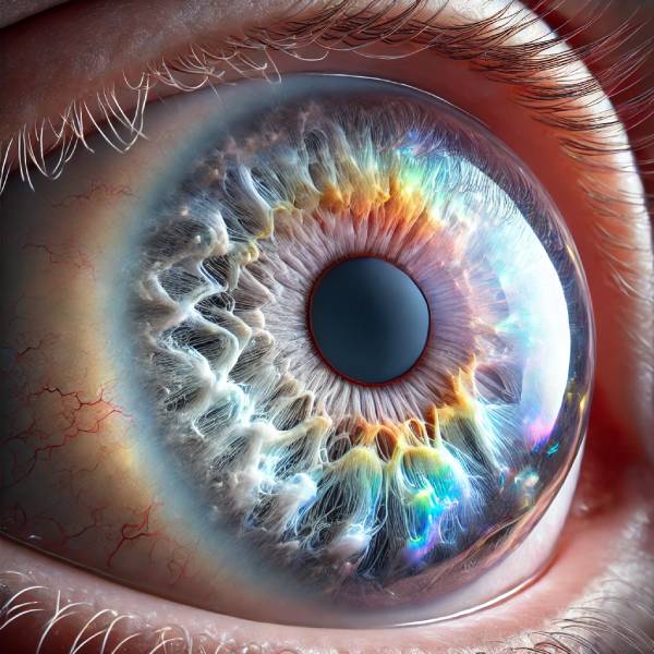
Polychromatic cataract is a unique type of cataract distinguished by its multicolored appearance, which frequently manifests as a rainbow of colors within the eye’s lens. Cataracts are opacities that form in the crystalline lens, causing a decrease in vision. Cataracts are commonly associated with aging, but they can also be caused by genetics, trauma, or systemic diseases. The polychromatic cataract is notable for its distinct appearance and underlying causes.
Pathophysiology
The lens of the eye is normally clear and helps to focus light on the retina. A cataract forms when proteins in the lens clump together, resulting in cloudy areas. Polychromatic cataracts have opacities that reflect and refract light, making different colors visible. This multicolored appearance is due to the varying densities and arrangements of the protein clumps, as well as potential interactions with different wavelengths of light.
Etiology
There are several factors that can cause polychromatic cataracts:
- Genetic Factors: Certain genetic mutations and inherited conditions can increase the risk of developing cataracts at a young age. These genetic factors can also influence the type and appearance of cataracts, including polychromatic cataracts.
- Trauma: A physical injury to the eye can disrupt the normal structure of the lens, resulting in cataract formation. Traumatic cataracts can have a polychromatic appearance due to the irregular way in which lens fibers heal and reorganize following injury.
- Systemic Diseases: Diabetes, metabolic disorders, and other systemic illnesses can all affect the eye’s lens, potentially resulting in cataract formation. These diseases’ biochemical changes can give polychromatic cataracts their distinct multicolored appearance.
- Medications: Prolonged use of certain medications, such as corticosteroids, can raise the risk of cataract formation. These drug-induced cataracts can occasionally show polychromatic characteristics.
- Radiation Exposure: Exposure to radiation, whether from medical treatments or environmental sources, can harm the lens and cause cataract formation. Polychromatic cataracts can have a distinct appearance due to their specific pattern of damage.
Clinical Presentation
Patients with polychromatic cataracts frequently report a variety of visual symptoms, including:
- Blurry Vision: As with other types of cataracts, the main symptom is gradual blurring of vision, which can interfere with daily activities like reading, driving, and recognizing faces.
- Multicolored Halos: One of the distinguishing features of polychromatic cataracts is the perception of multiple colors around lights. This is due to how the irregular lens fibers scatter light.
- Glare Sensitivity: Increased sensitivity to bright lights and glare is common, especially in situations with high contrast lighting, such as night driving.
- Reduced Contrast Sensitivity: Patients may struggle to distinguish between objects of similar color and brightness, making it difficult to see fine details.
Epidemiology
Cataracts are one of the most common causes of visual impairment worldwide, affecting millions of people. While age-related cataracts are the most common, polychromatic cataracts occur less frequently. However, determining their exact prevalence is difficult due to the variety of underlying causes and overlap with other types of cataracts. Depending on the underlying causes, they can affect people of any age.
Impact on Vision
The effect of polychromatic cataracts on vision varies greatly depending on the size, location, and density of the opacities within the lens. Small, peripheral cataracts may cause minor visual disturbances, whereas larger, central opacities can severely impair vision. The unique polychromatic nature of these cataracts can complicate the visual symptoms, as patients may experience unusual visual artifacts that are not present in other types of cataracts.
Differential Diagnosis
Differentiating polychromatic cataracts from other types of cataracts and ocular conditions is critical for proper diagnosis and treatment. The differential diagnosis can include:
- Nuclear Sclerotic Cataracts: The most common type of age-related cataracts, with yellowing and hardening of the central lens. They do not usually have a multicolored appearance like polychromatic cataracts.
- Cortical Cataracts: These cataracts affect the lens cortex and frequently appear as spoke-like opacities. While they can cause visual disturbances, they typically lack the distinguishing polychromatic characteristic.
- Posterior Subcapsular Cataracts: These cataracts develop at the back of the lens and can cause significant vision loss, particularly in bright light. They are not usually polychromatic.
- Keratoconus is a condition in which the cornea thins and bulges into a cone shape, causing visual distortion. Unlike cataracts, keratoconus affects the cornea rather than the lens, and it does not produce the multicolored halos associated with polychromatic cataract.
Complications
While polychromatic cataracts themselves are harmless, they can cause complications if left untreated. Progressive visual impairment can have an impact on an individual’s quality of life by making it difficult to perform daily tasks. In severe cases, untreated cataracts can cause complete vision loss. Polychromatic cataracts can also be associated with other eye conditions that require treatment, such as glaucoma or retinal detachment.
Pathologic Findings
Polychromatic cataracts show varying patterns of protein aggregation within the lens fibers. These protein clumps are what cause cataracts to scatter light and appear multicolored. Understanding the specific pathological changes can provide information about the underlying mechanisms and potential therapeutic targets.
Ongoing Research
Polychromatic cataract research is ongoing, with a focus on understanding the genetic, molecular, and environmental factors that influence their development. Advances in imaging techniques and molecular biology are shedding light on the pathophysiology of these distinct cataract types. This study holds the promise of improved diagnostic methods and targeted treatments in the future.
Methods to Diagnose Polychromatic Cataracts
Polychromatic cataracts are diagnosed using a combination of clinical examination, imaging studies, and, in some cases, laboratory tests. Each diagnostic method makes a unique contribution to the accurate identification and differentiation of cataracts from other ocular conditions.
Clinical Examination
A thorough clinical examination is the first step in the diagnosis of polychromatic cataracts. This includes a thorough patient history to better understand the onset and progression of symptoms, as well as any potential risk factors like trauma, systemic diseases, or medication use. During the physical examination, an ophthalmologist will use a slit-lamp microscope to look for the distinctive multicolored opacities on the lens. The slit-lamp exam provides a detailed view of the anterior segment of the eye, which includes the cornea, iris, and lens.
Visual Acuity Test
A visual acuity test is performed to determine the effect of cataracts on the patient’s vision. This test measures vision sharpness at various distances and aids in determining the severity of visual impairment. The results of this test can provide useful information about the severity of cataracts, guiding future diagnostic and therapeutic decisions.
Imaging Studies
Imaging studies are necessary for a thorough evaluation of polychromatic cataracts.
- Optical Coherence Tomography (OCT): OCT is a non-invasive imaging technique for obtaining high-resolution cross-sectional images of the eye’s structures. It can be used to determine the size and location of lens opacities, as well as the health of the retina and optic nerve.
- Ultrasound Biomicroscopy (UBM): This technique creates detailed images of the eye’s anterior segment using high-frequency ultrasound waves. It is especially useful for inspecting the lens and identifying any associated abnormalities, such as zonular dehiscence or posterior capsular opacification.
- Scheimpflug Imaging: This technique employs a rotating camera to obtain detailed images of the anterior segment, including the cornea and lens. It can provide quantitative measurements of lens opacity and help to track the progression of cataracts over time.
Lab Tests
In some cases, laboratory tests may be necessary to identify underlying systemic conditions that could contribute to the development of polychromatic cataracts. Blood tests, for example, may be performed to assess blood glucose levels in patients suspected of having diabetes, while genetic testing may be considered in cases with a strong family history of cataracts.
Differential Diagnosis
Differentiating polychromatic cataracts from other types of cataracts and ocular conditions is critical for proper diagnosis and treatment. To rule out other possible causes of visual impairment, this process combines clinical evaluation, imaging studies, and, in some cases, laboratory tests.
Genetic Testing
Genetic testing is especially useful when a hereditary component is suspected. Identifying specific genetic mutations linked to cataract formation can provide useful information for diagnosis and may also have implications for family members who are at risk.
Polychromatic Cataract Management
Managing polychromatic cataracts requires a comprehensive approach that takes into account the patient’s overall health, the severity of the cataract, and the impact on vision and daily life. The primary goal is to restore and maintain vision, which frequently necessitates surgical intervention. Here are the primary strategies for managing polychromatic cataracts:
Non-surgical Management
- Monitoring and Regular Eye Exams: In the early stages, if the cataract is not significantly impairing vision, regular monitoring and eye exams are required. This allows for the assessment of progression and timely intervention as needed.
- Lifestyle Changes: Patients are frequently advised to make lifestyle changes to alleviate symptoms. This may include using brighter lighting at home, wearing sunglasses to reduce glare, and using magnifying lenses for reading and other close-up tasks.
- Prescription Glasses or Contact Lenses: Updating the prescription for glasses or contact lenses may temporarily improve vision. These visual aids do not treat cataracts, but they can help manage symptoms until surgery is necessary.
Surgical Management
Surgical intervention is the most effective treatment for cataracts, including polychromatic ones. The standard procedure is cataract extraction and intraocular lens (IOL) implantation.
- Phacoemulsification is the most popular cataract surgery technique. It entails breaking up the cloudy lens into smaller pieces with ultrasound waves before removing them through a small incision. An artificial intraocular lens (IOL) is implanted to replace the missing lens. Phacoemulsification is popular because of its small incision size, which promotes faster healing and lowers the risk of complications.
- Extracapsular Cataract Extraction (ECCE): When the cataract is too dense or difficult to phacoemulsify, ECCE may be used. This involves removing the cloudy lens in one piece using a larger incision. The IOL is then implanted. While effective, ECCE typically has a longer recovery time and a higher risk of complications than phacoemulsification.
- Laser-Assisted Cataract Surgery: This advanced technique employs femtosecond lasers to perform specific steps of cataract surgery with extreme precision. The laser can make corneal incisions, open the lens capsule, and soften the cataract before removing it with traditional phacoemulsification. Laser-assisted surgery provides greater accuracy and may improve surgical outcomes, but it is typically more expensive.
Post-operative Care and Follow-Up
Postoperative care is critical for achieving successful results and avoiding complications. Key features include:
- Medications: Typically, patients are given antibiotic and anti-inflammatory eye drops to prevent infection and reduce inflammation. It is critical to adhere to the ophthalmologist’s medication regimen.
- Follow-Up Visits: Regular follow-up visits are scheduled to monitor the healing process, check the IOL’s positioning, and evaluate visual acuity. These visits aid in the early detection and treatment of complications such as infection, raised intraocular pressure, and lens displacement.
- Activity Restrictions: Patients should avoid strenuous activities, heavy lifting, and rubbing their eyes during the initial healing period. Protective eyewear may be prescribed to protect the eye from injury and bright light.
Treating Complications and Secondary Cataracts
- Posterior Capsular Opacification (PCO): Also referred to as secondary cataract, PCO can appear months or years after cataract surgery. It happens when the back of the lens capsule, which is left in place to support the IOL, turns cloudy. The treatment involves a quick and painless laser procedure known as YAG laser capsulotomy, which creates a clear opening in the capsule to restore vision.
- Intraocular Lens (IOL) Issues: Complications with the IOL, such as dislocation or incorrect lens power, are possible but rare. To address these issues, the IOL may require surgical adjustment or replacement.
- Infections and Inflammation: Endophthalmitis, or severe eye infection, is a rare but serious complication. Prompt medical treatment is critical to preventing vision loss. Persistent inflammation may necessitate prolonged use of anti-inflammatory medications.
Trusted Resources and Support
Books
- “Cataracts: A Patient’s Guide to Treatment” by Robert S. Feder, MD
- This comprehensive guide provides detailed information on cataract types, treatment options, and postoperative care, making it an invaluable resource for patients.
- “Ophthalmology” by Myron Yanoff and Jay S. Duker
- A detailed textbook that offers in-depth coverage of various eye conditions, including cataracts, with the latest research and treatment methodologies.
Organizations
- American Academy of Ophthalmology (AAO)
- Website: www.aao.org
- The AAO provides extensive resources on cataracts, including patient education materials, research updates, and professional guidelines.
- National Eye Institute (NEI)
- Website: www.nei.nih.gov
- The NEI offers comprehensive information on eye health, research on eye diseases, and support for patients and families affected by cataracts.


