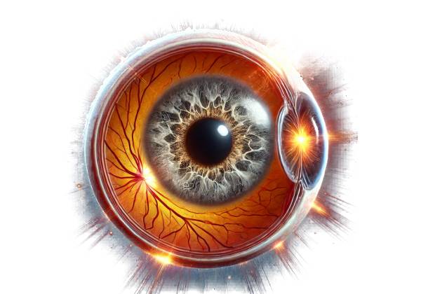
What is Solar Retinopathy (SR)?
Solar retinopathy is an ocular condition caused by direct exposure of the retina to intense sunlight, especially when viewing the sun without proper eye protection. This condition is most commonly associated with solar eclipses, in which people may be tempted to stare at the partially obscured sun for extended periods. However, solar retinopathy can also result from other forms of sun exposure or exposure to bright artificial lights, such as lasers. The condition is defined by retinal damage, specifically to the macula, which is the central part of the retina responsible for sharp, detailed vision.
Retina Anatomy and Macula Function
Understanding solar retinopathy requires a basic understanding of the retina and macula. The retina is a thin layer of tissue in the back of the eye that receives light and converts it into neural signals, which the brain interprets as visual images. The macula is a small, specialized area of the retina that provides the central field of vision, allowing us to see fine details clearly, such as when reading text or recognizing faces.
The macula has a high concentration of photoreceptor cells, specifically cones, which are responsible for color vision and sharp central vision. These photoreceptors are very sensitive to light. When the retina is exposed to excessive light, especially ultraviolet (UV) and visible light, the photoreceptors and underlying retinal pigment epithelium (RPE) can be damaged, resulting in solar retinopathy.
Pathophysiology Of Solar Retinopathy
Solar retinopathy occurs when the retina, particularly the macula, is exposed to a large amount of sunlight or other intense light sources. This exposure causes photochemical injury, in which light energy triggers toxic reactions in the retinal tissue. The photoreceptors and RPE absorb the intense light, which produces reactive oxygen species (ROS) and other free radicals. These reactive molecules cause oxidative stress, which harms cellular structures like photoreceptor cells, RPE, and surrounding retinal tissue.
Solar retinopathy frequently causes damage to the fovea, the central part of the macula responsible for high-acuity vision. The fovea lacks blood vessels, making it especially vulnerable to photochemical injury. Furthermore, because the fovea is rich in pigments that absorb blue light, it is particularly vulnerable to damage from this part of the light spectrum.
Solar retinopathy injury is usually symmetrical, affecting both eyes in the same way, because people use both eyes when looking at a bright light source. Several factors influence the extent of the damage, including the duration of exposure, the intensity of the light, and the use (or lack thereof) of protective eyewear.
Clinical Features and Symptoms
Solar retinopathy symptoms typically appear within hours to a day of exposure to an intense light source. The symptoms can vary depending on the severity of the retinal damage, but they commonly include the following:
- Central Scotoma: One of the most common symptoms of solar retinopathy is the appearance of a central scotoma, or dark spot, in the field of vision. This scotoma corresponds to the area of retinal damage and can make it difficult to see objects directly in front of the person, interfering with activities such as reading or driving.
- Blurred Vision: Patients with solar retinopathy frequently experience blurred or distorted vision. This blurring usually affects the central vision, making it difficult to focus on small details.
- Metamorphopsia: A visual distortion that causes straight lines to appear wavy or bent. This symptom occurs as a result of macula damage, which impairs normal visual perception.
- Photophobia: Another common symptom is increased light sensitivity, or photophobia. The damaged retina becomes more light sensitive, making bright environments uncomfortable.
- Color Vision Changes: Some patients may experience changes in their color vision, including difficulty distinguishing between certain colors. This happens because the cones responsible for color vision are concentrated in the macula and are frequently damaged in solar retinopathy.
- Afterimages: Some people have reported seeing afterimages, or residual images, after looking away from the light source. These afterimages are frequently negative (dark) or positive (bright), and they can last for some time after the initial exposure.
Prognosis and Long-Term Effects
The prognosis of solar retinopathy varies according to the extent of the damage and the patient’s healing response. In many cases, symptoms may improve over several weeks or months as the retina heals. However, some degree of permanent visual impairment may occur, especially if the exposure was prolonged or severe.
- Recovery: For many patients, the scotoma and other visual disturbances may fade with time, with significant improvement in vision occurring within the first few months. However, complete recovery is not guaranteed, and some patients may continue to experience residual symptoms, such as mild scotomas or changes in color vision.
- Permanent Damage: Severe exposure can result in irreversible damage to the photoreceptors and RPE. This can result in permanent central vision loss, making it difficult to perform detailed vision tasks like reading or recognizing faces.
- Macular Scarring: In some cases, retinal damage can result in the formation of scar tissue in the macula. This scarring can worsen central vision and lead to a more severe and permanent central scotoma.
Risk Factors
Several factors can raise the risk of developing solar retinopathy.
- Solar Eclipses: Viewing a solar eclipse without proper eye protection is one of the leading causes of solar retinopathy. During a solar eclipse, the sun is partially obscured, reducing overall brightness and making it tempting to look directly at it without realizing the dangers.
- Prolonged Sun Gazing: Intentional or unintentional prolonged exposure to the sun, such as during meditation, sunbathing, or while under the influence of drugs, can increase the risk of developing solar retinopathy.
- Lack of Protective Eyewear: Failure to wear adequate eye protection, such as eclipse glasses or proper filters, when viewing the sun significantly increases the risk of retinal damage.
- Younger Age: Younger people, especially children and teenagers, may be more vulnerable because they are more likely to engage in risky behaviors like sun-gazing during a solar eclipse.
- High-Altitude Locations: Being at a high altitude, where the atmosphere is thinner and less UV radiation is absorbed, can increase the risk of developing solar retinopathy from sun exposure.
Differential Diagnosis
Solar retinopathy is distinct from other conditions that cause central vision loss or scotomas. These conditions include the following:
- Age-Related Macular Degeneration (AMD): AMD is a common condition that affects the macula and causes central vision loss. Unlike solar retinopathy, AMD usually develops slowly and is associated with aging.
- Central Serous Chorioretinopathy (CSCR): CSCR is characterized by the accumulation of fluid beneath the retina, which causes the retinal pigment epithelium to detach. It can cause similar visual disturbances but uses different mechanisms.
- Toxic Maculopathy: Certain medications or toxins, such as chloroquine or hydroxychloroquine, can damage the macula, resulting in symptoms similar to solar retinopathy.
- Macular Hole: A macular hole is a tiny break in the macula that can result in central vision loss and other visual symptoms. It is frequently associated with aging or trauma, rather than sun exposure.
- Optic Neuritis: Inflammation of the optic nerve can cause sudden vision loss and the development of scotomas. However, this condition is usually associated with other neurological symptoms and does not result in direct retinal damage from light exposure.
Differentiating solar retinopathy from these other conditions is critical for determining the best treatment and prognosis. A thorough clinical evaluation and history-taking are required to make an accurate diagnosis.
Effective Diagnosis of Solar Retinopathy
Solar retinopathy is diagnosed using a combination of patient history, clinical examination, and specialized imaging techniques. Accurate diagnosis is critical for determining the extent of retinal damage and making appropriate management decisions.
Clinical Examination
A thorough eye exam is the first step in diagnosing solar retinopathy. The exam typically includes:
- Visual Acuity Testing: This test measures the sharpness of the patient’s vision and is required to determine the impact of solar retinopathy on central vision. Patients with solar retinopathy frequently exhibit decreased visual acuity, particularly in the central field of vision.
- Amsler Grid Test: The Amsler grid test can detect distortions in the central visual field, such as scotomas or metamorphopsia. Patients with solar retinopathy may notice dark spots, wavy lines, or other visual distortions when viewing the grid.
- Fundus Examination: The ophthalmologist uses an ophthalmoscope or a slit-lamp with a special lens to examine the retina, particularly the macula, for signs of damage. In the early stages of solar retinopathy, the macula may appear normal or have minor changes. Yellowish spots, foveal granularity, and changes to the retinal pigment epithelium may appear over time.
Imaging Techniques
Advanced imaging techniques are frequently used to provide a more thorough assessment of retinal damage in solar retinopathy. These imaging methods aid in the visualization of structural changes in the retina that would otherwise be difficult to detect during a standard clinical examination.
- Optical Coherence Tomography (OCT): OCT is a non-invasive imaging technique for obtaining high-resolution cross-sectional images of the retina. It is one of the most useful tools for diagnosing solar retinopathy because it enables detailed visualization of the macular structure. In cases of solar retinopathy, OCT may reveal photoreceptor layer disruptions, retinal layer thinning, and changes in the retinal pigment epithelium (RPE). OCT can also help monitor the condition’s progression and determine whether it is improving or worsening over time.
- Fundus Photography: Fundus photography involves taking detailed images of the retina to document and compare over time. Fundus photographs may not show significant changes in the early stages of solar retinopathy, but as the disease progresses, they may reveal changes such as small yellowish-white spots at the fovea or changes in the RPE.
- Fluorescein Angiography (FA): Fluorescein angiography is a technique that involves injecting a fluorescent dye into the bloodstream and taking a series of photographs as the dye travels through the retinal blood vessels. This test can detect abnormalities in the retinal circulation, but it is less commonly used in solar retinopathy unless there is a suspicion of associated vascular damage.
- Autofluorescence Imaging: This technique detects naturally occurring fluorescence in the retinal pigment epithelium (RPE). In solar retinopathy, areas of RPE damage may exhibit altered autofluorescence, assisting in determining the extent and location of retinal injury.
- Electroretinography (ERG): Measures the electrical responses of retinal cells to light stimuli. While ERG is not commonly used to diagnose solar retinopathy, it can be useful in assessing the overall function of the retina, especially if there is concern about widespread retinal damage beyond the macula.
- Visual Field Testing: Visual field testing maps the visual field and detects scotomas or areas of vision loss. In solar retinopathy, this test can help quantify the size and location of central scotomas, which is useful for both diagnosis and monitoring.
Immediate care and observation
In the immediate aftermath of sun exposure, patients are frequently advised to rest their eyes and avoid further bright light exposure. This includes wearing sunglasses outdoors and limiting screen time or activities that require intense visual focus. Immediate care is primarily observational, as the severity of retinal damage and the body’s natural healing response can take several days to manifest.
Patients are usually closely monitored in the days and weeks after an incident. Follow-up visits may include additional visual acuity tests, Amsler grid testing, and imaging studies such as optical coherence tomography (OCT) to assess retinal changes and monitor for signs of improvement or deterioration.
Pharmaceutical Interventions
Although there is no direct pharmacological treatment for solar retinopathy, the following medications may be prescribed to manage symptoms or prevent complications:
- Anti-inflammatory Medications: Nonsteroidal anti-inflammatory drugs (NSAIDs) or corticosteroids may be prescribed to reduce eye inflammation, particularly if there is significant discomfort or retinal edema. Topical corticosteroid eye drops can also be used to reduce intraocular inflammation, but their use is typically limited due to potential side effects.
- Antioxidant Supplements: Antioxidant supplements such as vitamins C and E, beta-carotene, and zinc are occasionally recommended to improve retinal health and reduce oxidative stress. While there is limited evidence to support their efficacy in solar retinopathy, these supplements may help promote retinal healing.
- Vasodilators: In some cases, vasodilators may be considered to improve blood flow to the retina and aid in healing. However, the use of these agents is not universally accepted, and their benefits in solar retinopathy are unknown.
Observations and Follow-Up
Given the possibility of gradual improvement in solar retinopathy, regular follow-up appointments are required. These visits typically include:
- Monitoring Visual Acuity: Observing changes in visual acuity over time can help determine whether the retina is healing and whether the patient’s vision is stabilizing or improving.
- OCT and Other Imaging: Repeated OCT scans and other imaging studies allow for close observation of the retinal layers, which aids in detecting subtle improvements or signs of further deterioration.
- Symptom Management: Patients are monitored for persistent symptoms, such as scotomas, blurred vision, or photophobia, and receive ongoing support to manage them.
Vision Rehabilitation
Vision rehabilitation may be an important part of a patient’s treatment plan if they have persistent visual impairment as a result of solar retinopathy. Vision rehabilitation services assist patients in adapting to vision changes while also maintaining their quality of life. This may include:
- Low Vision Aids: Magnifiers, specialized reading glasses, and electronic reading aids can assist patients with central vision loss in continuing to perform daily tasks like reading and writing.
- Occupational Therapy: Occupational therapists who specialize in vision impairment can train patients on how to use low vision aids and teach strategies for navigating the environment with limited vision.
- Adaptive Techniques: Patients may be taught adaptive techniques to compensate for central vision loss, such as improving peripheral vision or changing the lighting and contrast in their living spaces.
Preventative Measures
Preventing solar retinopathy is critical, particularly in vulnerable populations such as those who may be exposed to solar eclipses or other intense light sources. Prevention measures include:
- Public Education: Educating the public about the dangers of looking directly at the sun, particularly during solar eclipses, is crucial. Emphasizing the importance of wearing proper eye protection, such as ISO-certified eclipse glasses, can significantly reduce the occurrence of solar retinopathy.
- Protective Eyewear: Recommending the use of protective eyewear, such as UV-protected sunglasses, for people who are exposed to bright sunlight or work in environments with intense artificial light can help prevent retinal damage.
- Avoiding Risky Behaviors: It is critical to advise patients to avoid behaviors that increase their risk of developing solar retinopathy, such as intentional sun-gazing or using unfiltered optical devices.
Emerging Therapies
Research into treatments for solar retinopathy is ongoing, and emerging therapies may provide new management options in the future. This may include:
- Stem Cell Therapy: Although still in the experimental stage, stem cell therapy has the potential to repair retinal damage by regenerating photoreceptor cells or the retinal pigment epithelium.
- Neuroprotective Agents: Another area of interest is the development of neuroprotective drugs that protect retinal neurons from damage and promote survival following injury. These therapies have the potential to limit the extent of damage in solar retinopathy while also promoting recovery.
- Gene Therapy: While gene therapy has primarily been studied in inherited retinal diseases, it may one day be used to treat damage in conditions such as solar retinopathy, particularly if specific genetic pathways influencing susceptibility or recovery are identified.
Overall, solar retinopathy management consists of a combination of supportive care, symptom management, and preventative strategies. While there is currently no definitive treatment for solar retinopathy, ongoing research and emerging therapies may provide hope for improved outcomes in the future.
Trusted Resources and Support
Books
- “The Retina: An Atlas of Key Diseases” by Joseph W. S. Patrick: This comprehensive atlas provides detailed images and descriptions of various retinal conditions, including solar retinopathy. It’s a valuable resource for ophthalmologists and eye care professionals.
- “Retinal Disorders: A Clinical Guide to Diagnosis” by Jerry A. Shields: This guide covers a range of retinal disorders, offering insights into their diagnosis and management. It includes a section on solar retinopathy, making it a useful reference for both students and practitioners.
Organizations
- American Academy of Ophthalmology (AAO): The AAO is a leading resource for information on eye conditions, including solar retinopathy. They provide educational materials for both patients and healthcare providers, as well as updates on the latest research and treatment options.
- National Eye Institute (NEI): The NEI offers extensive resources on eye health and vision research, including information on solar retinopathy. Their website includes educational content, clinical trial information, and patient resources.
- Prevent Blindness: Prevent Blindness is a national nonprofit organization dedicated to preventing blindness and preserving sight. They offer public education on eye health, including resources on protecting eyes from solar damage and understanding solar retinopathy.


