What is vitreous hemorrhage?
Vitreous hemorrhage is a medical condition in which blood leaks into the vitreous humor, a clear, gel-like substance that fills the space between the lens and retina in the eye. Depending on the severity and extent of the bleeding, this condition can cause a variety of visual disturbances, ranging from mild blurring to total vision loss. Vitreous hemorrhage is not a disease in and of itself, but rather a symptom of underlying ocular or systemic conditions that impair the integrity of blood vessels within or near the eye.
Anatomy of the Vitreous Body and Retina
Understanding vitreous hemorrhage requires a basic understanding of the eye’s anatomy, particularly the vitreous body and retina. The vitreous humor is a transparent, gelatinous substance that accounts for approximately 80% of the eye’s volume. It helps keep the eye’s spherical shape, serves as a medium for light to pass from the lens to the retina, and supports the retina by exerting a slight pressure that keeps it in place against the back of the eye.
The retina is a thin layer of light-sensitive cells that covers the inner surface of the back of the eye. It converts light into electrical signals, which are then transmitted to the brain via the optic nerve to enable vision. The retina is well-supplied with blood vessels that deliver oxygen and nutrients. However, these blood vessels are fragile and easily damaged, resulting in bleeding inside the eye, including into the vitreous humor.
The causes of vitreous hemorrhage
A variety of conditions can cause vitreous hemorrhage, including those that affect the retina, its blood vessels, or other structures in the eye. The most common causes of vitreous hemorrhage are:
- Diabetic Retinopathy: Diabetic retinopathy is the most common cause of vitreous hemorrhage, especially in people who have poorly controlled diabetes. Diabetes can eventually damage the blood vessels in the retina, resulting in the formation of abnormal, fragile blood vessels (neovascularization). These vessels are susceptible to rupture, allowing blood to leak into the vitreous humor. Proliferative diabetic retinopathy, an advanced stage of the disease, is frequently associated with vitreous hemorrhage.
- Retinal Tears and Detachments: Retinal tears or detachments are a common cause of vitreous hemorrhage. When the retina tears, the blood vessels within it may break, resulting in bleeding into the vitreous. Retinal detachment is a more serious condition in which the retina is pulled away from its normal position, usually due to the vitreous humor tugging on the retina. This can also lead to significant hemorrhage.
- Trauma: Ocular trauma, including blunt force, penetrating injury, and surgical procedures, can result in vitreous hemorrhage. Trauma can directly damage blood vessels in the retina or other parts of the eye, causing bleeding. Traumatic vitreous hemorrhage can occur immediately after an injury or as a complication.
- Posterior Vitreous Detachment (PVD): PVD occurs when the vitreous humor separates from the retina, which is a common occurrence as we age. However, in some cases, PVD can cause retinal tears or damage to the retinal blood vessels, resulting in vitreous hemorrhage. This is more likely to happen in people who have pre-existing retinal conditions or have significant myopia.
- Age-Related Macular Degeneration (AMD): Neovascular (wet) AMD occurs when abnormal blood vessels develop beneath the retina and leak blood or fluid. If these vessels break through the retina and bleed into the vitreous, they can cause a vitreous hemorrhage. AMD is a common cause of vision loss in older adults and can lead to serious complications if not treated promptly.
- Retinal Vein Occlusion: This condition occurs when one of the veins that drain blood from the retina becomes clogged, causing increased pressure in the retinal blood vessels and subsequent hemorrhage. If the pressure is high enough, blood can leak into the vitreous humor.
- Blood Disorders: Sickle cell disease, leukemia, and severe anemia can all increase the risk of vitreous hemorrhage. These conditions can impair the blood’s ability to clot properly or cause abnormal blood vessel formation, both of which can cause bleeding inside the eye.
Symptoms of Vitreous Hemorrhage
The symptoms of vitreous hemorrhage can vary greatly depending on how much blood enters the vitreous humor and what caused the bleeding. Common symptoms include:
- Sudden Onset of Floaters: Patients frequently report a sudden increase in floaters, which are small, dark shapes that move across their visual field. The shadows cast by the blood clumps in the vitreous humor are what these floaters are. The floaters can appear as spots, cobwebs, or thread-like strands and can be especially bothersome when viewed against a bright background.
- Blurred Vision: Blurred vision is another common sign of vitreous hemorrhage. The presence of blood in the vitreous humor can obstruct the passage of light to the retina, resulting in reduced visual clarity. The amount of blood present determines the degree of blurring; mild hemorrhages may result in only minor blurring, whereas severe hemorrhages can cause significant visual impairment.
- Dark Shadows or Red Tint in Vision: Patients who have had moderate to severe vitreous hemorrhage may notice dark shadows or a red tint in their vision. Larger blood clots within the vitreous humor prevent light from reaching the retina, resulting in these shadows. The red tint results from the blood’s ability to absorb and scatter light differently than the clear vitreous gel.
- Sudden Loss of Vision: In severe cases, when a large amount of blood enters the vitreous humor, the patient may suffer a sudden and dramatic loss of vision. This can be particularly alarming and frequently necessitates immediate medical attention. In such cases, the vitreous hemorrhage may be so extensive that it completely blocks the passage of light to the retina, leaving the patient effectively blind in the affected eye until the blood clears.
- Photopsia (Flashes of Light): Some patients with vitreous hemorrhage may experience photopsia, which are flashes of light. This symptom is frequently associated with retinal tears or detachment, which can occur alongside vitreous hemorrhage. The flashes occur as a result of the vitreous humor pulling on the retina, stimulating the retinal cells and producing the sensation of light even in the absence of external light sources.
Risk Factors of Vitreous Hemorrhage
Several factors can raise the risk of developing vitreous hemorrhage, including:
- Diabetes: Diabetes, especially when poorly controlled, is a significant risk factor for vitreous hemorrhage because it increases the likelihood of developing diabetic retinopathy. Regular monitoring and management of blood sugar levels are critical in lowering this risk.
- High Myopia: People with high myopia (severe nearsightedness) have a higher risk of retinal tears, detachment, and vitreous hemorrhage. The elongated shape of the myopic eye adds stress to the retina and vitreous, increasing the risk of complications.
- Age: Aging is another significant risk factor. As people age, they are more likely to develop conditions like PVD, AMD, and retinal vein occlusion, all of which can cause vitreous hemorrhage.
- History of Ocular Trauma: Previous eye injuries or surgeries can increase the risk of vitreous hemorrhage, especially if the retinal blood vessels have been severely damaged.
- Blood Clotting Disorders: People who have blood clotting disorders, such as hemophilia, or who take anticoagulants may be more likely to bleed into the vitreous humor.
Potential complications of vitreous hemorrhage
While some cases of vitreous hemorrhage heal on their own as the blood is gradually reabsorbed by the body, others can result in serious complications that threaten vision. Possible complications include:
Retinal detachment is one of the most serious complications of vitreous hemorrhage. If the hemorrhage causes or results in a retinal tear, there is a high risk that the retina will detach, resulting in permanent vision loss if not treated promptly.
- Proliferative Vitreoretinopathy (PVR): PVR is a condition in which scar tissue forms on the surface of the retina, leading to additional complications such as retinal detachment. PVR can develop as a result of recurrent or severe vitreous hemorrhages, especially in proliferative diabetic retinopathy.
- Neovascular Glaucoma: Neovascular glaucoma is a type of glaucoma that develops when abnormal blood vessels grow and obstruct the eye’s drainage angle, causing increased intraocular pressure. This condition may develop in response to the same factors that cause vitreous hemorrhage, particularly in diabetic retinopathy.
- Vision Loss: Persistent or recurrent vitreous hemorrhages, particularly when combined with complications like retinal detachment or PVR, can cause significant and sometimes permanent vision loss.
Diagnostic methods
To diagnose vitreous hemorrhage, a patient history, clinical examination, and advanced imaging techniques are all required. The diagnostic process aims to determine the source and extent of the hemorrhage, as well as the underlying cause and potential complications.
Clinical Examination
The first step in diagnosing vitreous hemorrhage is usually a thorough patient history and eye examination. The clinician will inquire about the onset, duration, and nature of the patient’s symptoms, such as floaters, blurred vision, or sudden loss of vision. A thorough understanding of the patient’s medical history, particularly any history of diabetes, high myopia, ocular trauma, or previous eye surgeries, is also required to guide the diagnostic process.
- Visual Acuity Test: The clinician will typically start with a visual acuity test to determine the degree of visual impairment. This test involves reading letters on a chart from a specific distance to determine how much the hemorrhage has impacted the patient’s vision. Depending on the severity of the vitreous hemorrhage, visual acuity can range from slightly reduced to severely impaired.
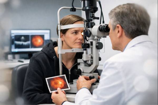
- Slit-Lamp Examination: A slit-lamp examination allows the clinician to examine the anterior structures of the eye, such as the cornea and lens, as well as check for blood in the anterior chamber. Although the slit-lamp is primarily used to examine the front of the eye, special lenses can be used to view the vitreous humor and detect any visible blood clots or other abnormalities.
- Dilated Fundus Examination: To thoroughly examine the retina and vitreous humor, the clinician will perform a dilated fundus examination. Eye drops dilate the pupil, allowing the clinician to look into the back of the eye with an ophthalmoscope. This examination can help determine the source of the hemorrhage, which could be retinal tears, detachments, or abnormal blood vessels, as well as the extent of the bleeding.
Imaging Techniques
If the clinical examination is inconclusive, or if the severity of the hemorrhage prevents a clear view of the retina, imaging techniques are used to provide a more detailed assessment.
- B-Scan Ultrasonography: B-scan ultrasonography is an important imaging tool in the diagnosis of vitreous hemorrhage, particularly when the hemorrhage is dense enough to obscure the clinician’s view of the retina. This non-invasive technique employs sound waves to generate cross-sectional images of the eye, allowing the clinician to see the vitreous cavity and retina behind it. B-scan can detect vitreous opacities, retinal detachment, and other abnormalities that would not be visible during a standard eye exam.
- Optical Coherence Tomography (OCT): OCT is another advanced imaging technique for evaluating the retina and vitreous humor. OCT generates high-resolution, cross-sectional images of the retina, allowing for the detection of subtle changes like macular edema, retinal tears, or a macular hole. Although OCT is less effective in the presence of dense vitreous hemorrhage, it can be useful when the hemorrhage is minor or resolving.
- Fluorescein Angiography: When retinal or choroidal neovascularization (abnormal blood vessel growth) is suspected, fluorescein angiography may be used. This test involves injecting a fluorescent dye into the bloodstream and photographing it as it travels through the retina’s blood vessels. Fluorescein angiography can detect areas of leakage, blocked blood vessels, or abnormal vessel growth that may be causing the vitreous hemorrhage.
Additional Diagnostic Tests
In some cases, additional diagnostic tests may be required to determine the cause and severity of the vitreous hemorrhage.
- Blood Tests: Blood tests may be performed to look for underlying systemic conditions that could cause vitreous hemorrhage, such as diabetes, hypertension, or blood clotting disorders. These tests are especially useful when the hemorrhage is unexplained or recurring.
- Electrophysiological Tests: Electrophysiological tests, such as electroretinography (ERG), can be used to evaluate the retina’s functional status, especially when retinal detachment or other retinal disorders are suspected. ERG measures the retina’s electrical responses to light stimuli and can reveal important information about retinal health.
Treatment and Management of Vitreous Hemorrhage
The severity of vitreous hemorrhage, the underlying cause of the bleeding, and the presence of any complications all influence treatment. Treatment options range from conservative observation to more invasive surgical procedures. The primary goals of management are to stop the hemorrhage, treat the underlying cause, and avoid recurrence or complications like retinal detachment.
Observation and Conservative Management
In many cases, especially when the hemorrhage is minor and the underlying cause is not immediately life-threatening, conservative treatment with close monitoring is appropriate. Over time, the body can absorb small amounts of blood from the vitreous humor, resulting in gradual improvement in vision. During this time, patients are usually advised to avoid activities that could aggravate their condition, such as heavy lifting, strenuous exercise, or any actions that raise intraocular pressure.
Patients are typically closely monitored with frequent follow-up visits to assess the resolution of the hemorrhage and detect any signs of complications. The duration of observation varies, but it can take several weeks or months for the blood to completely clear from the vitreous. If the hemorrhage persists or vision does not improve, additional treatment may be required.
Medical Management
Medical treatment focuses on determining the root cause of the vitreous hemorrhage. For example, if diabetic retinopathy is the cause, strict blood sugar control and retinopathy treatment are required to prevent future hemorrhages. This may include administering anti-VEGF (vascular endothelial growth factor) injections, which can help reduce neovascularization (abnormal blood vessel growth) in the retina, lowering the risk of future bleeding.
If the hemorrhage is caused by retinal vein occlusion or other vascular issues, treatment may include blood pressure control, anticoagulation therapy (in cases where clotting disorders exist), or laser photocoagulation to seal leaking blood vessels and prevent further bleeding.
Patients with underlying systemic conditions, such as blood clotting disorders, require proper management. This may entail the use of blood thinners or other medications to control clotting and prevent further eye bleeding.
Laser Photocoagulation
Laser photocoagulation may be used to treat vitreous hemorrhage in the presence of retinal tears, neovascularization, or other retinal abnormalities. This procedure uses a focused laser beam to create small burns on the retina, sealing off abnormal or leaking blood vessels and preventing further bleeding.
Laser photocoagulation is frequently used in combination with other treatments, such as anti-VEGF injections, especially in patients with proliferative diabetic retinopathy or retinal vein occlusion. The procedure is usually performed as an outpatient and is effective in lowering the risk of recurring hemorrhages.
Vitrectomy
When vitreous hemorrhage is severe, persistent, or associated with complications such as retinal detachment, a surgical procedure known as vitrectomy may be necessary. Vitrectomy is the removal of the vitreous gel and the blood within it from the eye. The vitreous is then replaced with a saline solution, gas bubble, or silicone oil to keep the eye in shape and support the retina.
Vitrectomy is a more invasive procedure that is usually reserved for cases where conservative treatment has failed or there is a high risk of permanent vision loss due to complications. The procedure is typically performed under local or general anesthesia and necessitates a period of recovery during which patients may need to avoid certain activities and maintain specific head positions to promote proper healing.
The results of vitrectomy are generally positive, with many patients reporting significant improvements in vision after the blood is removed from the eye. However, there are risks associated with any surgical procedure, such as infection, cataract formation, retinal detachment, and hemorrhage recurrence.
Addressing Underlying Causes
Long-term management of vitreous hemorrhage entails identifying and controlling the underlying causes to avoid recurrence. Patients with diabetic retinopathy must maintain strict blood glucose control, have regular eye exams, and receive prompt treatment for any retinal complications. It is critical for those with conditions like retinal vein occlusion to manage cardiovascular risk factors such as hypertension and hyperlipidemia.
Patients with a history of vitreous hemorrhage should also be educated on the signs and symptoms of complications, such as retinal detachment, and encouraged to seek immediate medical attention if they notice any sudden changes in vision.
Overall, managing vitreous hemorrhage is a multi-step process that includes conservative observation, medical treatment, and, in some cases, surgical intervention to preserve vision and prevent future episodes.
Trusted Resources and Support
Books
- “Vitreous: In Health and Disease” by J. Sebag
- This book offers a comprehensive exploration of the vitreous body, including detailed discussions on vitreous hemorrhage, its causes, and management strategies. It is an invaluable resource for both medical professionals and patients interested in understanding this condition.
- “The Diabetic Retinopathy Book” by David McCulloch
- A practical guide for managing diabetic retinopathy, which is a leading cause of vitreous hemorrhage. This book provides insights into prevention, treatment, and the importance of controlling blood sugar to avoid ocular complications.
Organizations
- American Academy of Ophthalmology (AAO)
- The AAO provides extensive resources on vitreous hemorrhage and related eye conditions, including educational materials, treatment guidelines, and the latest research. Their website is a trusted source of information for both patients and healthcare providers.
- National Eye Institute (NEI)
- As part of the U.S. National Institutes of Health, the NEI offers comprehensive information on eye health, including detailed explanations of vitreous hemorrhage, ongoing research initiatives, and support resources for those affected by this condition.


