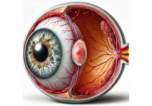
Uveitis-Glaucoma-Hyphema (UGH) syndrome is a complex and potentially blinding ocular condition that usually develops as a complication of cataract surgery, particularly after the implantation of an intraocular lens (IOL). UGH syndrome, first described by Dr. Robert Ellingson in 1978, is characterized by three symptoms: uveitis (uveal inflammation), glaucoma (increased intraocular pressure), and hyphema. Mechanical irritation or malposition of the IOL causes chronic damage to the delicate structures within the eye, resulting in the syndrome.
Pathophysiology of UGH Syndrome
The mechanical interaction between the IOL and the surrounding ocular tissues is the primary cause of UGH syndrome. This interaction can occur for a variety of reasons, including improper IOL positioning, the use of specific lens designs, or complications during or after cataract surgery. The syndrome is more frequently associated with anterior chamber intraocular lenses (ACIOLs) and rigid lenses, which were more common in previous cataract surgeries. However, it can also happen with posterior chamber intraocular lenses (PCIOLs) if they become displaced or if the haptics (the lens’s support arms) come into contact with the ciliary body or iris.
Mechanisms Contributing to UGH Syndrome
- **Mechanical Trauma:
Improper placement or dislocation of the IOL can cause direct mechanical trauma to the surrounding ocular structures. For example, the haptics of an IOL can rub against the ciliary body or iris, resulting in chronic inflammation (uveitis) and microhemorrhages (hyphema). Repeated friction can also cause secondary glaucoma by obstructing the trabecular meshwork, the eye’s drainage system, resulting in high intraocular pressure (IOP). - Uveal irritation:
Continuous irritation of the uvea by the IOL causes uveitis, which is characterized by redness, pain, and light sensitivity. This inflammation can exacerbate the risk of glaucoma by increasing aqueous humor production or clogging the trabecular meshwork, resulting in elevated IOP. - Hyphema formation:
Another distinguishing feature of UGH syndrome is hyphema, or blood in the anterior chamber of the eye. It is the result of repeated trauma to the iris or ciliary body, which causes small blood vessels to rupture. The presence of blood in the anterior chamber can obstruct aqueous humor outflow, causing secondary glaucoma. - Secondary Glaucoma:
Glaucoma in UGH syndrome is usually secondary and results from a combination of uveal inflammation, hyphema, and mechanical blockage of the trabecular meshwork by the IOL or inflammatory and hemorrhagic debris. If not treated promptly, increased intraocular pressure can cause optic nerve damage and irreversible vision loss.
Risk Factors for UGH Syndrome
Several factors can increase the likelihood of developing UGH syndrome. This includes:
- Type of intraocular lens:
The type of IOL used during cataract surgery significantly influences the risk of UGH syndrome. Rigid anterior chamber lenses are more prone to mechanical irritation than modern flexible posterior chamber lenses. Similarly, lenses with sharp edges or that are too large for the eye can increase the risk of injury to the surrounding tissues. - Lens malposition:
Incorrect IOL positioning during surgery, or dislocation after surgery, can result in direct contact between the lens and the uveal structures. This is more likely to happen when the IOL is not securely anchored, there is zonular weakness (weakness of the fibers that hold the lens in place), or the eye has anatomical abnormalities. - Surgical complications:
Complications during cataract surgery, such as capsular tears, vitreous prolapse, or improper wound closure, can increase the risk of IOL malposition and, as a result, UGH. - Pre-existing Eye Conditions:
Patients with pre-existing ocular conditions like uveitis, glaucoma, or a history of ocular trauma are more likely to develop UGH syndrome following cataract surgery. These conditions can make the eye more prone to inflammation, increased intraocular pressure, and complications from IOL placement. - Advanced age:
Older patients may be more susceptible to UGH syndrome due to factors such as weaker ocular structures, the presence of pre-existing ocular conditions, and a higher likelihood of needing cataract surgery.
Symptoms of UGH Syndrome
The symptoms of UGH syndrome are frequently non-specific and overlap with other postoperative complications, making diagnosis difficult. The presentation varies according to the severity of the syndrome and its underlying cause. Common symptoms include:
- Ocular Pain
Patients with UGH syndrome frequently experience eye pain, which can range from mild discomfort to severe throbbing pain. Inflammation of the uvea and increased intraocular pressure are usually the causes of pain. - Redness:
Uveitis is the most common cause of eye redness. The inflammation causes blood vessels in the eye to dilate, resulting in a red, irritated appearance. - Blurry Vision:
Blurred vision is a common complaint among patients with UGH syndrome. This could be due to the presence of hyphema, elevated IOP, or corneal edema (swelling) caused by underlying inflammation and increased pressure. - Light sensitivity (photophobia):
Patients with UGH syndrome frequently report light sensitivity, a common symptom of uveitis. The inflammation of the iris and surrounding tissues increases the eye’s sensitivity to light. - Hyphema:
The presence of blood in the anterior chamber is indicative of UGH syndrome. Patients may notice a reddish tint in their vision, particularly when looking at bright backgrounds. In severe cases, the hyphema may appear as a layer of blood at the bottom of the iris when the patient stands upright. - Elevated intraocular pressure:
Elevated IOP is a critical feature of UGH syndrome, causing symptoms such as headache, nausea, and halos around lights, particularly in low-light conditions. If left untreated, the increased pressure can irreversibly damage the optic nerve, resulting in glaucoma and permanent vision loss.
Complications of UGH Syndrome
If not diagnosed and treated promptly, UGH syndrome can cause severe complications, including permanent vision loss. Some of the most significant complications are:
- Chronic Uveitis:
Persistent uveal inflammation can progress to chronic uveitis, which may necessitate long-term immunosuppressive treatment. Chronic uveitis increases the risk of cataract formation, cystoid macular edema (central retina swelling), and posterior synechiae (iris-lens adhesions). - Glaucoma:
Secondary glaucoma is one of UGH syndrome’s most serious complications. Chronically elevated intraocular pressure can cause progressive optic nerve damage, resulting in irreversible vision loss. In severe cases, patients may require surgery to relieve pressure and prevent further damage. - Corneal edema:
Elevated IOP and inflammation can cause corneal edema, which is when the cornea becomes swollen and cloudy. This can cause significant vision impairment and may necessitate medical or surgical intervention to resolve. - Recurring Hyphema:
Recurrent bleeding in the anterior chamber can cause long-term visual problems and raise the risk of angle-closure glaucoma. Recurrent hyphema may require surgical intervention, such as anterior chamber washout or IOL revision. - ** Retinal Detachment:**
Although rare, severe cases of UGH syndrome can cause retinal detachment, which occurs when the retina separates from the underlying tissue. Retinal detachment is a medical emergency that necessitates prompt surgical intervention to avoid permanent vision loss.
Epidemiology of UGH Syndrome
UGH syndrome is a rare condition, but it is worth considering for patients who have had cataract surgery with IOL implantation. UGH syndrome has become less common in recent years as IOL design and surgical techniques have advanced. However, it remains a significant risk in patients with older lens designs, anterior chamber lenses, or misaligned lenses.
The exact prevalence of UGH syndrome is difficult to determine due to its rarity and variable clinical presentation. It is more common in older patients who had cataract surgery with anterior chamber lenses, particularly those with rigid or oversized IOLs. The condition is also more common in patients who already have ocular conditions like uveitis or glaucoma.
Diagnostic methods
Diagnosing UGH syndrome necessitates a thorough patient history, an ocular examination, and the use of diagnostic imaging techniques. The primary goal of the diagnostic process is to confirm the presence of UGH syndrome, identify the underlying cause, and determine the condition’s severity.
Clinical Evaluation
During the slit-lamp examination, the ophthalmologist carefully examines the eye’s anterior segment, paying special attention to the position and condition of the intraocular lens (IOL), the presence of anterior chamber inflammation, and the presence of hyphema. Specific signs that may indicate UGH syndrome are:
- IOL Malposition: The ophthalmologist looks for indications that the IOL is misaligned, such as tilted or dislocated lenses. They also determine whether the IOL’s haptics (support arms) are in contact with the iris or ciliary body, which could cause mechanical irritation.
- Anterior Chamber Reaction: The presence of inflammatory cells or a flare in the anterior chamber indicates uveitis, which is a major component of UGH syndrome. These findings are evaluated under a slit lamp and graded according to severity.
- Hyphema: Hyphema is defined as a layering of blood in the anterior chamber. Even a small amount of blood is significant and should raise concerns about UGH syndrome, especially if the patient has had recent cataract surgery with IOL implantation.
- Corneal Edema: The ophthalmologist inspects the cornea for signs of edema, which can manifest as swelling or cloudiness. Elevated intraocular pressure or chronic inflammation, both of which are associated with UGH syndrome, can cause corneal edema.
Intraocular Pressure Measurement
Intraocular pressure (IOP) measurement is a critical step in the diagnosis of UGH syndrome. Elevated IOP may indicate the presence of secondary glaucoma, a common complication of UGH syndrome. IOP is typically measured with a tonometer, with normal values ranging from 10 to 21 mmHg. IOP readings above this range are concerning and indicate the need for further investigation and potential intervention.
When IOP rises, the ophthalmologist will determine whether the cause is mechanical blockage of the trabecular meshwork by the IOL, inflammatory debris, or blood from hyphema. Identifying the cause of elevated IOP is critical for developing an appropriate management strategy.
Gonioscopy
Gonioscopy is a specialized technique for inspecting the angle of the anterior chamber, where the cornea and iris meet. This area is critical for aqueous humor drainage, and its examination is required in patients with suspected UGH syndrome. During gonioscopy, the ophthalmologist examines the angle structure with a gonioscope, looking for abnormalities that could lead to glaucoma, such as synechiae (adhesions), angle closure, or IOL obstruction.
Gonioscopy can also tell you whether the IOL haptics are encroaching on the angle, which could cause mechanical irritation and contribute to the syndrome’s pathogenesis. This examination provides important information about the potential causes of elevated IOP and helps guide secondary glaucoma treatment.
Ultrasound Biomicroscopy (UBM)
Ultrasound biomicroscopy (UBM) is a high-resolution imaging technique that produces detailed images of the anterior segment of the eye, such as the IOL, ciliary body, and angle structures. UBM is especially useful when the diagnosis of UGH syndrome is unclear or when other diagnostic methods have yielded insufficient results.
UBM can show the exact position of the IOL, the haptics’ relationship with the surrounding ocular tissues, and any abnormal contact between the IOL and the uveal structures. This imaging modality can also detect any ciliary body abnormalities, such as cysts or detachment, that may be causing the patient’s symptoms.
Anterior Segment Optical Coherence Tomography (AS-OCT)
Another advanced imaging technique is anterior segment optical coherence tomography (AS-OCT), which produces cross-sectional images of the anterior segment of the eye. AS-OCT can help visualize the position of the IOL as well as detect subtle abnormalities in the anterior chamber angle, cornea, and iris. This non-invasive imaging method is particularly useful for tracking the progression of UGH syndrome and assessing treatment efficacy.
AS-OCT is frequently used in conjunction with UBM to provide a comprehensive assessment of the anterior segment, allowing the ophthalmologist to make informed decisions about the diagnosis and treatment of UGH.
Ancillary tests
In some cases, additional tests may be required to rule out other conditions that can mimic UGH syndrome or to determine the extent of inflammation and its effect on the eye. These tests can include:
- Fluorescein Angiography: This imaging technique evaluates the blood vessels in the retina and choroid. It can assist in detecting any vascular abnormalities or leakage that may be contributing to the patient’s symptoms.
- Visual Field Testing: This test evaluates the patient’s peripheral vision and can detect glaucoma-related field loss. It is especially important to assess the functional impact of elevated IOP in UGH syndrome.
- Blood Work: If systemic inflammation or an autoimmune condition is suspected, blood tests may be performed to identify underlying causes of uveitis.
Uveitis-Glaucoma-Hyphema (UGH) Syndrome Management
Managing Uveitis-Glaucoma-Hyphema (UGH) syndrome necessitates a multifaceted approach that addresses the underlying cause, reduces inflammation, regulates intraocular pressure (IOP), and prevents complications. The severity of the condition, the presence of complications, and the patient’s overall ocular health all influence the management strategy. Here are the primary methods for managing UGH syndrome:
1. Medical management
a. Anti-inflammatory Drugs:
The first line of treatment for UGH syndrome is frequently the use of anti-inflammatory medications to control uveitis. Topical corticosteroids, such as prednisolone acetate, are commonly used to reduce inflammation in the eye. In more severe cases, oral corticosteroids or nonsteroidal anti-inflammatory drugs (NSAIDs) may be required to control systemic inflammation. The goal of anti-inflammatory therapy is to relieve pain, redness, and photophobia while reducing the risk of chronic uveitis and its complications.
b. Intraocular Pressure Control:
Elevated IOP is a major concern in UGH syndrome, as it can cause optic nerve damage and irreversible vision loss. Topical ocular hypotensive agents, such as beta-blockers (e.g., timolol), alpha agonists (e.g., brimonidine), carbonic anhydrase inhibitors (e.g., dorzolamide), or prostaglandin analogs (e.g., latanoprost), are commonly used to treat elevated IOP. In some cases, oral carbonic anhydrase inhibitors, such as acetazolamide, may be prescribed to reduce pressure more quickly. The severity of the pressure elevation and the patient’s response to treatment determine the appropriate IOP-lowering medication.
**c. **Mydriatic and cycloplegic Agents:
These medications are used to dilate the pupil and alleviate pain caused by ciliary spasm, which is a common symptom of uveitis. Mydriatic agents, such as atropine or cyclopentolate, aid in the prevention of posterior synechiae (adhesions between the iris and lens) and increase patient comfort.
2. Surgical management
**a. **Repositioning or removing an intraocular lens (IOL)
If medical treatment fails to control UGH syndrome, surgical intervention may be required. The primary surgical option is to reposition or remove the offending IOL. If the IOL is misaligned and causing mechanical irritation, repositioning it within the eye may relieve symptoms and prevent further complications. However, if the IOL is severely dislocated or its design causes irritation, removal may be required. Following IOL removal, a different type of lens, such as a scleral-fixated IOL or a more advanced anterior chamber IOL, may be implanted.
b. Anterior Chamber Washout:
In cases of recurrent or severe hyphema, an anterior chamber washout may be performed to remove blood from the anterior chamber and lower the risk of angle-closure glaucoma. This procedure, which clears the drainage angle, can help restore normal vision and lower IOP.
c. Glaucoma Surgery:
If medications do not control elevated IOP, glaucoma surgery may be necessary. Trabeculectomy (creating a new drainage pathway for aqueous humor), glaucoma drainage device placement (e.g., tube shunt), and laser procedures such as cyclophotocoagulation are among the surgical options. The severity of glaucoma, the anatomy of the eye, and the patient’s overall health all influence the decision to undergo surgery.
3. Long-term Monitoring and Follow-up
Long-term monitoring is frequently required for UGH syndrome management to ensure that inflammation is under control, IOP is within a safe range, and no new complications develop. Regular follow-up visits with an ophthalmologist are essential for assessing treatment efficacy, making necessary adjustments, and detecting any signs of recurrence early.
Patients who have undergone surgical intervention for UGH syndrome will require ongoing monitoring to assess the procedure’s success and manage any post-operative complications. This may include periodic imaging studies, visual field testing, and an examination of the optic nerve to detect any signs of glaucoma.
4. Patient education and support
Educating patients about UGH syndrome, its potential complications, and the importance of treatment adherence is critical for successful management. Patients should be informed of the symptoms of high IOP, such as headaches, halos around lights, and blurred vision, and advised to seek immediate medical attention if they occur.
Support groups and counseling services may also be useful, especially for patients dealing with the anxiety and uncertainty that comes with chronic ocular conditions. Giving patients access to trusted resources can encourage them to take an active role in managing their health and improve their overall quality of life.
Trusted Resources and Support
Books
- “Cataract Surgery: Technique, Complications, and Management” by Roger F. Steinert
This comprehensive textbook provides in-depth information on the surgical techniques and potential complications of cataract surgery, including UGH syndrome. - “Glaucoma: A Patient’s Guide to the Disease” by Graham E. Trope
This book offers a detailed explanation of glaucoma, its relationship with UGH syndrome, and the various management options available.
Organizations
- American Academy of Ophthalmology (AAO)
The AAO provides a wealth of resources for patients and professionals, including information on UGH syndrome, its management, and recent research findings. AAO Website - Glaucoma Research Foundation
This organization offers educational materials and support for individuals affected by glaucoma, including those dealing with secondary glaucoma as a result of UGH syndrome. Glaucoma Research Foundation Website


