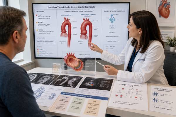
A thoracic aortic aneurysm develops when part of the aorta within the chest enlarges and its wall becomes vulnerable to tearing or rupture. Genetic testing can identify heritable thoracic aortic disease in some people, including conditions caused by FBN1, TGFBR1, TGFBR2, ACTA2, and other genes. A molecular diagnosis may change how much of the arterial system is imaged, how often surveillance occurs, when preventive surgery is discussed, and which relatives require testing. It does not replace accurate aortic measurements or emergency evaluation. Many aneurysms are multifactorial, age-related, associated with hypertension or bicuspid aortic valve, or remain unexplained after a high-quality panel. Conversely, a person with a high-risk pathogenic variant may dissect at a smaller diameter than the threshold used for a typical sporadic aneurysm. The most useful approach combines gene-specific evidence with age, body size, growth rate, family history, pregnancy plans, valve anatomy, and findings throughout the arterial tree.
- Genetic testing is especially relevant with young onset, syndromic features, or a family history of aneurysm, dissection, or sudden death.
- FBN1 and TGF-β pathway genes often affect the aortic root but can have broader arterial consequences.
- Smooth-muscle genes may cause dissection with few outward physical features.
- A pathogenic result can alter surveillance and preventive-surgery planning, but diameter and clinical factors still matter.
- A variant of uncertain significance should not determine surgery or predictive testing in healthy relatives.
- First-degree relatives often need aortic imaging even when the affected person’s genetic test is negative.
Table of Contents
- Why the thoracic aorta enlarges
- When to suspect heritable aortic disease
- How genes shape the arterial pattern
- From imaging to test selection
- What each result category means
- Using genetics in aortic care
- Relatives, pregnancy, and long-term planning
Why the thoracic aorta enlarges
The aorta carries blood from the heart to the body. Its thoracic segments include the aortic root, ascending aorta, arch, and descending thoracic aorta. An aneurysm is abnormal enlargement of one or more segments. The clinical danger is not simply size: weakened tissue may split within the wall, creating an aortic dissection, or may rupture. Either event can be rapidly fatal and requires emergency care.
Thoracic aortic disease is often silent. An aneurysm may be discovered on an echocardiogram, computed tomography scan, magnetic resonance study, or imaging obtained for another reason. Some people develop chest or back discomfort, compression symptoms, aortic valve leakage, or heart failure, but symptoms are unreliable markers of stability. Sudden severe chest, back, neck, or abdominal pain; fainting; new weakness; difficulty speaking; loss of a pulse; or shock requires immediate emergency assessment, not genetic testing.
Aortic size must be interpreted in context. The same diameter may represent different degrees of enlargement in a small child, a short adult, and a very tall adult. Specialists may index measurements to height or body surface area and track change using the same imaging method and measurement convention when possible. Growth rate, segment involved, blood pressure, valve anatomy, family history, and gene all contribute to risk.
The causes form a spectrum. Hypertension, aging, smoking-related vascular injury, bicuspid aortic valve, prior dissection, inflammatory disease, and trauma may contribute. Heritable thoracic aortic disease, or HTAD, includes syndromic conditions with recognizable features and nonsyndromic forms in which the aorta may be the main organ affected. Some families have clear inheritance even though no causal variant is found.
An abdominal aortic aneurysm has a different epidemiology and genetic architecture from many root and ascending thoracic aneurysms. A standard HTAD panel is not a general test for every aneurysm anywhere in the body. The location, age at diagnosis, family pattern, and associated findings determine whether monogenic testing is likely to be informative.
The phrase “connective-tissue disorder” is also incomplete. Some HTAD genes affect extracellular matrix proteins, while others alter transforming growth factor beta signaling, smooth-muscle contraction, or cellular stress pathways. These mechanisms help explain why two people with similar aortic diameters can have different risks and why a gene-specific diagnosis can add information beyond anatomy.
When to suspect heritable aortic disease
Genetic evaluation is strongly considered when thoracic aortic aneurysm or dissection occurs at a relatively young age, when a first- or second-degree relative has thoracic aortic disease, or when physical findings suggest a syndromic aortopathy. Current US guidance uses onset before age 60 as one important risk feature, but age is not an absolute boundary. A dissection at 62 with several affected relatives may be more suspicious than an isolated mild aneurysm at 58 with decades of uncontrolled hypertension.
Syndromic clues can include tall stature with disproportion, pectus deformity, scoliosis, lens dislocation, characteristic craniofacial findings, translucent or unusually fragile skin, easy bruising, clubfoot, joint contractures or hypermobility, bifid uvula, arterial tortuosity, hypertelorism, or spontaneous organ rupture. No single feature establishes a molecular diagnosis, and absence of external signs does not exclude one. ACTA2-, MYLK-, MYH11-, LOX-, or PRKG1-related disease may present with little that is visible outside the vascular system.
The family history should extend beyond known “aneurysm.” Sudden unexplained death, fatal “heart attack” without coronary confirmation, stroke at a young age, aortic valve surgery, emergency chest surgery, pregnancy-associated death, intracranial aneurysm, and arterial rupture may all be relevant. Medical records, operative reports, imaging, death certificates, and autopsy findings can correct family descriptions that were incomplete or inaccurate.
Bicuspid aortic valve commonly coexists with ascending aortic enlargement. Most people with bicuspid valve do not have an identifiable single-gene HTAD syndrome, but family clustering and syndromic features may still justify genetics referral. Turner syndrome, coarctation, congenital heart defects, and other developmental findings require their own diagnostic framework. A broad panel should not be ordered reflexively without deciding which phenotype is being investigated.
Testing is also considered after an apparently spontaneous dissection, even if the aorta was not previously known to be enlarged. Certain genes predispose to dissection at modest diameters. A normal external examination and lack of prior family diagnosis do not make the event sporadic: relatives may be young, mildly affected, or undiagnosed, and a pathogenic variant may have arisen de novo.
A genetics visit typically constructs a three-generation pedigree and performs or coordinates a targeted physical examination. The goal is not merely to decide “genetic or not.” It is to identify the most plausible disease group, choose the correct assay, and determine what imaging relatives need now rather than waiting for DNA results.
How genes shape the arterial pattern
FBN1 encodes fibrillin-1, an extracellular matrix protein. Pathogenic variants cause Marfan syndrome and can also present with a less complete Marfan phenotype. Aortic root aneurysm is characteristic, often accompanied by lens dislocation, skeletal features, dural ectasia, or lung findings. Not every FBN1 variant has the same consequence, and a variant associated with an unrelated fibrillin disorder should not automatically be used to diagnose Marfan syndrome.
TGFBR1 and TGFBR2 encode receptors in the transforming growth factor beta pathway. Pathogenic variants can cause Loeys–Dietz syndrome, which may involve aggressive root aneurysm, arterial tortuosity, branch-vessel aneurysms, craniofacial features, skeletal findings, allergic or inflammatory disease, and other manifestations. SMAD2, SMAD3, TGFB2, and TGFB3 are related pathway genes. SMAD3 disease may combine arterial aneurysm with early-onset osteoarthritis.
COL3A1 causes vascular Ehlers–Danlos syndrome, in which medium and large arteries, bowel, and uterus may be fragile. The management approach differs from Marfan or Loeys–Dietz syndrome because invasive procedures themselves can carry unusual risk. COL3A1 belongs on appropriate differential diagnoses but should be interpreted according to its distinctive mechanism and phenotype.
ACTA2 encodes smooth-muscle alpha-actin and is a leading cause of nonsyndromic familial HTAD. Some pathogenic variants are associated with coronary artery disease, early stroke, moyamoya-like cerebrovascular disease, or characteristic iris findings. Aortic dissection may occur below conventional sporadic-disease thresholds. MYH11 and MYLK affect smooth-muscle contraction; MYH11 disease can be associated with patent ductus arteriosus, while MYLK-related dissection may occur with limited preceding enlargement.
LOX encodes lysyl oxidase, which contributes to cross-linking of elastin and collagen. PRKG1 affects smooth-muscle signaling and can cause early, aggressive dissection. Additional well-supported genes may include EFEMP2, FBLN5, FLNA, MAT2A, MFAP5, SKI, SLC2A10, and others depending on the phenotype and evidence framework. Some are primarily associated with recessive syndromes or distinctive developmental findings.
Gene lists change as evidence develops. A commercial aortopathy panel may contain dozens of genes, but not all have equal proof for thoracic aortic disease. Large panels increase the chance of uncertain findings. A useful report identifies the exact variant, gene-disease validity, inheritance mechanism, and whether the variant type is known to cause disease in that gene.
The gene can influence which arteries are imaged. Root-predominant disease may still require assessment beyond the root, while TGF-β pathway and COL3A1 disorders can involve extensive branch vessels. ACTA2 can have cerebrovascular and coronary implications. Surveillance should follow the established gene-associated pattern and the individual family history rather than a one-size-fits-all “annual echo” rule.
From imaging to test selection
Genetic testing is most valuable after the aortic phenotype is documented precisely. The ordering team records the largest diameter, segment, measurement method, rate of growth, valve anatomy, prior dissection, surgical history, and imaging of other arteries. Echocardiography is excellent for the root and proximal ascending aorta in many people, but computed tomography angiography or magnetic resonance angiography may be needed when the entire thoracic aorta or branch vessels must be assessed.
The most clearly affected living relative is usually tested first. If a family includes a person with early dissection and another with only mild enlargement late in life, the early severe case is often more informative. Testing an unaffected relative before a family variant is known may yield a negative result that cannot answer whether the family’s disease is genetic.
A multigene panel is generally favored because clinical features overlap. It should include genes with definitive or strong evidence and detect sequence variants plus clinically relevant deletions and duplications. Some pathogenic mechanisms, deep intronic changes, structural rearrangements, or mosaic variants may escape a standard assay. Exome or genome sequencing can be considered when a high-quality panel is negative and the phenotype remains strongly syndromic or familial, but broader sequencing also increases incidental and uncertain findings.
Pretest counseling addresses the possible results, implications for relatives, privacy and insurance rules, secondary findings, and the limits of prediction. A molecular result cannot specify the exact day or diameter at which a dissection will occur. It may, however, move a patient into a gene-specific risk group that changes surveillance or surgical discussions.
Testing may also be performed after death if stored blood, tissue, or another suitable specimen is available. A molecular autopsy can be particularly valuable after unexplained type A dissection at a young age. DNA quality varies by specimen, and a rare variant should be interpreted with autopsy findings and segregation in living relatives.
An urgent aneurysm or acute aortic syndrome is managed immediately on clinical grounds. Treatment should not wait for a panel result. Genetics becomes part of the subsequent plan for extent of repair, residual-aorta surveillance, syndromic evaluation, and family screening.
What each result category means
A pathogenic or likely pathogenic result can establish the molecular cause when the gene and phenotype fit. It may confirm Marfan syndrome, Loeys–Dietz syndrome, vascular Ehlers–Danlos syndrome, or a nonsyndromic HTAD. The report can guide targeted testing of relatives and may influence imaging territory, follow-up interval, blood-pressure strategy, pregnancy counseling, and the diameter at which preventive surgery is discussed.
The exact variant matters. A truncating variant may cause disease in one gene but not through the same mechanism in another. Some genes have regions associated with different phenotypes or severity. The laboratory should use accepted classification standards, population data, segregation, functional evidence, and disease-specific expertise. “Mutation detected” without a modern classification and evidence summary is not enough.
A variant of uncertain significance, or VUS, is not a positive diagnosis. It should not determine prophylactic surgery, trigger prenatal testing, or be used to tell healthy relatives that they are safe or affected. Family studies may help reclassification when chosen strategically, but testing every relative for a VUS without a defined segregation question can create more confusion.
A negative panel does not eliminate heritable disease. The family may carry a variant outside the assay’s reach, a gene not yet associated with HTAD, or a mechanism that remains undiscovered. The tested person may also have been the wrong family member, or the condition may be multifactorial. When the phenotype or pedigree remains compelling, clinical surveillance of the patient and imaging of relatives continue.
A report may identify a pathogenic variant unrelated to the aneurysm, especially after exome or genome sequencing. That finding needs a separate evaluation rather than being forced into the aortic explanation. A result may also establish carrier status for a recessive disorder without explaining dominant familial disease.
Variant classification evolves. Patients should keep the complete report, laboratory name, and accession information and should know whether updated reports are issued automatically. Reanalysis is reasonable when new relatives become affected, the phenotype changes, or several years have passed since an unresolved test. Clinical decisions in the meantime remain anchored to actual aortic disease and family history.
Using genetics in aortic care
Aortic surveillance is individualized by gene, segment, diameter, growth, age, and prior events. After baseline imaging, follow-up may occur in months rather than years when a new aneurysm is near an intervention threshold or appears to be growing. Stable smaller aneurysms may be imaged less frequently. Cross-sectional imaging can be extended from head to pelvis in disorders associated with widespread arterial aneurysm or tortuosity, while other conditions use a more targeted plan.
Blood-pressure control reduces mechanical stress on the aortic wall. Beta-blockers and angiotensin receptor blockers are commonly used in Marfan syndrome and selected other HTAD conditions, but drug choice depends on blood pressure, tolerance, pregnancy status, and comorbidities. Smoking cessation and treatment of standard cardiovascular risk factors remain important. Medication does not make a high-risk aneurysm immune to dissection or replace imaging.
Preventive surgery aims to repair the aorta before dissection while avoiding premature major surgery. Thresholds are not identical across diagnoses. In typical Marfan root aneurysm, discussion often centers around 5.0 cm and may move toward 4.5 cm with high-risk features or at an experienced center. Loeys–Dietz, ACTA2, PRKG1, and other gene-specific disease may justify intervention at smaller dimensions depending on the exact gene, variant, body size, growth, valve function, family dissection diameters, and surgical expertise. These numbers are not do-it-yourself cutoffs.
Rapid growth, family dissection at a small diameter, desire for pregnancy, severe valve disease, and marked body-size indexing can alter timing. Measurements from different modalities are not always directly interchangeable. Decisions should be made by a multidisciplinary aortic team that can review images rather than relying on a single number copied into a report.
Exercise advice seeks to limit abrupt blood-pressure surges without eliminating healthy movement. Heavy isometric lifting, straining, and high-intensity collision activity may be restricted, especially with a substantial aneurysm. Aerobic activity can often be tailored safely. The plan should account for current dimensions, blood pressure, gene, prior repair, and sport rather than applying the same weight limit to every patient.
After surgery, genetic risk remains relevant because unrepaired segments can enlarge or dissect. Lifelong surveillance is necessary. Repair of the root does not cure a systemic arteriopathy, and prior dissection creates its own chronic imaging needs.
Relatives, pregnancy, and long-term planning
Most major HTAD conditions are autosomal dominant. A person with a causal variant has a 50% chance of passing it to each child. Disease expression can differ greatly within a family, so a mildly affected parent can have a child with earlier or more extensive disease. Some conditions are recessive, X-linked, or de novo; the report and counselor should explain the mechanism for the exact gene.
When a familial pathogenic or likely pathogenic variant is known, targeted testing identifies which relatives inherited it. Carriers receive gene-specific vascular evaluation even if asymptomatic. Relatives who test negative for the established family variant can usually stop variant-based aortic surveillance, unless they have an independent clinical indication. A VUS cannot provide that reassurance.
When no variant is found, first-degree relatives of a person with root or ascending aneurysm or aortic dissection should still receive aortic imaging. If one relative is found to have enlargement, the screening circle may expand. Normal imaging at one age may not exclude later disease, so the specialist sets follow-up according to family onset and severity.
Pregnancy creates major hemodynamic and hormonal stress. People with HTAD need preconception assessment of the entire relevant aorta, medication review, and coordinated planning among aortology, cardiology, maternal-fetal medicine, anesthesia, surgery, and genetics. Some blood-pressure drugs are unsafe in pregnancy. The risk of dissection varies by gene and aortic size and continues into the postpartum period. Preventive surgery may be discussed before pregnancy at dimensions lower than those used for someone not planning pregnancy.
Reproductive options include natural conception, prenatal diagnosis, and preimplantation genetic testing when a clearly causal familial variant is known. Genetic testing can determine whether a fetus inherited the variant but usually cannot predict severity. These decisions require nondirective counseling and should not be based on a VUS.
Long-term planning includes maintaining copies of imaging and genetic reports, using medical identification when appropriate, and ensuring emergency information is available. Family members should recognize symptoms of acute aortic syndrome and tell clinicians about the inherited diagnosis before invasive procedures or pregnancy. The central benefit of genetic testing is not the label itself; it is converting an otherwise hidden familial risk into a coordinated, lifelong prevention plan.
References
- 2022 ACC/AHA Guideline for the Diagnosis and Management of Aortic Disease. Clinical practice guideline, 2022.
- 2024 ESC Guidelines for the management of peripheral arterial and aortic diseases. Clinical practice guideline, 2024.
- Heritable Thoracic Aortic Disease Overview. GeneReviews clinical reference, updated 2023.
- Hereditary Thoracic Aortic Diseases. Clinical and genetic review, 2024.
- Association Between Genetic Diagnosis and Clinical Outcomes in Patients With Heritable Thoracic Aortic Disease. Cohort study, 2023.
- Genetics of Heritable Thoracic Aortic Disease. Clinical review, 2022.
Disclaimer
This article is for general education and does not diagnose an aneurysm or establish an individual surgical threshold. Genetic results and aortic measurements should be interpreted by qualified cardiovascular genetics and aortic specialists using the full clinical and family context. Sudden severe chest, back, neck, or abdominal pain, fainting, stroke symptoms, or shock requires immediate emergency care.