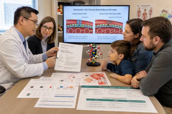
A Duchenne muscular dystrophy genetic test analyzes the DMD gene to identify the molecular cause of progressive muscle weakness and dystrophin loss. A confirmed result can establish diagnosis without muscle biopsy in most people, distinguish a Duchenne-pattern dystrophinopathy from related conditions, identify female relatives who carry the familial variant, and determine whether a variant-specific treatment or clinical trial may apply. DMD is exceptionally large, and pathogenic changes include multi-exon deletions, duplications, small sequence variants, splice alterations, and rarer deep intronic or complex rearrangements. Testing must therefore include both copy-number and sequence analysis. An out-of-frame variant often predicts Duchenne muscular dystrophy because it disrupts production of functional dystrophin, but the reading-frame rule has exceptions. Results are interpreted with age at symptom onset, creatine kinase, motor findings, cardiac evaluation, family history, and sometimes dystrophin studies. A negative routine test does not end the evaluation when the clinical evidence remains strong.
- Duchenne muscular dystrophy is an X-linked dystrophinopathy caused by pathogenic DMD variants.
- Multi-exon deletions and duplications require copy-number testing; small variants require sequencing.
- The exact exon structure and reading-frame effect can influence phenotype and treatment eligibility.
- A pathogenic result confirms a dystrophinopathy, but clinical findings determine whether it is Duchenne, intermediate, or another form.
- Female heterozygotes can have cardiomyopathy or muscle symptoms and need appropriate follow-up.
- Unresolved cases may require RNA studies, genome testing, or muscle dystrophin analysis.
Table of Contents
- When Duchenne Should Be Suspected
- What the Genetic Test Must Establish
- Stepwise DMD Testing
- Reading Frame and Phenotype
- How to Interpret the Report
- X-Linked Inheritance and Carrier Results
- Newborn Screening and Result-Guided Care
- Negative or Unresolved Testing
When Duchenne Should Be Suspected
Duchenne muscular dystrophy usually becomes clinically apparent in early childhood, although biochemical evidence is present much earlier. The first clues may be delayed walking, frequent falls, difficulty running or jumping, toe walking, enlarged calves, a waddling gait, or using the hands to “climb” up the thighs when rising from the floor. Speech or developmental concerns can precede obvious weakness in some children.
Weakness is typically proximal, affecting the hips and thighs before the hands and feet. A child may struggle with stairs, playground activity, getting up from a chair, or keeping pace with peers. Symptoms are progressive rather than episodic. Sensation is generally preserved.
Serum creatine kinase, or CK, is usually markedly elevated. Aspartate aminotransferase and alanine aminotransferase may also rise because they are present in muscle, which can lead to an incorrect liver workup if CK is not checked. A creatine kinase test guide explains why a high value points to muscle injury but does not identify the cause.
Duchenne should also be considered after an elevated CK-MM newborn screen, a family history of affected males linked through maternal relatives, an unexpectedly high CK found during another evaluation, or an X-chromosome finding that disrupts DMD. Some boys are identified before symptoms through family testing.
Cardiac disease can be an early or prominent feature. Dilated cardiomyopathy, rhythm abnormalities, or myocardial fibrosis may occur even before severe mobility loss. Rarely, a DMD variant presents mainly as X-linked cardiomyopathy. This broad spectrum means that cardiologists as well as neurologists may initiate testing.
Several conditions can resemble Duchenne, including limb-girdle muscular dystrophies, spinal muscular atrophy, congenital myopathies, metabolic muscle disease, and inflammatory myopathy. Genetic confirmation prevents assumptions based on CK and gait alone. It also matters because management, family risk, and treatment options differ.
A child with rapidly worsening weakness, breathing difficulty, dark urine after exertion, chest pain, fainting, or palpitations needs direct medical evaluation. Genetic testing is important but should not delay care for an active complication.
What the Genetic Test Must Establish
The DMD gene lies on the X chromosome and encodes dystrophin, a structural protein that stabilizes muscle-cell membranes during contraction. Without adequate dystrophin, repeated mechanical stress damages skeletal and cardiac muscle fibers. Duchenne usually reflects near-absence of functional dystrophin, while Becker muscular dystrophy and milder dystrophinopathies retain more.
A useful genetic result should answer four questions:
- What exact variant is present? The report should name the deleted or duplicated exons or provide standardized DNA and protein notation for a sequence variant.
- Does the finding disrupt DMD function? Pathogenicity must be supported by established evidence, not prediction alone.
- What is the expected effect on the reading frame and dystrophin? This helps estimate whether the phenotype is likely Duchenne, Becker, intermediate, or uncertain.
- Can the result guide family testing or variant-specific therapy? Exon structure, phase, and transcript details must be accurate.
The diagnosis in a male is established by a hemizygous pathogenic or likely pathogenic DMD variant when the clinical context is appropriate. “Hemizygous” means there is one X-chromosome copy of the gene. In a female, a heterozygous pathogenic variant may indicate carrier status, symptomatic dystrophinopathy, or a risk that requires cardiac surveillance.
The result should not be summarized only as “DMD positive.” Duchenne muscular dystrophy and the DMD gene share the same abbreviation, which can cause confusion. A DMD gene variant may produce Duchenne, Becker, an intermediate phenotype, isolated CK elevation, or cardiomyopathy. The molecular and clinical labels must be kept separate.
The laboratory should use a current DMD transcript and standardized exon numbering. Accurate notation is essential for exon-skipping eligibility, prenatal diagnosis, PGT-M, and testing relatives. Families should retain the original report rather than relying on a clinic note that says “deletion near exon 50.”
A pathogenic result can also reduce the need for biopsy. Dystrophin protein analysis remains valuable when genetics is negative or the predicted phenotype conflicts with the child’s course, but most straightforward cases can be diagnosed molecularly.
Stepwise DMD Testing
The DMD gene contains 79 coding exons and spans about 2.2 million DNA bases. Its size and variant diversity require more than one detection strategy. Many laboratories now combine methods in a single next-generation sequencing assay, while others use a tiered workflow.
First: deletion and duplication analysis. Multi-exon deletions are the most common pathogenic change, and duplications account for another substantial group. Multiplex ligation-dependent probe amplification, validated next-generation sequencing copy-number analysis, or array-based methods can assess exon dosage across the gene. A single-exon finding often requires confirmation because technical dropout can mimic a deletion.
Second: sequence analysis. If copy-number testing is negative, sequencing looks for nonsense variants, frameshifts, small insertions or deletions, splice-site changes, and rare missense variants. A modern comprehensive assay may perform both steps simultaneously.
Third: clarification of structure or phase. Duplication orientation and insertion location can change interpretation. A reported duplication may be tandem within DMD, inserted elsewhere, or part of a larger rearrangement. Family studies or genome-level methods can clarify complex findings.
Fourth: advanced testing when routine analysis is negative. Deep intronic variants can create abnormal pseudoexons. Regulatory changes, inversions, mosaic variants, and complex structural rearrangements may escape standard testing. RNA analysis, genome sequencing, long-read sequencing, or optical mapping may identify them.
The specimen is usually blood, saliva, or a cheek swab, and fasting is not required. Blood may be preferred when mosaicism or multiple confirmatory studies are possible. A muscle sample is not required for DNA testing.
A multigene neuromuscular panel may be selected when the phenotype is not clearly dystrophinopathy. The clinician should verify that the panel has robust DMD copy-number performance. Exome sequencing alone can miss exon-level deletions or duplications if its laboratory pipeline is not validated for them.
Older negative results deserve review. Multiplex PCR once tested only common deletion hotspots and did not reliably detect duplications or many sequence variants. A family tested decades ago may still lack a fully characterized molecular diagnosis despite being told that “Duchenne genetic testing was negative.”
Turnaround time varies from one to several weeks. Urgent family or prenatal testing may be faster once the exact familial variant is known. Sponsored testing programs may improve access, but families should understand how samples and results are handled and whether confirmatory testing is included.
Reading Frame and Phenotype
The reading-frame rule explains many genotype–phenotype relationships in dystrophinopathy. Protein-coding DNA is read in three-letter codons. If a deletion joins the remaining exons without shifting those triplets, it is “in frame” and may produce a shorter dystrophin protein. In-frame deletions are often associated with Becker muscular dystrophy. If the deletion shifts the reading frame, an early stop signal usually prevents production of functional dystrophin, making Duchenne more likely.
The rule is powerful but not absolute. An in-frame deletion can remove an essential dystrophin domain and cause severe disease. An out-of-frame change can be partly rescued by alternative splicing, downstream translation, or low-level production of a functional transcript. Duplication effects can be especially difficult to predict if orientation is unknown.
Nonsense and frameshift sequence variants usually create a premature stop signal and lead to Duchenne. Splice variants vary according to how much normal transcript remains. A “leaky” splice change may allow enough dystrophin for an intermediate or Becker phenotype. RNA testing can sometimes measure the actual effect.
Missense variants are uncommon causes of classic Duchenne. A novel missense finding should not be called causative without strong evidence, such as a critical domain effect, functional data, segregation, and absence from population databases. Another genetic cause may be present.
Age matters when assigning phenotype. A very young boy with an out-of-frame variant may be labeled “DMD-associated dystrophinopathy” before progression confirms a classic Duchenne course. Conversely, corticosteroids and modern multidisciplinary care can extend ambulation beyond historical cutoffs, so age at wheelchair use is no longer a perfect diagnostic boundary.
The genotype may indicate whether an exon-skipping drug could restore the reading frame. For example, certain deletions are theoretically amenable to skipping a neighboring exon. Eligibility depends on the exact variant and the current approved label; it is not determined simply by the child’s phenotype. A laboratory report should avoid promising treatment.
Dystrophin immunohistochemistry or western blot can add functional evidence. Near-complete absence supports Duchenne, while reduced quantity or abnormal size can support Becker or intermediate disease. Biopsy is considered when genetics remains unresolved or the molecular prediction does not fit the clinical course.
How to Interpret the Report
The result category and molecular consequence should be read together. Common patterns include:
| Finding | Typical interpretation | Important caution |
|---|---|---|
| Out-of-frame multi-exon deletion | Usually Duchenne muscular dystrophy | Exceptions occur through alternative splicing or translation rescue |
| Truncating sequence variant | Usually severe loss of dystrophin | Confirm classification and transcript effect |
| In-frame deletion | Often Becker or intermediate dystrophinopathy | Some in-frame changes cause Duchenne |
| Pathogenic splice variant | Phenotype depends on residual normal transcript | RNA evidence may refine prediction |
| Pathogenic variant in a female | Heterozygous carrier or symptomatic dystrophinopathy | Cardiac risk exists even without weakness |
| VUS | Inconclusive | Do not use alone for diagnosis, prenatal testing, or predictive family testing |
| No reportable variant | DMD cause not found by the assay | Review copy-number coverage and advanced options |
A pathogenic or likely pathogenic result supports a clinical diagnosis. A variant of uncertain significance does not. The laboratory may seek family segregation, RNA studies, dystrophin staining, population data, or prior case evidence. The general distinctions are covered in a genetic variant classification guide.
A copy-number result should list all affected exons and the predicted frame. Ask whether a duplication is known to be tandem. If the report says “exons 45–50 deletion,” the same wording must be used when discussing treatment or reproductive testing; one-exon errors can change interpretation.
Mosaicism means the variant is present in only a proportion of cells. A male with low-level somatic mosaicism may have a milder or patchy phenotype. A mother can have germline mosaicism even if blood testing is negative, leaving a recurrence risk above the population baseline.
A negative result does not negate a strongly elevated CK and classic examination. It may redirect the evaluation toward advanced DMD analysis or another neuromuscular gene. Repeating the same assay at another laboratory is less useful than identifying what the first method could not detect.
The report can be reinterpreted over time. A VUS may be reclassified, structural breakpoints may be resolved, or treatment criteria may change. Families should maintain contact with a neuromuscular or genetics clinic and keep the laboratory report accessible.
X-Linked Inheritance and Carrier Results
DMD is on the X chromosome. A person with one X chromosome who inherits a pathogenic variant has no second DMD copy to compensate and is usually affected. A person with two X chromosomes can be heterozygous and may have variable expression because each cell inactivates one X chromosome.
A heterozygous female has a 50% chance of passing the variant in each pregnancy. A son who inherits it is at risk for a dystrophinopathy. A daughter who inherits it is heterozygous and may be asymptomatic, have elevated CK or muscle symptoms, or develop cardiomyopathy. An affected male passes his DMD variant to all daughters and no sons in typical X-linked inheritance.
The family history can appear negative. A variant may arise de novo in the child, be present as low-level mosaicism in the mother, or pass through women who had no recognized symptoms. Small families and early deaths can conceal the pattern.
Carrier testing should target the known familial variant. If no affected relative has a molecular diagnosis, comprehensive DMD testing in an unaffected woman can be difficult to interpret, especially for copy-number changes or VUS findings. Testing an affected male first is preferred.
Female heterozygotes need a personal health discussion, not only reproductive counseling. Cardiac surveillance is recommended because dilated cardiomyopathy can develop without skeletal weakness. Some women have exercise intolerance, cramps, weakness, or elevated CK and should be evaluated by a neuromuscular specialist.
When a mother’s blood test is negative after a son’s de novo result, recurrence risk is reduced but not zero because of germline mosaicism. A genetic counselor can explain empiric estimates and testing options. The broader transmission pattern is described in an X-linked inheritance guide.
Reproductive options include natural conception, prenatal diagnosis with chorionic villus sampling or amniocentesis, IVF with PGT-M, donor eggs, donor embryos, adoption, and pregnancy without genetic testing. Fetal sex screening alone does not diagnose DMD, and a female fetus can still inherit the variant and have future health implications.
Newborn Screening and Result-Guided Care
Duchenne newborn screening is evolving. Programs typically measure CK-MM in a dried blood spot. CK-MM can be elevated by birth trauma, prematurity, or other muscle disorders, so a positive screen requires repeat or confirmatory testing and genetic analysis. A normal screen does not replace evaluation when symptoms or family history are present.
Early diagnosis can end a prolonged diagnostic search and allow baseline cardiac, developmental, and neuromuscular assessment. It also provides time for family testing and treatment discussions before major functional loss. Screening implementation differs by jurisdiction, so families should confirm local practice rather than assume every newborn is screened.
Once DMD is genetically confirmed, care should be coordinated through a multidisciplinary neuromuscular program. Evaluations typically address motor function, contractures, bone health, nutrition, breathing, sleep, cardiac status, learning and behavior, immunizations, and psychosocial needs. The molecular result is one part of the care plan.
The exact variant can affect therapy discussions. Exon-skipping therapies target selected out-of-frame deletions. Nonsense-variant approaches, gene transfer, gene editing, and other strategies have different molecular requirements and evidence. Approvals and safety information can change; eligibility must be checked against current regulatory labeling and specialist guidance.
A pathogenic variant does not predict how an individual will respond to treatment. Age, disease stage, antibodies, organ function, and other clinical factors can matter. Families should distinguish an approved indication from a clinical trial and from a theoretical genetic match.
Cardiac surveillance should start early even if there are no symptoms. Respiratory and orthopedic planning evolve with disease stage. Corticosteroid regimens and other therapies require individualized benefit–risk discussion. Do not start, stop, or change treatment based solely on an online interpretation of the variant.
A result also has immediate implications for the mother, sisters, maternal aunts, and cousins. Cascade testing can identify women who need cardiac monitoring and families who want reproductive counseling. The diagnostic appointment should include a plan for communicating the finding.
Negative or Unresolved Testing
When routine DMD testing is negative, first document exactly what was done. Confirm that the laboratory assessed both exon-level deletions/duplications and small sequence variants across the gene. Check whether the assay could detect mosaicism, deep intronic variants, inversions, and complex rearrangements.
If the phenotype remains highly suggestive, options include:
- Reanalysis of raw sequencing and copy-number data.
- RNA analysis to identify abnormal splicing or pseudoexons.
- Genome or long-read sequencing for structural and intronic changes.
- Muscle biopsy with dystrophin immunostaining and western blot.
- A broader neuromuscular panel to evaluate other genes.
- Testing relatives to clarify segregation or mosaicism.
Muscle biopsy can show whether dystrophin is absent, reduced, or abnormal in size. If dystrophin is normal, another muscular dystrophy becomes more likely. If it is absent but DNA testing is negative, the result strengthens the case for advanced DMD analysis.
A single VUS should not close the search. The clinician should ask whether its predicted mechanism makes sense for DMD, whether it affects RNA, and whether it tracks with disease. Novel missense variants deserve particular caution.
Other diagnoses can produce very high CK. A broad differential protects the child from years of incorrect counseling. The examination, muscle MRI pattern, cardiac findings, developmental profile, and inheritance clues can guide the next test.
Families should also be protected from therapeutic delay. Physical therapy, cardiac evaluation, immunization planning, and symptom management can proceed based on clinical need while the molecular search continues. Research enrollment may be considered for unresolved dystrophinopathy, but it should complement standard care.
The final goal is a result that explains the phenotype, supports accurate family risk, and is technically precise enough for treatment decisions. “Negative” is not a diagnosis; it is a description of what a particular assay did not find.
References
- Dystrophinopathies 2022 (GeneReviews)
- Genetic counseling for the dystrophinopathies—Practice resource of the National Society of Genetic Counselors 2024 (Practice Resource)
- Newborn Screening for Duchenne Muscular Dystrophy: The Time to Start Is Now 2024 (Consensus White Paper)
- Newborn screening for Duchenne muscular dystrophy 2024 (Review)
- Genetic diagnosis of Duchenne and Becker muscular dystrophy using next-generation sequencing technology: comprehensive mutational search in a single platform 2022 (Research Article)
- Advances in Duchenne Muscular Dystrophy: Diagnostic and Therapeutic Approaches 2025 (Review)
Disclaimer
This article provides general education and does not replace evaluation by a neuromuscular specialist, cardiologist, geneticist, or genetic counselor. DMD results must be interpreted with clinical findings, CK, cardiac assessment, test coverage, and current treatment guidance. Seek urgent care for new breathing difficulty, chest pain, fainting, severe weakness, or dark urine.