
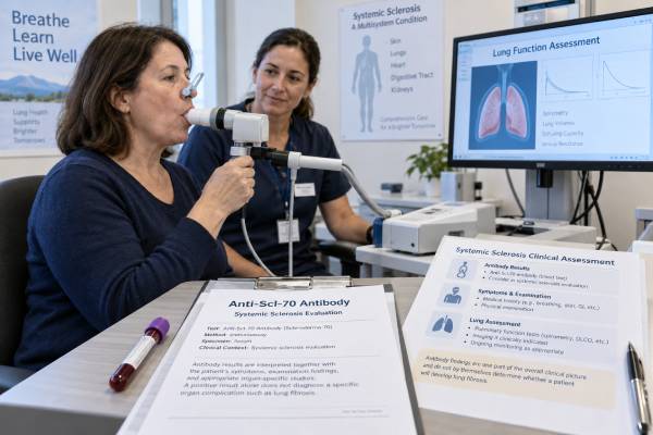
The anti-Scl-70 antibody test looks for an autoantibody strongly associated with systemic sclerosis, especially the diffuse cutaneous form and interstitial lung disease. Anti-Scl-70 is also called anti-topoisomerase I because it targets an enzyme inside the cell nucleus. A positive result can strengthen the case for systemic sclerosis when a person has compatible symptoms, but it does not diagnose the disease by itself. False-positive results occur, particularly with some automated laboratory methods, and the antibody can occasionally appear before a person meets full diagnostic criteria.
The most useful interpretation combines the result with the antinuclear antibody pattern, skin and blood-vessel findings, lung testing, and examination by a rheumatologist. A confirmed positive result should prompt careful assessment for lung involvement, even when breathing feels normal, because early interstitial lung disease may cause few symptoms. The result is mainly a risk and classification marker; repeated antibody levels usually do not track treatment response or day-to-day disease activity.
- A positive anti-Scl-70 result supports systemic sclerosis most strongly when symptoms and examination findings also fit the disease.
- The antibody is linked to a higher likelihood of interstitial lung disease, but it does not prove that lung fibrosis is present or predict exactly how fast it will progress.
- No universal “normal range” applies; laboratories commonly report the test as negative, equivocal, or positive using method-specific cutoffs.
- Fasting is usually unnecessary, and common medications generally do not need to be stopped unless the ordering clinician gives specific instructions.
- A newly confirmed positive result often leads to pulmonary function tests and high-resolution chest CT rather than treatment based on the antibody alone.
Table of Contents
- What the Anti-Scl-70 Test Detects
- Why the Test Is Ordered
- Understanding Positive, Negative, and Equivocal Results
- What Anti-Scl-70 Means for Lung Fibrosis Risk
- Testing Methods, Confirmation, and False Positives
- Next Steps After a Positive Result
- Common Questions and Practical Takeaways
What the Anti-Scl-70 Test Detects
Anti-Scl-70 is an autoantibody directed against DNA topoisomerase I, an enzyme involved in managing the structure of DNA. The name “Scl-70” came from an older laboratory observation involving a roughly 70-kilodalton protein fragment. Modern reports may use any of these names:
- Anti-Scl-70 antibody
- Anti-topoisomerase I antibody
- ATA, meaning antitopoisomerase antibody
- Scl-70 IgG
The test uses a blood sample. It is often included in an extractable nuclear antigen panel, a systemic autoimmune rheumatic disease panel, or a dedicated scleroderma antibody panel. A clinician may also order it as a stand-alone follow-up test after a positive antinuclear antibody result.
Systemic sclerosis, also called scleroderma, is an autoimmune connective tissue disease involving abnormal immune activity, blood-vessel injury, and fibrosis. It can affect the skin, lungs, digestive tract, heart, kidneys, and other tissues. Anti-Scl-70 is one of several disease-associated antibodies that help identify a clinical pattern. Other important markers include anticentromere antibodies and anti-RNA polymerase III antibodies.
These antibody groups are often described as “mutually exclusive,” meaning most people with systemic sclerosis have no more than one major disease-specific antibody. Exceptions occur, especially when broad automated panels detect low-level reactivity. A result therefore needs clinical and laboratory context rather than automatic acceptance as proof of a particular subtype.
Anti-Scl-70 is not a fibrosis measurement. It does not show the amount of scar tissue in the skin or lungs, and the numeric antibody value is not a direct severity score. Its main value is identifying an immune signature associated with systemic sclerosis and a greater probability of certain organ complications.
Why the Test Is Ordered
A clinician usually orders anti-Scl-70 testing when symptoms or examination findings raise concern for systemic sclerosis. It is not a useful general-population screening test because the disease is uncommon and false-positive results become more likely when testing people with a very low pretest probability.
Features that may lead to testing include:
- Raynaud phenomenon, in which fingers turn white, blue, or red with cold or stress
- Puffy fingers that later become tight, thickened, or tapered
- Skin tightening beyond the fingers
- Fingertip ulcers, pits, or scars
- Dilated small blood vessels called telangiectasias
- Abnormal nailfold capillaries
- Reflux, swallowing difficulty, or reduced movement of the esophagus
- Unexplained dry cough, shortness of breath, or reduced exercise tolerance
- Crackling sounds at the lung bases
- A positive antinuclear antibody test with a pattern or symptoms suggestive of connective tissue disease
The test may also help distinguish systemic sclerosis from other conditions that cause Raynaud phenomenon, muscle weakness, inflammatory arthritis, or overlapping autoimmune symptoms. It contributes to the 2013 ACR/EULAR classification criteria for systemic sclerosis as one of three disease-specific antibody groups. Classification criteria support research consistency and can assist clinical reasoning, but clinicians do not use them as a substitute for diagnosis.
A positive result is especially meaningful when accompanied by characteristic systemic sclerosis findings. For example, a person with Raynaud phenomenon, puffy fingers, abnormal nailfold capillaries, and confirmed anti-Scl-70 has a very different probability of disease from someone with no symptoms whose broad wellness panel shows a weak positive result.
Testing may also be appropriate in “very early” systemic sclerosis. Some people have Raynaud phenomenon and disease-specific antibodies years before definite skin thickening or internal-organ disease develops. In this setting, the antibody identifies increased risk but cannot say whether progression will occur or when. Follow-up focuses on symptoms, examination, capillaroscopy, lung function, and other organ assessments.
Understanding Positive, Negative, and Equivocal Results
Laboratories do not use one universal anti-Scl-70 scale. Depending on the platform, the report may give an index value, antibody units, units per milliliter, or a simple qualitative category. Always use the reference interval printed on the report.
| Result | What it usually means | What it does not mean |
|---|---|---|
| Negative | No antibody was detected above that method’s cutoff. This lowers support for an anti-topoisomerase I systemic sclerosis pattern. | It does not rule out systemic sclerosis or interstitial lung disease. |
| Equivocal or borderline | The signal is near the laboratory cutoff. Repeat or confirmatory testing may be useful if clinical suspicion is meaningful. | It is not a diagnosis and should not be treated as clearly positive without context. |
| Positive | The antibody was detected. A confirmed result supports systemic sclerosis, particularly a diffuse or lung-involvement phenotype, when clinical findings agree. | It does not prove current lung fibrosis, define its extent, or determine treatment by itself. |
How specific is a positive result?
Anti-Scl-70 is considered a relatively specific systemic sclerosis antibody when detected with a well-validated method in the right clinical setting. Specificity is not the same as certainty. Test performance varies by assay, and a weak positive from a multiplex panel may have a lower positive predictive value than a result confirmed by a more specific method.
The antibody can occasionally be reported in people with other autoimmune diseases or no confirmed connective tissue disease. Some apparent cross-disease positives reflect assay limitations rather than true anti-topoisomerase I autoimmunity. This is one reason clinicians may question an isolated result when the ANA is negative and the person has none of the expected symptoms.
Does a higher number mean more severe disease?
Usually, no. Some research has explored whether antibody concentration or change over time correlates with disease features, but routine clinical practice does not use anti-Scl-70 titers as a reliable activity gauge. A high value does not automatically mean severe fibrosis, and a falling value does not prove that treatment is working.
Doctors monitor systemic sclerosis using organ-specific information: skin examination, symptom changes, blood pressure, kidney function, pulmonary function tests, chest imaging, echocardiography, and other targeted studies. Repeating anti-Scl-70 without a specific reason rarely adds more value than monitoring the organs at risk.
What does a negative result mean?
A negative result cannot exclude systemic sclerosis. Many patients have anticentromere antibodies, anti-RNA polymerase III antibodies, less common antibodies, or no identifiable disease-specific antibody on a standard panel. The clinical pattern remains central.
A negative anti-Scl-70 also does not guarantee low lung risk. Interstitial lung disease occurs in antibody-negative systemic sclerosis and in people with other antibody profiles. Symptoms, disease subtype, ethnicity, inflammatory markers, lung function, and imaging all influence risk.
What Anti-Scl-70 Means for Lung Fibrosis Risk
Anti-Scl-70 is associated with systemic sclerosis–related interstitial lung disease, often shortened to SSc-ILD. Interstitial lung disease affects the supporting tissue around the air sacs. In systemic sclerosis, inflammation and fibrosis can make the lungs stiffer, reduce oxygen transfer, and cause cough or breathlessness.
The association is clinically important, but it must be interpreted correctly:
- The antibody raises the probability of ILD. People with anti-Scl-70 are more likely to develop lung involvement than those with some other systemic sclerosis antibody profiles.
- The antibody does not detect ILD. High-resolution computed tomography, not the blood test, shows whether interstitial abnormalities are present.
- The antibody does not reliably forecast an individual course. Some antibody-positive people have mild, stable changes for years. Others develop clinically important or progressive fibrosis.
- Risk is shaped by multiple factors. Diffuse skin disease, early disease duration, lower baseline lung function, greater CT extent, certain demographic factors, and evidence of progression all matter.
Why screening may be needed before symptoms appear
Early SSc-ILD can be silent. A person may have normal daily activity and no obvious breathlessness while high-resolution CT already shows ground-glass or reticular changes. Pulmonary function tests may also remain within the laboratory reference range despite a meaningful decline from that person’s prior baseline.
Current guidance supports screening people with systemic sclerosis who are at risk using pulmonary function tests and high-resolution chest CT. Pulmonary function testing usually includes forced vital capacity, or FVC, and diffusing capacity for carbon monoxide, or DLCO. FVC estimates how much air a person can exhale after a full breath. DLCO estimates how well gases move from the lungs into the blood.
The typical CT pattern in systemic sclerosis is nonspecific interstitial pneumonia, often with lower-lung ground-glass change, fine reticulation, traction bronchiectasis, and variable fibrosis. A usual interstitial pneumonia pattern can occur but is less typical.
Progression matters more than antibody status alone
Once ILD is identified, the central questions are how extensive it is and whether it is progressing. Clinicians compare symptoms, FVC, DLCO, oxygen levels during activity, and CT findings over time. A meaningful pattern may include worsening breathlessness, declining lung function, or increasing fibrosis on imaging.
No single threshold fits every patient. A change must be interpreted alongside test quality, baseline values, infection, anemia, pulmonary hypertension, effort during spirometry, and other factors. A small one-time fluctuation may be measurement noise; a sustained decline across several measures is more concerning.
Treatment decisions can include mycophenolate, cyclophosphamide, rituximab, tocilizumab, nintedanib, or other approaches depending on disease pattern, progression, comorbidities, pregnancy plans, local approvals, and specialist judgment. The antibody may influence vigilance, but it does not choose the medication.
Testing Methods, Confirmation, and False Positives
Anti-Scl-70 can be measured by enzyme immunoassay, chemiluminescent immunoassay, multiplex bead assay, line immunoassay, immunoblot, immunodiffusion, and other laboratory techniques. These methods do not perform identically.
Older immunodiffusion methods are often considered highly specific, but they are less widely available and slower. Automated methods are efficient and support broad panels, yet some platforms produce more low-level positives in people who do not have systemic sclerosis. Assay antigen design, cutoff selection, and the population being tested all affect the result.
Confirmation deserves consideration when all of the following are true:
- The result is weak or just above the cutoff.
- The test came from a broad multiplex panel.
- The ANA by indirect immunofluorescence is negative or shows an unrelated pattern.
- There are no systemic sclerosis symptoms or examination findings.
- A major diagnosis, repeated imaging, or long-term follow-up would depend heavily on the result.
A clinician may repeat the test at a laboratory using another platform or request a more specific confirmatory method. The goal is not to chase every minor laboratory difference. It is to avoid labeling someone with a serious chronic disease based on an unexpected isolated signal.
ANA context
Most people with systemic sclerosis have a positive ANA. Anti-topoisomerase I reactivity can produce a nuclear pattern on indirect immunofluorescence, but pattern recognition alone cannot identify the antibody with certainty. The ANA titer and pattern should be viewed as supporting context, not as a replacement for antigen-specific testing.
An isolated anti-Scl-70 positive with a negative ANA does not make systemic sclerosis impossible, but it should increase attention to assay validity and clinical fit. Conversely, a strongly positive ANA with compatible features and confirmed anti-Scl-70 creates a more coherent result pattern.
Preparation and sample issues
The test generally requires no fasting. Blood is drawn from a vein, and the main risks are brief pain, bruising, or lightheadedness. Biotin can interfere with some immunoassays, though the effect depends on the platform. Anyone taking high-dose biotin for hair, nail, neurologic, or metabolic reasons should tell the laboratory and clinician. Do not stop prescribed medicine or supplements without instructions.
Recent infection or immune stimulation can alter some autoantibody results, but there is no standard waiting period for anti-Scl-70 testing. When a result is unexpected, repeating it after the acute illness and using a different method may be more informative than assuming it represents systemic sclerosis.
Next Steps After a Positive Result
A positive result calls for structured evaluation, not panic. The next steps depend on symptoms, physical findings, ANA results, and whether systemic sclerosis has already been diagnosed.
1. Confirm that the result fits the clinical picture
A rheumatology assessment may review Raynaud phenomenon, finger swelling, skin texture, fingertip injury, nailfold capillaries, reflux, swallowing, joint or muscle symptoms, and cardiopulmonary complaints. The clinician may order or review a broader connective tissue disease blood test panel to look for a coherent antibody pattern and alternative diagnoses.
2. Establish baseline organ screening
For suspected or confirmed systemic sclerosis, baseline evaluation often includes:
- Pulmonary function tests with FVC and DLCO
- High-resolution CT of the chest to detect ILD
- Echocardiography and other screening for pulmonary hypertension
- Blood pressure monitoring
- Serum creatinine and urinalysis
- Complete blood count and metabolic testing
- Assessment of reflux, swallowing, nutrition, and gastrointestinal symptoms
The exact plan varies. High-resolution CT uses radiation, so follow-up imaging is individualized rather than performed automatically at every visit.
3. Monitor trends, not just single numbers
If no ILD is found, the clinician may repeat pulmonary function tests periodically, with timing based on risk and disease duration. If ILD is present, follow-up is usually more frequent. The same laboratory and testing technique can make trends easier to interpret.
Call the treating team sooner for a new persistent dry cough, increasing breathlessness, reduced exercise tolerance, chest discomfort, fainting, or lower oxygen readings. Sudden severe shortness of breath, chest pain, coughing blood, blue lips, confusion, or very low oxygen requires urgent medical assessment because infection, blood clot, pneumothorax, heart disease, or another emergency may be responsible.
4. Do not treat the antibody itself
There is no medication whose goal is simply to make anti-Scl-70 negative. Treatment targets active or threatening disease manifestations. A person with a positive antibody but no definite systemic sclerosis may need observation rather than immunosuppression. A person with progressive SSc-ILD may need prompt therapy even if the antibody value is unchanged.
Lifestyle measures support overall lung and vascular health but do not replace disease-specific care. Avoid smoking and inhaled irritants, keep recommended vaccines current, stay physically active within safe limits, manage reflux, and discuss pulmonary rehabilitation when exercise capacity is reduced.
Common Questions and Practical Takeaways
Can anti-Scl-70 be positive before systemic sclerosis develops?
Yes. Disease-specific autoantibodies can precede a full clinical diagnosis. The result is most concerning when Raynaud phenomenon, puffy fingers, or abnormal nailfold capillaries are also present. Some people remain in an early or incomplete stage for years, so follow-up should be proportionate rather than based on a fixed prediction.
Does anti-Scl-70 mean diffuse systemic sclerosis?
It is associated more often with diffuse cutaneous systemic sclerosis, in which skin thickening extends above the elbows or knees or involves the trunk. However, it can appear in limited cutaneous disease and in systemic sclerosis without obvious skin thickening. Antibody profile informs probability; the physical distribution of skin disease defines the cutaneous subtype.
Is it the same as the Scl-70 gene?
No. Anti-Scl-70 is an antibody, not a genetic test. It measures an immune response against topoisomerase I. It does not determine inherited risk and does not mean the person has a mutation in the TOP1 gene.
Can someone have anti-Scl-70 without lung disease?
Yes. Many antibody-positive people do not have clinically important ILD at a given point in time. That is why the result should lead to appropriate screening, not an assumption that fibrosis already exists.
How does it compare with other scleroderma antibodies?
The anticentromere antibody is more often associated with limited cutaneous disease and later pulmonary arterial hypertension, while anti-RNA polymerase III is associated with rapidly progressive skin involvement and a higher risk of scleroderma renal crisis. These are broad risk patterns, not guarantees. Every antibody group still requires comprehensive organ screening.
What is the most important action after an unexpected positive?
Verify the result’s strength, method, ANA context, and clinical fit. If systemic sclerosis is plausible, arrange rheumatology evaluation and baseline lung assessment. If the test is an isolated weak positive in someone without symptoms, confirmation may prevent unnecessary worry and testing.
The practical bottom line is that anti-Scl-70 is a valuable systemic sclerosis marker and an important warning sign for lung involvement, but it is not a stand-alone diagnosis, fibrosis scan, or prognosis calculator. The safest interpretation joins accurate laboratory methods with symptoms, examination, pulmonary function, imaging, and longitudinal follow-up.
References
- The 2024 British Society for Rheumatology guideline for management of systemic sclerosis 2024 (Guideline)
- 2023 American College of Rheumatology (ACR)/American College of Chest Physicians (CHEST) Guideline for the Screening and Monitoring of Interstitial Lung Disease in People with Systemic Autoimmune Rheumatic Diseases 2024 (Guideline)
- 2023 American College of Rheumatology (ACR)/American College of Chest Physicians (CHEST) Guideline for the Treatment of Interstitial Lung Disease in People with Systemic Autoimmune Rheumatic Diseases 2024 (Guideline)
- EULAR recommendations for the treatment of systemic sclerosis: 2023 update 2025 (Guideline)
- Diagnosis and monitoring of systemic sclerosis-associated interstitial lung disease using high-resolution computed tomography 2022 (Review)
- Expert consensus on the management of systemic sclerosis-associated interstitial lung disease 2023 (Position Statement)
Disclaimer
This article provides general education about anti-Scl-70 testing and cannot diagnose systemic sclerosis or interstitial lung disease. Results should be interpreted by a qualified clinician using the laboratory method, symptoms, examination, pulmonary testing, and imaging. Seek urgent care for sudden or severe breathing difficulty, chest pain, fainting, coughing blood, confusion, or blue lips.





