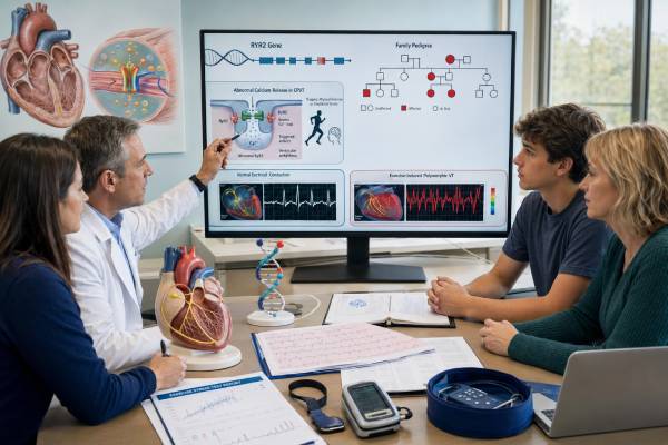
Catecholaminergic polymorphic ventricular tachycardia (CPVT) is an inherited rhythm disorder in which physical exertion or intense emotion can trigger dangerous ventricular arrhythmias. The resting electrocardiogram is often normal, and the heart may appear structurally normal, so the condition can be missed after fainting or seizure-like episodes. Exercise testing is central because progressively faster heart rates may reveal characteristic premature beats, bidirectional ventricular tachycardia, or polymorphic ventricular tachycardia. Genetic testing most often examines RYR2, the principal gene associated with autosomal dominant CPVT. A pathogenic RYR2 variant can confirm the molecular cause, help identify relatives who need treatment and surveillance, and support testing in children before symptoms occur. A negative test does not exclude CPVT, and a rare RYR2 change is not automatically disease-causing. Results must be interpreted with exercise-induced rhythm findings, symptoms, family history, variant location and mechanism, and laboratory evidence. Prompt specialist evaluation matters because appropriate medication, activity planning, and family screening can substantially reduce risk.
- CPVT commonly causes exertional or emotion-triggered fainting despite a normal resting ECG.
- RYR2 variants disrupt calcium handling inside heart muscle cells and account for many—but not all—cases.
- Exercise testing usually provides the most important clinical evidence for diagnosis and treatment response.
- A pathogenic familial variant enables targeted testing before symptoms appear in relatives.
- Beta-blockers and sometimes flecainide are central treatments; an implanted defibrillator is not a stand-alone solution.
- A variant of uncertain significance should not be used by itself to diagnose CPVT or restrict an unaffected relative.
Table of Contents
- RYR2 and the CPVT mechanism
- Recognizing the clinical pattern
- Who should consider genetic testing
- How the test is performed
- Interpreting RYR2 results
- Treatment and monitoring
- Exercise, emotion, and daily safety
- Family screening and reproductive care
RYR2 and the CPVT mechanism
RYR2 encodes the cardiac ryanodine receptor, a large calcium-release channel in the membrane of the sarcoplasmic reticulum inside heart muscle cells. During each heartbeat, a small influx of calcium through the cell membrane prompts RYR2 channels to release a much larger stored calcium signal. That signal allows the muscle fiber to contract. Calcium is then pumped back into storage so the cell can relax and prepare for the next beat.
Most classic RYR2-related CPVT variants make the channel abnormally prone to releasing calcium during adrenergic stimulation. When exercise, fear, excitement, or another stress raises catecholamine levels, calcium can leak from storage between normal beats. This may create delayed afterdepolarizations and premature ventricular contractions. As the heart rate increases, the ectopy can become repetitive, alternate in direction, and progress to bidirectional or polymorphic ventricular tachycardia. Ventricular fibrillation may follow.
The heart can be structurally normal because CPVT is primarily a calcium-handling disorder, not initially a disease of weakened or thickened muscle. A normal echocardiogram does not make exertional syncope safe, and a normal resting ECG does not exclude the condition. The dangerous electrical behavior often appears only when adrenergic stress reaches a threshold.
RYR2-associated CPVT is generally autosomal dominant. One pathogenic variant is sufficient to create susceptibility, and each child of a carrier has a 50% chance of inheriting it. Penetrance is incomplete: some carriers have clear exercise-induced arrhythmia, while others remain asymptomatic or develop findings later. Lack of symptoms in a parent therefore does not prove that a variant arose for the first time in a child.
Other genes can produce CPVT or CPVT-like phenotypes. Biallelic variants in CASQ2, TRDN, and TECRL are important examples, and selected CALM genes may be considered in specialized testing. Some RYR2 variants cause calcium-release deficiency syndrome, which may have a different mechanism and less typical exercise-test findings. This is why testing is usually performed with a curated CPVT or inherited arrhythmia panel when the phenotype is not already tied to a known familial RYR2 variant.
Not every rare RYR2 variant causes disease. RYR2 is a very large gene, and rare missense changes are found in healthy people. Disease-associated variants cluster in recognized functional regions, but location alone is not proof. Interpretation requires population frequency, functional evidence, prior cases, segregation, phenotype fit, and expert classification.
Recognizing the clinical pattern
CPVT often presents in childhood or adolescence, but diagnosis can occur in infancy or adulthood. The most characteristic symptom is fainting during running, swimming, competitive sport, a sudden fright, intense excitement, anger, or another adrenergic trigger. Some episodes are misdiagnosed as epilepsy because loss of cerebral blood flow can cause stiffening or jerking. A careful history asks what the person was doing, whether warning symptoms occurred, how quickly consciousness returned, and whether similar events or sudden deaths occurred in relatives.
The resting ECG is frequently normal. A slow resting heart rate may occur, but there is usually no diagnostic QT prolongation or Brugada pattern. Echocardiography and cardiac magnetic resonance imaging are used to exclude cardiomyopathy or other structural causes when indicated. Routine blood tests can identify electrolyte or metabolic contributors.
Exercise stress testing is the principal provocative assessment. The rhythm may begin with isolated ventricular premature beats at a reproducible heart-rate threshold. With increasing workload, ectopy can become bigeminal, couplets, bidirectional ventricular tachycardia, or polymorphic ventricular tachycardia. The arrhythmia often improves as exercise stops and catecholamine levels fall. A test must be performed where malignant arrhythmia can be treated immediately.
A single negative exercise test does not always exclude CPVT. Arrhythmias can vary between days, medication may suppress the phenotype, and young carriers may not yet express clear findings. Repeat testing, ambulatory monitoring during ordinary activity, or carefully selected pharmacologic provocation may be considered by an inherited arrhythmia specialist. An exercise test should not be deliberately attempted outside medical supervision to “see what happens.”
Differential diagnosis includes long QT syndrome, arrhythmogenic cardiomyopathy, anomalous coronary arteries, myocarditis, supraventricular tachycardia, vasovagal syncope, epilepsy, and other causes of exertional collapse. Andersen-Tawil syndrome can also cause bidirectional ventricular tachycardia but usually has additional ECG or physical findings. The clinical evaluation must establish that catecholamine-triggered ventricular ectopy is the likely mechanism.
A clinical diagnosis may be made when a structurally normal heart and normal resting ECG are accompanied by otherwise unexplained exercise- or catecholamine-induced bidirectional or polymorphic ventricular tachycardia. A pathogenic variant in RYR2 or another definitive CPVT gene can also establish a molecular diagnosis, particularly in a relative, but the exact phenotype still needs assessment.
Who should consider genetic testing
Testing is recommended when a cardiologist or electrophysiologist has established a clinical diagnosis or strong suspicion of CPVT. This includes people with characteristic ventricular ectopy during exercise testing, resuscitated cardiac arrest during exertion or emotion without a structural explanation, or recurrent exertional syncope with supportive rhythm findings. Testing is also valuable after sudden unexplained death when circumstances suggest an inherited arrhythmia and a suitable DNA sample is available.
The first person tested in a family should ideally be the most clearly affected living person. A positive result in that index case creates a precise marker for relatives. Starting with an asymptomatic relative before the familial cause is known has lower interpretive value because RYR2 contains many rare benign and uncertain changes.
A focused RYR2 test may be reasonable when the phenotype is classic, but many laboratories use a curated CPVT panel that includes CASQ2, TRDN, TECRL, and selected additional genes. A broader inherited arrhythmia panel may be appropriate if the ECG or history could represent long QT syndrome, Brugada syndrome, conduction disease, or another channelopathy. Very broad panels should be used cautiously because they increase uncertain findings.
Testing a healthy relative is strongly informative after a pathogenic or likely pathogenic familial variant has been identified. The targeted test asks only whether that exact change is present. A positive result prompts clinical evaluation and preventive treatment planning; a negative result usually removes the relative from surveillance for that familial variant, unless symptoms or a second diagnosis indicate otherwise.
Children are often offered targeted testing because CPVT can cause serious events in childhood and treatment can begin before symptoms. This differs from predictive testing for conditions that occur only in adulthood. Parents should receive counseling about both the potential benefit of early risk reduction and the uncertainty of severity among carriers.
Testing is less useful for an isolated fainting episode without an exertional or emotional trigger, normal cardiac evaluation, and no relevant family history. In that setting, broad genetic screening may produce a VUS that distracts from more likely causes. Genetic testing should follow a clinical assessment, not replace one.
Pretest counseling covers inheritance, possible outcomes, the chance of a negative result, implications for sports and relatives, reproductive options, and the possibility that a panel will find an unrelated or uncertain result. Families should understand that a molecular result can clarify susceptibility but cannot predict the exact timing or severity of an event.
How the test is performed
Testing usually uses a blood or saliva sample. Fasting is unnecessary, and taking a sample does not itself require stopping beta-blockers or other medications. Medication changes for exercise testing are a separate clinical decision and must be directed by the electrophysiology team because withdrawing therapy can increase risk.
The laboratory sequences RYR2 and any other genes included on the selected panel. It may also analyze exon-level deletions and duplications. Because RYR2 is large, high and consistent coverage is important. Reports should identify any regions that could not be analyzed reliably and state whether copy-number analysis was performed.
Clinical information should accompany the sample. Useful details include the exercise-test pattern, heart-rate threshold for ectopy, symptoms, age at onset, medication status, cardiac imaging, arrest circumstances, and family history. Laboratory scientists can interpret a rare RYR2 change more accurately when they know whether the patient has a classic CPVT phenotype.
Variants are generally classified as pathogenic, likely pathogenic, uncertain significance, likely benign, or benign. The classification considers whether the variant has been seen in affected people, whether it is absent or very rare in population databases, whether it lies in a functionally important region, whether laboratory studies show abnormal calcium release, and whether it segregates with disease in the family. Computational predictions are supporting evidence, not a final answer.
Results commonly take several weeks. If the test is urgent after a cardiac arrest or during a family evaluation, the laboratory may offer expedited analysis, but clinical precautions should not wait for the DNA report when suspicion is high. Treatment and activity advice are based on the observed risk while testing is pending.
The report should be reviewed by a clinician familiar with inherited arrhythmias. RYR2 variant interpretation is challenging because background rare variation is substantial. An expert may check ClinGen gene validity, current variant databases, original publications, functional evidence, and whether the laboratory applied disease-specific criteria. Reanalysis is appropriate when new evidence appears or the clinical picture changes.
Interpreting RYR2 results
A pathogenic or likely pathogenic RYR2 variant that fits a gain-of-function CPVT mechanism can confirm the molecular diagnosis in a person with a compatible phenotype. It supports targeted testing in relatives and can identify carriers whose resting ECG and initial examination are normal. The result does not quantify an individual’s exact future risk and does not show whether current medication is fully protective.
A positive result should be interpreted at the variant level. Many classic CPVT variants are missense changes in recognized RYR2 regions, but no single “hot spot” rule is sufficient. Some variants have direct functional evidence of abnormal calcium release; others are supported by repeated observations and strong segregation. Truncating variants require particular caution because simple loss of one copy is not the standard mechanism for classic RYR2-mediated CPVT.
A negative result means no reportable causal variant was identified with the test used. It does not rule out CPVT. The disease may be caused by a gene not included, a difficult-to-detect variant, an unresolved mechanism, or a variant that current knowledge cannot classify. A person with characteristic exercise-induced arrhythmia should continue treatment and follow-up despite a negative genetic test.
A VUS is an unresolved finding. It should not be treated as confirmation, and it should not be used alone to diagnose healthy relatives. Because RYR2 is large and rare variation is common, the potential for overdiagnosis is substantial. Family studies can sometimes help: if the variant is absent from several clearly affected relatives, causality becomes less likely; if it tracks with a strong phenotype across multiple informative relatives, evidence may strengthen. Those studies should be coordinated with the testing laboratory or genetics service.
A VUS may later be reclassified as benign, likely benign, likely pathogenic, or pathogenic. The patient should keep the exact report and ask how reanalysis is handled. The distinction between a confirmed result and an uncertain one is explained further in the overview of pathogenic, benign, and VUS results.
A benign or likely benign variant is not an explanation for CPVT and should not guide treatment. Consumer DNA raw data or third-party interpretations should be confirmed in a clinical laboratory before any medical decision, because technical false positives and unsupported classifications can occur.
Treatment and monitoring
Treatment aims to suppress catecholamine-triggered ventricular ectopy and prevent syncope, cardiac arrest, and sudden death. A nonselective beta-blocker without intrinsic sympathomimetic activity is generally the foundation. Nadolol is often preferred when available because of its long duration and clinical experience in CPVT. Dosing is individualized by weight, heart rate, tolerance, and exercise-test response. Missing doses can leave a dangerous gap in protection.
Exercise testing is repeated after treatment starts and after major dose changes. The aim is not simply to lower resting heart rate; it is to assess whether ventricular ectopy appears as adrenergic stress rises. Suppression may be incomplete even when the person feels well. Ambulatory monitoring can add information about daily-life rhythms and adherence.
Flecainide may be added when ventricular arrhythmias persist despite an adequately dosed beta-blocker or when the clinical situation remains high risk. It reduces triggered activity through effects that include inhibition of abnormal calcium release and sodium current. Dosing and monitoring require specialist oversight because flecainide is not appropriate for every rhythm or structural heart condition.
Left cardiac sympathetic denervation can be considered when arrhythmias remain uncontrolled, medications are not tolerated, or recurrent events occur despite treatment. The operation reduces sympathetic input to the heart but does not eliminate the genetic substrate. Medication often continues afterward.
An implanted cardioverter-defibrillator may be appropriate after cardiac arrest or for selected patients with recurrent life-threatening events despite optimized therapy. CPVT creates a special challenge: an ICD shock can be painful and provoke an adrenergic surge, which may trigger further arrhythmia and repeated shocks. Device programming, beta-blockade, flecainide, and sometimes sympathetic denervation are therefore essential. An ICD should not be viewed as a replacement for antiadrenergic treatment.
Follow-up should assess symptoms, adherence, side effects, growth-related dose changes in children, exercise-test findings, and device data when applicable. Significant bradycardia, fatigue, or exercise intolerance may require dose adjustment, but therapy should not be stopped abruptly. Some patients benefit from psychological support because fear of exertion, shocks, or another family death can become limiting.
Exercise, emotion, and daily safety
Because adrenergic stimulation triggers CPVT, exercise planning must be individualized. Unsupervised high-intensity and competitive sports are generally restricted until a specialist has assessed risk, established treatment, and evaluated rhythm control. Some people may participate in lower-intensity activity under a shared decision-making plan, but a normal feeling during exercise is not enough; objective testing is needed.
The plan should address swimming specifically because exertion, cold-water stress, and delayed rescue can combine dangerously. Children, schools, coaches, and caregivers need written instructions about allowed activity, medication timing, symptoms, and emergency response. A person with CPVT should not exercise alone in settings where collapse would go unnoticed.
Acute emotion can also trigger arrhythmia. This does not mean ordinary feelings should be suppressed or that a person must avoid normal life. It does mean medication adherence, sleep, stress management, and preparation for predictable high-adrenaline situations matter. Sudden fright-based activities, extreme amusement rides, and stimulant exposure may require discussion with the clinical team.
Decongestants, stimulants, energy products, illicit sympathomimetics, and certain attention-deficit medications can raise catecholamine activity. Prescribers and pharmacists should know the diagnosis. Medication decisions are individualized; a necessary medicine should not be stopped without discussing alternatives and monitoring. Electrolyte loss from vomiting, diarrhea, or severe dieting should be corrected promptly.
Family members and close contacts should learn cardiopulmonary resuscitation. Access to an automated external defibrillator may be advised at home, school, training sites, or organized activities depending on risk and local resources. Emergency plans should state the diagnosis, current medicines, ICD status, and electrophysiology contact information.
Pregnancy and delivery increase physiologic stress but can often be managed safely with coordinated obstetric and electrophysiology care. Medication choice and dose should be reviewed before conception rather than stopped independently. Postpartum sleep deprivation and stress also deserve planning.
Family screening and reproductive care
When a pathogenic RYR2 variant is identified, first-degree relatives should be offered targeted testing. Each child, sibling, or parent has a 50% chance of carrying an autosomal dominant variant. Testing can reveal an apparently healthy carrier before the first exertional faint or arrest. Positive relatives need an ECG, exercise testing when age and circumstances permit, and treatment planning by an inherited arrhythmia specialist.
A negative targeted result for a confirmed familial variant usually means the relative did not inherit that specific CPVT susceptibility and can avoid repeated variant-related surveillance. This is one of the clearest benefits of molecular diagnosis. The conclusion may differ if the relative has concerning symptoms or if the family appears to have more than one rhythm disorder.
If the index patient’s genetic test is negative, first-degree relatives still require clinical evaluation because no DNA marker is available to exclude disease. Screening may include history, resting ECG, exercise testing, and follow-up as advised. A normal childhood test may need repetition because expression can be age-dependent.
Children who carry a pathogenic familial variant are generally evaluated and treated before unrestricted sports participation. Doses must be adjusted as weight changes. Parents should understand that an asymptomatic child is not necessarily unaffected and that medication adherence remains important even when exercise tests improve.
A familial VUS should not be used for predictive testing of unaffected children or adults. Clinical screening continues while evidence is reassessed. If several relatives have clear exercise-induced arrhythmias, carefully planned segregation analysis may help classification, but it is not the same as routine predictive testing.
Adults with a confirmed variant can discuss reproductive options. Autosomal dominant inheritance gives each pregnancy a 50% chance of inheriting the variant, but severity cannot be predicted reliably. Options may include natural conception, prenatal diagnosis, or in vitro fertilization with preimplantation genetic testing. Decisions are personal and should be supported with nondirective counseling.
Families should keep copies of the original laboratory report and share the exact gene and variant—not only the phrase “heart gene”—with relatives. A genetics professional can prepare a family letter and help arrange testing across health systems. Early identification allows treatment before a preventable event, while negative targeted results spare noncarriers from unnecessary restrictions and anxiety.
References
- 2022 ESC Guidelines for the management of patients with ventricular arrhythmias and the prevention of sudden cardiac death (2022 Guideline)
- European Heart Rhythm Association (EHRA)/Heart Rhythm Society (HRS)/Asia Pacific Heart Rhythm Society (APHRS)/Latin American Heart Rhythm Society (LAHRS) Expert Consensus Statement on the state of genetic testing for cardiac diseases (2022 Consensus Statement)
- Catecholaminergic Polymorphic Ventricular Tachycardia (2022 GeneReviews)
- Prognosis and clinical management of asymptomatic family members with RYR2-mediated catecholaminergic polymorphic ventricular tachycardia: a review (2024 Review)
- Catecholaminergic Polymorphic Ventricular Tachycardia: Multiple Clinical and Cellular Insights (2025 Review)
Disclaimer
This article is for general education and does not provide personalized medical advice or emergency instructions. CPVT diagnosis, exercise testing, genetic interpretation, medication dosing, and activity recommendations require an inherited arrhythmia specialist and genetics professional. Fainting during exercise, seizure-like activity with exertion, sustained palpitations, or cardiac arrest signs require urgent medical evaluation.