
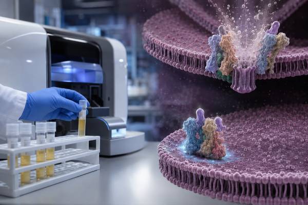
The complement C7 test assesses the function of complement component 7, one of the proteins that turns C5b-6 into a membrane-bound terminal complement complex. When C7 is absent or ineffective, the immune system cannot assemble a normal membrane attack complex, and susceptibility to invasive Neisseria infections rises substantially. The most revealing history is recurrent meningococcal meningitis or bloodstream infection, sometimes separated by years and occurring in a person who otherwise seems healthy.
C7 testing is specialized. Clinicians generally begin with CH50 and AH50, then measure individual terminal components only when both pathways fail or the infection history remains strongly suggestive. An abnormal C7 value can also result from complement consumption, liver dysfunction, protein loss, medication, or poor specimen handling, so confirmation is essential. This article explains how the C7 test fits into the diagnostic sequence, how inherited deficiency presents, why genetic findings vary by ancestry and family, and how a confirmed result changes vaccination, antibiotic planning, and emergency response.
- C7 allows the C5b-7 complex to associate with a microbial membrane before C8 and C9 complete the attack complex.
- Complete C7 deficiency typically causes absent CH50 and AH50 with preserved C3 and C4 concentrations.
- Functional testing detects both absent protein and rare nonfunctional C7 that an antigen test might miss.
- Recurrent or epidemiologically unusual meningococcal disease should prompt terminal complement screening.
- A confirmed diagnosis requires lifelong risk-based meningococcal prevention and rapid assessment of possible infection.
Table of Contents
- The specific step controlled by C7
- Why C7 is a second-line rather than screening test
- How to interpret C7 functional activity
- Recognizing inherited C7 deficiency
- Genetic confirmation, ancestry, and family testing
- Acquired and technical causes of low C7
- Management after C7 deficiency is confirmed
- Urgent care and questions for the specialist
The specific step controlled by C7
Complement activation can begin when antibodies recognize a target, when lectins bind microbial carbohydrates, or when the alternative pathway amplifies spontaneous surface activation. Despite different starts, all three routes can create a C5 convertase. Cleavage of C5 releases C5a and exposes C5b, which binds C6. C7 then joins the C5b-6 complex.
The arrival of C7 changes the physical behavior of the complex. C5b-7 exposes hydrophobic regions that permit association with lipid membranes. C8 binds next and begins membrane penetration; C9 molecules then polymerize to enlarge the pore. Without C7, the sequence stalls before stable membrane insertion, so the membrane attack complex cannot form normally.
This defect is selective. The person can still activate C3, deposit C3b on a bacterium, generate inflammatory mediators, and use antibodies and phagocytes. That preserved upstream immunity explains why many people with C7 deficiency do not have the broad pattern of childhood bacterial infections seen with complete C3 deficiency. The weakness becomes most apparent against Neisseria meningitidis and Neisseria gonorrhoeae, organisms for which terminal serum bactericidal activity is especially important.
C7 is part of the shared terminal pathway. A defect in C5, C6, C7, C8, or C9 can therefore produce the same broad screening result: classical-pathway activity and alternative-pathway activity are both absent. A C7 test identifies the failed component only after those pathway screens indicate where to look.
The routine C7 investigation is not a measurement of inflammation. Tests such as soluble C5b-9 assess terminal complement activation in selected diseases. C7 functional testing instead asks whether enough effective C7 is available to build a lytic complex. A low functional value does not tell the clinician how much inflammation exists in a kidney, blood vessel, or other tissue.
Likewise, C7 is not C-reactive protein and is not part of a routine “complement C3/C4” panel. A normal C3 test or C4 test does not prove that the terminal pathway works; in inherited C7 deficiency, both concentrations are commonly normal.
Why C7 is a second-line rather than screening test
Testing every individual complement protein is inefficient and can generate confusing borderline findings. The diagnostic sequence begins with the patient’s history and two pathway-level assays.
The CH50 test evaluates the classical pathway through the terminal complex. The AH50 test evaluates the alternative pathway through that same terminal complex. Their combination localizes the problem:
- Low CH50, normal AH50: an early classical component defect is more likely.
- Normal CH50, low AH50: an alternative-pathway component or regulator defect is more likely.
- Low CH50 and low AH50: consider C3, C5, C6, C7, C8, or C9 deficiency, severe consumption, drug effect, or sample degradation.
A terminal component test becomes particularly appropriate when both screens are repeatedly absent and upstream C3 and C4 concentrations are preserved. Clinicians may order C5 through C9 functions together or use sequential reconstitution testing, depending on laboratory availability.
The infection history can justify testing even when only one episode is documented. Recurrent meningococcal disease is the clearest signal, but a first episode may warrant screening when it occurs at an unusual age, involves a rare serogroup, follows prior unexplained meningitis, or clusters in a family. Disseminated gonococcal infection can also reveal terminal complement deficiency.
A C7 test is usually low yield for common colds, uncomplicated pneumonia, chronic fatigue, isolated joint pain, or a low C4 that points toward a different pathway. Antibody deficiency, anatomic asplenia, HIV, medication-related immunosuppression, and other conditions have their own diagnostic approaches. The immune evaluation should follow the actual infection phenotype.
Timing affects interpretation. During meningococcal sepsis, complement may be consumed. A pathway screen taken in the acute phase can be useful, but an abnormal result often needs repetition after recovery. Vaccination and infection education can begin while confirmation is pending; the clinician does not need to postpone prevention until genetic testing returns.
How to interpret C7 functional activity
Most clinical C7 assays are functional. A laboratory combines the patient’s serum with a test system in which C7 is the limiting component, then measures restoration of complement activity. A person whose C7 works normally supplies the missing step. A person with absent or dysfunctional C7 produces little or no restoration.
One current reference laboratory reports a normal value of at least 58 U/mL. That threshold belongs to its particular method. Another laboratory may use percent activity or a different unit. Results cannot be converted reliably between platforms, and the printed reference interval takes precedence over numbers found in articles or online catalogs.
A profoundly low or absent result is more concerning than a value just below the cutoff, but severity alone does not identify the mechanism. Interpretation should answer several questions:
- Were CH50 and AH50 also absent?
- Were C3 and C4 concentrations normal?
- Was the sample processed and frozen according to the laboratory protocol?
- Was the patient acutely ill when blood was collected?
- Is the patient taking a complement inhibitor?
- Does the clinical history include invasive neisserial disease?
Functional testing has an advantage over antigen testing: it detects a protein that is present but unable to work. Research and clinical reports describe rare C7 variants that produce detectable immunoreactive material with inadequate lytic activity. Measuring quantity alone could miss such a defect.
It also has a vulnerability. Complement function deteriorates in serum that is left at room temperature, separated late, thawed repeatedly, or shipped incorrectly. A sample-handling problem often lowers several functional assays simultaneously. The laboratory may reject visibly unsuitable specimens, but degradation is not always obvious. Repeating an unexpected result with carefully controlled collection is mandatory before diagnosing inherited deficiency.
A normal C7 function rules out complete C7 deficiency with reasonable confidence but does not explain an absent pathway screen. The next candidates include C3, C5, C6, C8, and C9, along with treatment and preanalytical causes. If both CH50 and AH50 are normal despite recurrent infection, clinicians may investigate properdin, factor D, factor H, the lectin pathway, antibody production, spleen function, or nonimmune explanations.
Recognizing inherited C7 deficiency
Inherited C7 deficiency is rare and usually autosomal recessive. Affected individuals have pathogenic changes in both C7 gene copies. The condition may not become apparent in infancy because upstream complement, immunoglobulins, and cellular immunity remain available.
The classic presentation is invasive meningococcal disease. This includes meningitis, meningococcemia, or both. Some people have repeated episodes; others have a single episode followed by diagnosis. Historical records may describe “bacterial meningitis” without a stored isolate, so a careful review of prior hospitalizations can reveal recurrence that was not initially recognized.
Meningitis symptoms can include fever, severe headache, neck stiffness, vomiting, photophobia, confusion, or seizures. Bloodstream infection can produce sudden malaise, severe muscle pain, rapid breathing, low blood pressure, cold extremities, and petechiae or purpura. In terminal complement deficiency, disease may be caused by meningococcal serogroups that are less common in the general population. Clinical severity is variable and can still be life-threatening.
Disseminated gonococcal infection is another clue. Patients may develop fever, scattered pustules, migratory joint pain, tenosynovitis, or septic arthritis. Genital symptoms may be absent. A recurrent disseminated infection, especially in someone with appropriate exposure and no other explanation, should prompt terminal complement testing.
C7 deficiency does not have one fixed age of onset. Reports include children, adolescents, and adults diagnosed after recurrent disease. Exposure patterns influence when infection occurs. Living in close quarters, military service, travel, outbreaks, and sexual exposure can reveal a preexisting immune vulnerability.
The absence of infections in a sibling does not exclude the same genotype. Penetrance of infection is incomplete because vaccination, antibody repertoire, bacterial exposure, antibiotic use, and chance all affect outcome. Conversely, frequent upper-respiratory infections alone are not a typical C7 phenotype and should prompt assessment for other causes.
Some individuals with terminal component deficiency may develop chronic or relapsing meningococcal syndromes with fever, skin lesions, and joint symptoms rather than fulminant meningitis. This presentation can mimic vasculitis or autoimmune disease. Blood cultures or molecular testing and a high index of suspicion are important before immunosuppression is considered.
Genetic confirmation, ancestry, and family testing
More than one pathogenic C7 variant exists, and variant frequencies differ among populations. Published systematic reviews describe nonsense, frameshift, splice, deletion, and missense changes. Some variants recur because of founder effects in particular ancestral groups, while others are private to one family.
This diversity matters in two ways. First, a targeted test for one common variant can miss C7 deficiency in a person from another background. Comprehensive sequencing and deletion/duplication analysis may be needed when function is absent. Second, ancestry can inform—but never replace—functional testing. A person should not be considered affected or unaffected based on ethnicity.
Genetic confirmation is strongest when it matches the phenotype:
- C7 function is repeatedly absent or severely reduced.
- CH50 and AH50 show a shared terminal defect.
- Other component studies localize the failure to C7.
- Two pathogenic or likely pathogenic C7 variants are identified in the expected configuration.
A variant of uncertain significance is not diagnostic by itself. Family segregation, protein studies, functional assays, population frequency, and laboratory classification help determine whether it is relevant. Results can be reclassified as evidence accumulates.
Parents of a person with autosomal recessive C7 deficiency are usually carriers. Full siblings have a one-in-four chance of being affected for each pregnancy when both parents carry the same familial variants, a one-in-two chance of being carriers, and a one-in-four chance of inheriting neither. These probabilities describe inheritance, not certainty about infection.
Once familial variants are known, relatives can receive targeted testing rather than broad panels. A sibling who is affected but has never had meningococcal disease still needs prevention. Carriers generally have sufficient activity and do not have the same risk, although a functional result may fall toward the lower end depending on the variant and assay.
Genetic counseling also addresses reproductive options, testing of minors, privacy, and communication with relatives. The objective is not to create alarm but to find people who can benefit from vaccination and emergency education before their first invasive infection.
Acquired and technical causes of low C7
Not every low C7 functional result represents a gene defect. Acquired explanations become more likely when several components are low or the abnormality is linked to illness or treatment.
Complement consumption: Severe infection, immune-complex disease, vasculitis, and complement-mediated kidney or blood disorders can use multiple proteins. Low C7 accompanied by low C3 and C4 supports consumption more than isolated inherited deficiency. Recovery of function after the illness resolves is additional evidence.
Reduced synthesis: C7 is produced largely in the liver. Advanced hepatic failure may lower a broad set of complement proteins. Albumin, prothrombin time or INR, bilirubin, and other evidence of liver synthetic failure help explain the pattern.
Protein loss: Nephrotic-range urinary loss, protein-losing enteropathy, extensive burns, plasma exchange, or major hemorrhage and dilution can reduce complement activity. These conditions usually affect albumin, immunoglobulins, and other proteins rather than selectively lowering C7.
Complement inhibitor therapy: Drugs that block C5 cleavage stop the cascade before C7 is recruited. Both CH50 and AH50 may be absent despite normal C7. Some specialized component assays can also be affected by the blocked upstream step or by drug carried into the test system. The laboratory needs the exact medication and dose timing.
Collection and shipping problems: Functional complement tests require strict handling. A delayed or warm specimen may lose activity. If all terminal components appear low or the result contradicts a normal pathway screen, technical error should be suspected.
Transient neonatal or age-related differences: Complement activity is lower in newborns than in older children and adults. Pediatric interpretation should use age-appropriate laboratory information. A child with a concerning infection still requires evaluation, but an adult reference threshold may not be appropriate.
The distinction can be summarized by pattern:
| Pattern | More likely explanation |
|---|---|
| Repeated absent C7, absent CH50/AH50, normal C3/C4 | Inherited C7 deficiency |
| Low C7 with low C3/C4 during sepsis | Acquired consumption |
| Low multiple complement proteins with liver synthetic failure | Reduced production |
| Absent pathway activity during C5 blockade | Expected drug effect, not C7 deficiency |
| Low result from one shipped sample, normal carefully handled repeat | Preanalytical degradation |
| Borderline C7 with normal CH50/AH50 and no neisserial history | Assay variation or clinically insignificant partial reduction |
Management after C7 deficiency is confirmed
There is no routine replacement infusion that permanently corrects inherited C7 deficiency. Management reduces exposure risk, strengthens antibody protection, and shortens the time from symptom onset to antibiotics.
Meningococcal vaccines
People with persistent complement component deficiency need risk-based MenACWY and MenB vaccination, not merely the routine adolescent schedule. The primary series depends on age and product. Boosters continue while risk persists. Current U.S. schedules generally use MenACWY boosters at five-year intervals for many older children and adults and MenB boosters beginning one year after the primary series and every two to three years thereafter; exact recommendations vary by age, prior product, and national policy. A pentavalent MenABCWY product may simplify visits when both components are due in eligible patients.
Vaccination does not eliminate risk. Terminal complement is needed for optimal antibody-mediated bacterial killing, and disease can be caused by strains not fully covered by a vaccine. Every vaccinated patient still needs emergency education.
Antibiotic planning
Some specialists recommend ongoing prophylactic antibiotics, especially after invasive disease, in younger patients, or when access to emergency care is limited. Others provide a standby prescription with instructions to take the first dose while seeking urgent assessment. Local resistance, allergy, adherence, and personal history influence the plan. Self-treatment without medical evaluation is unsafe because meningococcal disease requires urgent parenteral therapy and public-health action.
Routine and travel prevention
Pneumococcal and routine vaccinations should be reviewed. Before travel, a travel-medicine clinician should consider destination outbreaks, vaccine requirements, and access to care. The patient should carry documentation of C7 deficiency, vaccination history, prophylactic medicine, and specialist contacts.
Sexual-health prevention
Because disseminated gonococcal infection is possible, barrier methods, testing based on exposure, prompt evaluation of symptoms, and partner treatment are important. The diagnosis should not be stigmatized; it is a host-defense condition, not evidence of any particular behavior.
Follow-up
Once genetic and functional deficiency is established, repeatedly measuring C7 usually adds little. Follow-up focuses on vaccines, breakthrough infections, antibiotic strategy, family testing, and education. The care plan should be updated when public-health recommendations or vaccine products change.
Urgent care and questions for the specialist
A person with C7 deficiency should treat possible meningococcal symptoms as an emergency. Seek immediate care for fever plus severe headache, stiff neck, confusion, unusual drowsiness, vomiting, light sensitivity, rapidly worsening muscle pain, cold hands or feet, fast breathing, low blood pressure symptoms, or a new non-blanching rash. Do not wait for all symptoms or for the rash to appear.
At triage, state: “I have a terminal complement deficiency, C7 deficiency, with increased meningococcal risk.” Show the medical-alert card and antibiotic plan. Vaccination status should never be used to rule out disease.
After an initial abnormal test, patients can ask:
- Were CH50 and AH50 both absent on repeat testing?
- Was the C7 sample processed according to functional-assay requirements?
- Are C3 and C4 preserved, or is there evidence of broad consumption?
- Could my medication or acute illness explain the result?
- Were the other terminal components tested?
- Is molecular testing appropriate, and will it detect deletions as well as sequence variants?
- Which relatives should receive targeted testing?
- What MenACWY and MenB doses and boosters are due?
- Should I take daily or standby antibiotics?
- Which symptoms require emergency care rather than a routine appointment?
The C7 result is most valuable when it leads to a precise, documented diagnosis and a prevention plan. The laboratory number alone does not protect a patient; recognition, vaccination, access to antibiotics, and rapid emergency action do.
People diagnosed after an infection should also request a written discharge summary naming the organism, serogroup when available, antibiotic course, complement results, and public-health recommendations. These records can prevent future episodes from being misclassified as unrelated meningitis and help new clinicians maintain the correct booster and prophylaxis schedule.
References
- C7 Complement, Functional, Serum. 2026. Laboratory test guidance.
- Genetic bases of C7 deficiency: systematic review and report of a novel deletion determining functional hemizygosity. 2023. Systematic review and case report.
- The first case report of complement component 7 deficiency in the Arabian Gulf. 2023. Case report.
- Complement deficiencies and infections. 2026. Review article.
- Risk-based Indications for Meningococcal Vaccination. 2026. U.S. Centers for Disease Control and Prevention guidance.
- Immunodeficiency: Complement disorders. 2024. Clinical review.
Disclaimer
This article is educational and does not replace specialist diagnosis, vaccination planning, antibiotic advice, or emergency treatment. C7 functional results can be altered by acute illness, medications, and specimen handling and should be confirmed within a complete complement evaluation. Anyone with known or suspected terminal complement deficiency and symptoms of meningococcal disease should seek emergency care immediately, regardless of vaccination status.





