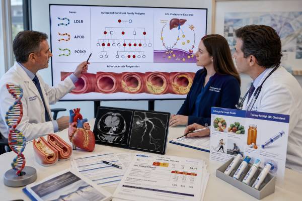
Familial hypercholesterolemia (FH) is an inherited disorder in which low-density lipoprotein cholesterol (LDL-C) is elevated from birth, creating decades of exposure that can cause premature coronary artery disease. Clinical diagnosis is based on the untreated LDL-C level, personal and family history, physical findings, and exclusion of secondary causes. Genetic testing most often analyzes LDLR, APOB, and PCSK9 to identify a monogenic cause. A pathogenic result confirms the molecular diagnosis, can strengthen risk assessment, and allows targeted testing of relatives—including children—before atherosclerosis develops. The result does not replace a lipid panel, and a negative genetic test does not rule out FH because some clinically affected people have variants that current tests cannot detect or a polygenic basis. The exact gene and variant matter: LDLR defects reduce LDL clearance, selected APOB variants impair LDL-receptor binding, and gain-of-function PCSK9 variants accelerate receptor removal. Treatment is guided by measured LDL-C, cumulative exposure, cardiovascular disease, age, and response to therapy rather than by genotype alone.
- FH is a lifelong LDL-clearance disorder, not simply cholesterol that rose after an unhealthy diet.
- LDLR is the most common causal gene; APOB and PCSK9 account for smaller proportions.
- A molecular diagnosis makes cascade testing of relatives more accurate than cholesterol screening alone.
- Children can inherit FH and benefit from diagnosis and treatment before arterial injury accumulates.
- Two pathogenic variants can cause a much more severe homozygous or compound-heterozygous phenotype.
- A negative panel does not exclude clinically diagnosed FH or remove the need for LDL-lowering treatment.
Table of Contents
- How familial hypercholesterolemia affects LDL
- LDLR, APOB, and PCSK9
- Clinical diagnosis and who to test
- How genetic testing is performed
- Understanding positive, negative, and uncertain results
- Treatment after a diagnosis
- Severe and biallelic FH
- Cascade screening and children
How familial hypercholesterolemia affects LDL
LDL particles transport cholesterol through the bloodstream. The liver normally removes many of these particles by binding apolipoprotein B-100 on the particle to LDL receptors on liver cells. The receptor-particle complex enters the cell, cholesterol is processed, and the receptor is usually recycled to the surface. FH disrupts this pathway, leaving more LDL in circulation.
The defining biological problem is cumulative exposure. A person with FH may have high LDL-C from infancy, so arterial walls experience decades more cholesterol than those of someone whose LDL rises later in life. Atherosclerotic plaque can therefore develop much earlier. Untreated adults may present with angina, heart attack, coronary revascularization, or peripheral arterial disease at ages when ordinary short-term risk calculators appear deceptively reassuring.
FH is often silent until vascular disease occurs. Some people develop tendon xanthomas, especially in the Achilles tendon or hand extensor tendons, or corneal arcus at a young age. Xanthelasma can occur but is not specific. Absence of these findings does not exclude FH, particularly in children or treated adults.
The diagnosis begins with an untreated or pretreatment lipid value whenever possible. LDL-C should be repeated if a result is unexpected, and secondary causes should be evaluated. Hypothyroidism, nephrotic syndrome, cholestatic liver disease, certain medicines, and severe dietary patterns can raise LDL. A secondary contributor can coexist with FH and make the level even higher.
Clinical scoring systems combine LDL-C, tendon findings, personal premature atherosclerotic disease, family history, and genetic results. Different countries use different criteria. A person can meet a definite or probable clinical diagnosis without a positive genetic test. Conversely, a pathogenic familial variant can identify a relative before the LDL level reaches a diagnostic threshold, especially in a child or someone already taking treatment.
FH is distinct from familial combined hyperlipidemia, sitosterolemia, lysosomal acid lipase deficiency, polygenic hypercholesterolemia, and elevated lipoprotein(a). These conditions may overlap in lipid patterns or cardiovascular risk. A thoughtful workup may include triglycerides, apolipoprotein B, lipoprotein(a), liver tests, and other targeted studies.
LDLR, APOB, and PCSK9
LDLR encodes the LDL receptor and accounts for most molecularly confirmed FH. Pathogenic variants can impair receptor production, transport, binding, internalization, or recycling. Some variants leave partial receptor function, while others produce little or no functional receptor activity. Laboratory reports may describe a variant as receptor-defective or receptor-negative when functional evidence is available, but clinical severity still varies.
LDLR testing must include sequence analysis and deletion-duplication analysis. Whole-exon or multiexon deletions and duplications are important causes that may be missed by sequencing alone. A report should state whether copy-number changes were assessed and which transcript was used.
APOB encodes apolipoprotein B, the main structural protein of LDL. Only selected gain-of-function variants in the LDL-receptor-binding region cause the classic familial defective apoB phenotype. These changes reduce how effectively an LDL particle binds the receptor. Many other APOB variants have different effects. Truncating loss-of-function variants typically lower apoB-containing lipoproteins and can cause familial hypobetalipoproteinemia rather than FH. A laboratory cannot label any rare APOB variant as an FH cause without mechanism-specific evidence.
PCSK9 encodes a protein that directs LDL receptors toward degradation. Gain-of-function variants increase receptor loss and raise LDL-C, causing autosomal dominant FH. Loss-of-function variants have the opposite effect and lower LDL-C. This biology inspired PCSK9-inhibiting therapies, but carrying a PCSK9 FH variant does not mean that only a PCSK9-directed drug will work.
Most LDLR-, APOB-, and PCSK9-related FH is autosomal dominant. A heterozygous carrier has one pathogenic variant and a 50% chance of passing it to each child. The phenotype is commonly called heterozygous FH. Severity is influenced by the gene, variant function, diet, smoking, blood pressure, diabetes, lipoprotein(a), sex, and access to early treatment.
Biallelic or digenic disease can occur when a person inherits two relevant pathogenic variants. The variants may be in the same gene on opposite chromosomes or in two different FH genes. Biallelic LDLRAP1 variants cause an autosomal recessive FH phenotype and may be included on comprehensive panels. ABCG5 and ABCG8 are often analyzed when sitosterolemia is a concern because it can mimic FH and has different dietary and treatment implications.
A panel should be curated. Adding weakly associated lipid genes can increase variants of uncertain significance without improving diagnosis. The laboratory should apply gene-specific interpretation rules, especially for APOB and PCSK9 where direction of effect is essential.
Clinical diagnosis and who to test
Genetic testing is most useful in a person with a definite or probable clinical FH phenotype, severe unexplained LDL elevation, tendon xanthomas, or premature atherosclerotic cardiovascular disease combined with a supportive family history. Identifying a molecular cause can make family screening much more efficient.
The first person tested should generally be an affected index patient with the highest untreated LDL-C or clearest clinical diagnosis. Testing an unaffected relative first lowers the chance of an interpretable answer. Once the familial variant is known, relatives need only targeted testing for that exact change.
Testing should be considered urgently when LDL-C is extremely high from childhood, xanthomas occur in a child, both parents appear to have FH, or premature coronary or aortic disease suggests homozygous FH. A severe phenotype may require an expanded panel that includes LDLRAP1 and mimic genes, along with assessment of whether two variants are located on different chromosomes.
Adults with LDL-C at or above commonly used severe-hypercholesterolemia thresholds may be evaluated even without known family history. Family history can be falsely negative because relatives were never tested, died young without a clear diagnosis, use lipid-lowering treatment, or come from a small family. Adoption and limited access to records can also obscure inheritance.
Genetic testing is not required before treatment. Markedly elevated LDL-C should be addressed promptly while the workup proceeds. Waiting for a result can prolong exposure, particularly in children or adults with established coronary disease.
Testing is less likely to be informative when LDL elevation is mild, begins late, and is well explained by secondary factors. A polygenic cause is common in these settings. Polygenic scores are not universally standardized for routine FH diagnosis, and a high score does not create the same cascade-testing pathway as a rare pathogenic variant.
Pretest counseling should explain detection limits, possible VUS findings, implications for relatives, and the possibility of identifying biallelic disease. Patients should be asked about current and historic lipid values, treatment response, cardiovascular events, tendon symptoms, and family diagnoses. A three-generation pedigree records premature heart attack, sudden death, bypass surgery, stents, stroke, very high cholesterol, and xanthomas.
How genetic testing is performed
The test usually uses blood or saliva. Fasting is not necessary for DNA collection, although a fasting or nonfasting lipid panel may be ordered at the same visit. Lipid-lowering medication does not alter the genetic result, but the clinician should document pretreatment LDL-C or estimate it from prior records when possible.
A typical panel sequences LDLR, APOB, and PCSK9 and analyzes LDLR copy-number changes. Broader tests may include LDLRAP1, APOE, ABCG5, ABCG8, and other genes selected to distinguish severe phenocopies. The report should specify gene coverage, transcripts, deletion-duplication methods, and technical limitations.
Clinical laboratories classify variants as pathogenic, likely pathogenic, uncertain significance, likely benign, or benign. Evidence includes population frequency, expected molecular effect, functional studies, segregation in families, prior affected cases, and gene-specific rules. LDLR loss-of-function variants often have a clear mechanism, but not every missense change is harmful. APOB and PCSK9 require proof that the direction of effect is compatible with high LDL.
Turnaround time is commonly several weeks. A positive result should be reviewed with the exact nomenclature, classification date, inheritance, and supporting evidence. Reports that say only “LDLR mutation” are inadequate for family testing; relatives need the specific DNA change.
When two pathogenic variants are found, the laboratory and family studies may determine whether they are in cis on the same chromosome or in trans on opposite chromosomes. Variants in trans can produce biallelic disease. Testing parents is often necessary to establish phase and understand recurrence risk.
A negative test should prompt review of methods. Was LDLR copy-number analysis included? Were severe mimics considered? Is the phenotype strong enough to justify reanalysis or broader sequencing? The answer may still be clinical FH with no detected monogenic cause.
Direct-to-consumer data are not sufficient for diagnosis. Consumer arrays test only selected sites, may miscall rare variants, and do not provide comprehensive LDLR analysis. Any medically relevant result should be confirmed in an accredited clinical laboratory.
Understanding positive, negative, and uncertain results
A pathogenic or likely pathogenic result confirms a molecular form of FH when it matches the phenotype. It may explain why LDL-C is high despite healthy habits and provides a definitive target for cascade testing. A molecular diagnosis can also indicate that cardiovascular risk is greater than suggested by a single LDL measurement because lifelong exposure began at birth.
The gene and variant influence interpretation but do not set an exact LDL value. Some LDLR variants produce minimal receptor activity and severe elevations; others retain partial function. APOB-related FH is often milder on average, but substantial overlap exists. PCSK9 gain-of-function variants vary in effect. Treatment response must be measured rather than predicted solely from the variant.
A result with two relevant pathogenic variants raises concern for homozygous, compound-heterozygous, or digenic FH. The phenotype may be severe, but the term “homozygous FH” is increasingly used as a clinical-genetic category that includes several biallelic combinations. Specialist assessment is needed rather than relying on the laboratory label alone.
A negative result means no reportable causal variant was detected. It does not exclude FH. The clinical diagnosis, LDL-C level, and need for treatment remain. Possible explanations include undiscovered genes, deep intronic or structural variants not detected, mosaicism, polygenic inheritance, or an acquired contributor layered on familial susceptibility.
A VUS is not confirmation. It should not be used as the sole reason to diagnose healthy relatives, intensify treatment beyond what LDL-C and risk require, or perform reproductive testing. Selected family studies may help classification, but only when carefully chosen. The difference between uncertain and actionable findings is reviewed in the guide to pathogenic, benign, and VUS results.
Likely benign and benign findings do not explain the phenotype. A low-LDL APOB or PCSK9 variant may be reported incidentally on broader sequencing, but it should not be misinterpreted as FH. Mechanism and direction matter.
Variant classifications can change. Patients should keep the original report and ask how reanalysis is handled. If a laboratory reclassifies a variant, management should be updated only after the new evidence is reviewed with the phenotype.
Treatment after a diagnosis
The goal is to reduce LDL-C early and maintain that reduction over decades. Lifestyle measures support cardiovascular health but are rarely sufficient for FH because the primary defect is LDL clearance. A heart-healthy eating pattern, regular physical activity, smoking avoidance, blood pressure control, healthy weight, and diabetes prevention remain important alongside medication.
A high-intensity or maximally tolerated statin is the foundation for many adults. Statins reduce hepatic cholesterol synthesis and increase LDL-receptor expression. Ezetimibe reduces intestinal cholesterol absorption and is often added when LDL-C remains above goal. PCSK9 monoclonal antibodies or small-interfering RNA therapy can produce substantial additional lowering in appropriate patients.
Other options include bempedoic acid and, in severe disease, therapies that work partly or entirely independently of LDL receptor function. The choice depends on age, pregnancy potential, cardiovascular disease, baseline LDL-C, variant function, access, side effects, and response. Treatment targets differ among guidelines and are often more intensive after atherosclerotic disease develops.
A genetic result does not replace repeated lipid measurement. LDL-C response should be assessed after each major therapy change. Adherence, secondary causes, drug interactions, and lipoprotein(a) may explain residual risk. Coronary artery calcium or other imaging may be used in selected adults, but a zero calcium score does not remove the lifelong need to treat a confirmed severe LDL disorder.
Children with heterozygous FH typically begin lifestyle counseling early and statin therapy during childhood according to age, LDL level, family history, and local guidance. The aim is to reduce cumulative exposure, not to wait for symptoms. Growth, development, liver enzymes when indicated, and treatment adherence are monitored.
Pregnancy planning requires medication review. Statins and several other lipid-lowering drugs are usually stopped or modified during pregnancy, while bile-acid sequestrants and LDL apheresis may be considered in selected cases. A woman with severe FH or established coronary disease should plan pregnancy with lipid and obstetric specialists.
Severe and biallelic FH
Homozygous FH is suspected when LDL-C is extremely elevated from early life, xanthomas develop in childhood, or aortic and coronary disease appears very young. Both parents may have heterozygous FH, but a parent can be unrecognized or carry a milder variant. Genetic confirmation can show two LDLR variants, two variants in another FH gene, variants in different genes, or recessive LDLRAP1 disease.
Severity depends partly on residual LDL-receptor activity. Receptor-negative LDLR combinations can be less responsive to treatments that rely on upregulating the receptor. Receptor-defective variants may retain more response. This distinction can help explain treatment patterns but should not delay therapy while functional classification is being clarified.
Children with suspected homozygous FH require urgent referral to a specialized lipid center. Treatment often combines multiple medicines at high intensity. Lomitapide, evinacumab, and other advanced therapies may be used according to age, country approvals, liver safety, and access. LDL apheresis physically removes apoB-containing lipoproteins and may begin in childhood when drug therapy is insufficient.
Aortic root and supravalvular aortic disease can occur in severe FH, in addition to coronary atherosclerosis. Assessment may include echocardiography, coronary imaging, vascular examination, and multidisciplinary planning. Symptoms are not a reliable gauge because advanced disease can develop silently.
The family evaluation is urgent because siblings may also have biallelic disease and each parent is often affected. Newborn or early-childhood lipid screening is appropriate when both parents have FH or when a prior child has homozygous FH. Early diagnosis can prevent irreversible vascular injury.
Liver transplantation is rarely considered in the most severe circumstances because the donor liver supplies functional LDL receptors, but it carries major lifelong risks. Modern receptor-independent therapies have expanded alternatives. Management should occur in an expert center familiar with rapidly evolving treatment options.
Cascade screening and children
Cascade screening begins with first-degree relatives of an index patient and extends outward through each newly identified carrier. When a pathogenic familial variant is known, targeted genetic testing gives a clear answer even when LDL levels overlap between affected and unaffected relatives. Lipid testing is still performed because it guides treatment.
For autosomal dominant FH, each child, sibling, or parent has a 50% chance of carrying the variant. A negative targeted result generally means the relative did not inherit that specific FH cause and can return to population-based lipid screening. A positive result prompts age-appropriate treatment and evaluation of that person’s branch of the family.
If the index patient has clinical FH but no identified variant, relatives are screened with lipid profiles and clinical criteria. A normal LDL value in a young child may need confirmation at an appropriate age, particularly when the family phenotype is strong. Treatment, illness, and laboratory variation can complicate comparisons.
Children should not wait until adulthood for evaluation. Cholesterol measurement is safe, and targeted testing can be performed from a blood or saliva sample. Identifying an affected child also creates an opportunity for reverse cascade screening, in which a parent and other relatives are diagnosed.
Family communication is often the limiting step. A concise letter should include the diagnosis, gene, exact variant, inheritance, and where relatives can request testing. The patient can share the letter without disclosing unrelated medical details. Health systems vary in whether clinicians may contact relatives directly.
Genetic diagnosis can affect reproductive planning. A heterozygous carrier has a 50% chance of transmission per pregnancy. When both partners have FH, the chance of a child with biallelic disease may be substantial and depends on the genes involved. Options include natural conception, prenatal diagnosis, or preimplantation genetic testing, supported by nondirective counseling.
The strongest reason to identify FH is that it is treatable. A molecular result turns one patient’s diagnosis into a prevention pathway for an entire family. Relatives who inherit the variant can lower LDL before disease develops, while noncarriers avoid unnecessary lifelong labeling.
References
- Familial Hypercholesterolemia (2025 GeneReviews)
- 2023 Update on European Atherosclerosis Society Consensus Statement on Homozygous Familial Hypercholesterolaemia: new treatments and clinical guidance (2023 Consensus Statement)
- Paediatric familial hypercholesterolaemia screening in Europe: public policy background and recommendations (2022 Consensus Statement)
- 2022 Consensus statement on the management of familial hypercholesterolemia in Korea (2022 Consensus Statement)
- Familial Hypercholesterolemia Panel, Sequencing (2025 Laboratory Resource)
Disclaimer
This article is for general education and does not replace evaluation by a lipid specialist, cardiologist, pediatric clinician, or genetics professional. Diagnosis and treatment require actual lipid measurements, cardiovascular history, medication review, and interpretation of the exact variant. Chest pressure, sudden breathlessness, neurologic symptoms, or other possible signs of an acute cardiovascular event require urgent medical care.