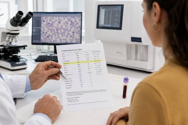
Hematocrit, often shortened to Hct, shows how much of your blood volume is made up of red blood cells. A result of 42% means that 42% of the blood sample is red blood cells, while the rest is mostly plasma, with white blood cells and platelets making up a much smaller part. Because red blood cells carry oxygen, hematocrit helps show whether your blood has enough red cell volume to support normal oxygen delivery.
Hematocrit is usually reported as part of a complete blood count, not as a stand-alone clue. A low result often points toward anemia, blood loss, iron deficiency, vitamin deficiency, chronic disease, kidney disease, or dilution from extra fluid. A high result can come from dehydration, smoking, high altitude, sleep apnea, lung or heart disease, testosterone use, or a red blood cell disorder. The safest interpretation comes from comparing hematocrit with hemoglobin, RBC count, MCV, RDW, reticulocytes, iron studies, symptoms, and prior results.
- Hematocrit measures the percentage of blood volume made up of red blood cells.
- Common adult reference ranges are about 40%–54% for men and 36%–48% for women, but each lab sets its own range.
- Low hematocrit usually means anemia, blood loss, low red blood cell production, hemolysis, pregnancy-related dilution, or excess body fluid.
- High hematocrit often means dehydration, low oxygen states, smoking, high altitude, sleep apnea, testosterone use, or polycythemia.
- No fasting is needed for hematocrit alone, but other blood tests ordered at the same time may require fasting.
- Urgent symptoms include chest pain, fainting, severe shortness of breath, stroke-like symptoms, heavy bleeding, or black stools.
Table of Contents
- What Hematocrit Measures
- Normal Hematocrit Range
- How Hematocrit Relates to Hemoglobin and RBC Count
- Low Hematocrit: Common Causes and Meaning
- High Hematocrit: Common Causes and Meaning
- Test Preparation and Factors That Can Change Results
- How to Interpret Hematocrit With Other CBC Markers
- When to Follow Up or Seek Urgent Care
What Hematocrit Measures
Hematocrit measures red blood cell volume as a percentage of total blood volume. Red blood cells contain hemoglobin, the oxygen-carrying protein that picks up oxygen in the lungs and delivers it to tissues. Plasma is the liquid part of blood. If the red cell portion rises or falls, hematocrit changes.
A hematocrit result can be written in two common ways:
- 42%, meaning 42% of the blood volume is red blood cells
- 0.42 L/L, meaning the same value expressed as a fraction of blood volume
In everyday lab reports, the percentage format is more familiar. A hematocrit of 45% is the same as 0.45 L/L.
Hematocrit can be measured directly by spinning a blood sample so red cells pack at the bottom of a tube. Many modern analyzers calculate it from two CBC values: red blood cell count and mean corpuscular volume, or MCV. In simple terms, hematocrit rises when there are more red blood cells or when red blood cells are larger. It falls when there are fewer red blood cells or when cells are smaller.
This is why hematocrit should not be read alone. Two people can have the same hematocrit for different reasons. One person may have many small red cells from iron deficiency. Another may have fewer but larger red cells from vitamin B12 or folate deficiency. The hematocrit number shows the red cell volume, but the surrounding CBC pattern explains why it changed.
Hematocrit is often ordered as part of a complete blood count. A CBC also includes hemoglobin, RBC count, white blood cells, platelets, and red blood cell indices such as MCV, MCH, MCHC, and RDW. Together, these markers help separate anemia, inflammation, infection, blood loss, bone marrow problems, and red blood cell disorders.
Normal Hematocrit Range
A typical adult hematocrit range is about 40%–54% for men and 36%–48% for women. Some laboratories use narrower ranges, such as about 41%–50% for men and 36%–47% for women. The range on your own report is the one your clinician usually uses first, because reference intervals can vary by analyzer, lab method, age, sex, altitude, pregnancy status, and local population.
| Group | Common reference range | How to read it |
|---|---|---|
| Adult men | About 40%–54% | Values below the range may suggest anemia or dilution; values above the range may suggest dehydration, low oxygen, or erythrocytosis. |
| Adult women | About 36%–48% | Lower average values are partly related to menstrual blood loss, body size, and hormonal differences. |
| Pregnancy | Often lower than nonpregnant adult ranges | Blood plasma expands during pregnancy, so hematocrit commonly falls even when red cell production increases. |
| Children | Varies by age | Newborns, infants, children, and adolescents need age-specific ranges. |
| High-altitude residents | Often higher than sea-level ranges | The body may produce more red blood cells to adapt to lower oxygen pressure. |
A value just outside the reference range is not automatically dangerous. Mild shifts can happen with hydration, recent exercise, a short illness, menstrual timing, pregnancy, recent blood donation, or lab variation. The meaning changes when the result is clearly abnormal, persistent, worsening, or paired with symptoms.
There is also no single “optimal” hematocrit that applies to everyone. A healthy endurance athlete living at altitude, a pregnant person in the second trimester, and an older adult with kidney disease may all have different expected values. Hematocrit is a context marker, not a stand-alone wellness score.
The reference range also does not replace medical thresholds used in specific diseases. For example, a person with polycythemia vera may have a treatment target for hematocrit that is stricter than a general lab range because clotting risk is part of that condition. On the other side, anemia is usually defined and graded using hemoglobin more than hematocrit, although both often move in the same direction.
When hematocrit is normal, it usually means the red blood cell portion of the blood is within the expected range. It does not rule out all blood or nutrient problems. Early iron deficiency, for example, can exist before hematocrit falls. That is why iron studies, ferritin, B12, folate, reticulocytes, kidney markers, and inflammation markers may be needed when symptoms or other CBC values raise concern.
How Hematocrit Relates to Hemoglobin and RBC Count
Hematocrit, hemoglobin, and RBC count are closely related, but they are not the same test.
Hemoglobin measures the oxygen-carrying protein inside red blood cells. RBC count measures how many red blood cells are present in a volume of blood. Hematocrit measures how much space red blood cells take up in the blood sample. For a deeper comparison, the difference between hemoglobin and hematocrit is especially useful when one result looks more abnormal than the other.
A simple pattern often holds true: hematocrit is roughly three times the hemoglobin value when hemoglobin is reported in g/dL. For example, a hemoglobin of 14 g/dL often goes with a hematocrit near 42%. This “rule of three” is only an estimate. It can break down when red cells are unusually small or large, when there are analyzer interferences, or when a sample problem affects one measurement more than another.
Hematocrit also depends on red blood cell size. If RBC count stays the same but red cells get larger, hematocrit can rise. If red cells become smaller, hematocrit can fall or look less impressive than the RBC count. This is why MCV often explains the pattern.
Three examples show how the same hematocrit change can have different meanings:
- Low hematocrit with low MCV often suggests microcytic anemia, commonly from iron deficiency or sometimes thalassemia trait.
- Low hematocrit with high MCV can suggest macrocytic anemia, often related to vitamin B12 deficiency, folate deficiency, alcohol use, liver disease, certain medicines, or bone marrow disorders.
- High hematocrit with high RBC count suggests true erythrocytosis more than simple dehydration, especially if it persists.
Hemoglobin is often the preferred marker for diagnosing anemia because it directly reflects oxygen-carrying capacity. Hematocrit still adds value because it reflects red cell volume and plasma volume changes. A low hematocrit from anemia and a low hematocrit from fluid overload can look similar on paper, but the rest of the CBC and clinical picture differ.
If your hematocrit is abnormal, compare it with the hemoglobin normal range, RBC count, MCV, and RDW before drawing conclusions. A single red blood cell marker rarely gives the full answer.
Low Hematocrit: Common Causes and Meaning
Low hematocrit means the red blood cell portion of blood is lower than expected. Most of the time, this points toward anemia, blood loss, reduced red blood cell production, increased red blood cell destruction, or dilution from extra fluid.
Symptoms depend on how low the hematocrit is, how fast it fell, and whether the person has heart, lung, kidney, or vascular disease. Mild anemia can cause no symptoms. A faster or more severe drop can cause fatigue, weakness, shortness of breath with activity, dizziness, headaches, pale skin, cold hands and feet, rapid heartbeat, chest discomfort, or reduced exercise tolerance.
Iron deficiency and blood loss
Iron deficiency is one of the most common reasons for low hematocrit. Red blood cells need iron to make hemoglobin. When iron supply runs low, the body often makes smaller, paler red cells. Hematocrit may fall, MCV may become low, MCH or MCHC may fall, and RDW often rises as red cell sizes become more mixed.
Common reasons include heavy menstrual bleeding, pregnancy, low iron intake, frequent blood donation, gastrointestinal bleeding, inflammatory bowel disease, celiac disease, bariatric surgery, or poor iron absorption. In adults, iron deficiency often deserves a search for blood loss, especially from the gastrointestinal tract when there is no obvious explanation.
A pattern such as low MCV with high RDW can strongly suggest iron deficiency, but ferritin and iron studies help confirm the cause. The relationship between CBC and ferritin is often more useful than hematocrit alone when checking iron status.
Vitamin B12, folate, and larger red blood cells
Vitamin B12 and folate help red blood cells mature normally. When either is low, red cells may become larger than usual, raising MCV. Hematocrit can still fall because the total number of healthy red cells drops.
Possible clues include high MCV, low hemoglobin, low RBC count, fatigue, mouth soreness, numbness or tingling, balance changes, memory problems, or a history of restricted diet, malabsorption, metformin use, acid-suppressing medicines, autoimmune gastritis, or gastrointestinal surgery. B12 deficiency can cause nerve damage, so it should not be ignored even when anemia is mild.
Chronic disease, kidney disease, and bone marrow response
Chronic inflammation, autoimmune disease, chronic infection, cancer, and kidney disease can lower hematocrit by reducing red blood cell production or changing how iron is used. The kidneys make erythropoietin, a hormone that tells the bone marrow to make red blood cells. When kidney function declines, erythropoietin may be too low for the body’s needs.
Bone marrow problems can also reduce red blood cell production. These may affect only red cells or may also lower white blood cells and platelets. When hematocrit is low together with low WBC and low platelets, the pattern needs prompt medical review.
Reticulocyte count helps show whether the bone marrow is responding. Reticulocytes are young red blood cells. A low reticulocyte response with anemia suggests underproduction. A high reticulocyte response suggests the marrow is trying to replace red cells after bleeding or hemolysis. The combination of reticulocyte count and hemoglobin can help clarify whether anemia is recovering or ongoing.
Dilution from pregnancy, IV fluids, or fluid overload
Low hematocrit does not always mean red blood cell production is failing. During pregnancy, plasma volume expands more than red cell mass, so hematocrit commonly falls. This is sometimes called dilutional or physiologic anemia of pregnancy, although true iron deficiency can also occur during pregnancy.
A similar dilution effect can happen after receiving intravenous fluids or with fluid overload from heart, kidney, or liver disease. In these cases, the red cell percentage falls because the plasma portion rises. The person may still need evaluation, but the treatment is different from iron deficiency or blood loss.
High Hematocrit: Common Causes and Meaning
High hematocrit means the red blood cell portion of blood is above the expected range. The increase may be relative, because plasma volume is low, or absolute, because the body has made too many red blood cells.
A relative increase is common with dehydration. Vomiting, diarrhea, heavy sweating, inadequate fluid intake, burns, or diuretic medicines can reduce plasma volume. The red cell amount may be normal, but the blood becomes more concentrated. After hydration and recovery, hematocrit may return toward baseline.
An absolute increase is called erythrocytosis or polycythemia. In this situation, red blood cell mass is truly increased. The body may make more red cells as a response to low oxygen, or the bone marrow may produce too many red cells because of a blood disorder.
Low oxygen signals
The body raises red blood cell production when tissues sense low oxygen. This can happen with chronic lung disease, congenital heart disease, severe sleep apnea, long-term smoking, carbon monoxide exposure, or living at high altitude. The hormone erythropoietin rises and stimulates the bone marrow to make more red blood cells.
Sleep apnea is a common and often missed contributor. A person may have high hematocrit along with loud snoring, witnessed pauses in breathing, morning headaches, daytime sleepiness, high blood pressure, or obesity. Treating the oxygen problem is more important than simply trying to lower the number.
Smoking can raise hematocrit through several pathways, including carbon monoxide exposure and lower oxygen delivery. Even when oxygen saturation looks acceptable during a brief office check, smoking-related carbon monoxide can affect oxygen transport.
Medicines, hormones, and erythropoietin
Testosterone therapy and other androgenic steroids can raise hematocrit by stimulating red blood cell production. This effect can become clinically important, especially with higher doses, injections, older age, sleep apnea, smoking, or underlying heart and lung disease. People using testosterone usually need periodic CBC monitoring.
Erythropoietin-stimulating agents can also raise hematocrit. These medicines may be used in some kidney disease, chemotherapy-related anemia, or other specific settings. They require careful monitoring because pushing red cell levels too high can increase risk.
Polycythemia vera and other blood disorders
Polycythemia vera is a bone marrow disorder in which the marrow makes too many red blood cells, often because of a JAK2 mutation. White blood cells and platelets may also be high. Symptoms can include headaches, dizziness, blurred vision, itching after a warm shower, burning pain in the hands or feet, redness of the face, unusual bleeding, enlarged spleen, or blood clots.
Persistent high hematocrit with high hemoglobin and high RBC count deserves medical evaluation, especially when there is no clear dehydration or altitude explanation. Evaluation may include repeat CBC, oxygen saturation, erythropoietin level, JAK2 testing, kidney and liver tests, sleep apnea assessment, smoking or carbon monoxide review, and sometimes hematology referral.
A high hematocrit can make blood more viscous, but clot risk depends on the cause. Polycythemia vera is managed differently from dehydration, sleep apnea, or testosterone-related erythrocytosis. General advice about “thick blood” can be misleading unless the cause is known. A dedicated discussion of high hematocrit causes can help separate temporary concentration from true red cell overproduction.
Test Preparation and Factors That Can Change Results
No special preparation is usually needed for a hematocrit test by itself. A healthcare professional draws blood from a vein, usually into a tube containing EDTA, which keeps the blood from clotting before analysis. The draw usually takes only a few minutes.
Fasting is not required for hematocrit alone. However, a CBC may be ordered with glucose, lipid, iron, kidney, liver, or hormone tests. Some of those may have timing or fasting instructions. Follow the instructions for the full lab order, not only the hematocrit portion.
Several ordinary factors can shift hematocrit:
- Hydration: Dehydration can raise hematocrit by lowering plasma volume. Overhydration or recent IV fluids can lower it.
- Altitude: Higher altitude can raise hematocrit as the body adapts to lower oxygen pressure.
- Smoking: Smoking can raise hematocrit, partly through oxygen delivery stress and carbon monoxide exposure.
- Pregnancy: Plasma expansion commonly lowers hematocrit.
- Recent bleeding or donation: Hematocrit can fall after blood loss, though very acute bleeding may not show the full drop immediately.
- Recent transfusion: Transfusion can raise hematocrit and temporarily change the pattern.
- Intense exercise or heat exposure: Fluid shifts and sweating can temporarily concentrate the blood.
- Sample handling: Clotting, hemolysis, delayed processing, or analyzer issues can affect results.
A surprising result is often repeated before major conclusions are made, especially if it does not match symptoms or prior CBCs. Repeating the CBC after hydration, recovery from illness, or review of medicines can prevent overreacting to a temporary shift.
It also helps to compare with older results. A hematocrit of 50% may be new and important for one person but stable for another who lives at altitude. A hematocrit of 35% may be normal during part of pregnancy but abnormal in a nonpregnant adult man. Trend, context, and symptoms matter as much as the single number.
How to Interpret Hematocrit With Other CBC Markers
Hematocrit becomes much more useful when it is read as part of a pattern. The surrounding CBC markers show whether the issue is red cell number, red cell size, hemoglobin concentration, bone marrow response, inflammation, or a broader blood cell problem.
| Pattern | Possible meaning | Common follow-up tests or checks |
|---|---|---|
| Low Hct, low Hgb, low MCV, high RDW | Often iron deficiency, especially from blood loss | Ferritin, serum iron, TIBC, transferrin saturation, bleeding history |
| Low Hct, low Hgb, high MCV | Possible B12 or folate deficiency, alcohol effect, liver disease, medication effect, or marrow disorder | B12, methylmalonic acid, folate, liver tests, medication review |
| Low Hct with normal MCV | Possible chronic inflammation, kidney disease, early blood loss, mixed deficiencies, or marrow underproduction | Reticulocyte count, kidney function, ferritin, inflammation markers |
| High Hct, high Hgb, high RBC | Possible erythrocytosis or polycythemia | Repeat CBC, oxygen saturation, EPO level, JAK2 testing when appropriate |
| High Hct with normal RBC pattern after fluid loss | Possible dehydration or hemoconcentration | Hydration review, repeat CBC, electrolytes when clinically needed |
| Low Hct plus low WBC and low platelets | Possible bone marrow suppression, severe illness, medication effect, nutritional deficiency, or marrow disease | Prompt clinical review, smear, reticulocytes, B12, folate, marrow evaluation when needed |
MCV and RDW are especially helpful in anemia patterns. MCV shows average red blood cell size. RDW shows how much red cell size varies. A combined MCV and RDW pattern can point toward iron deficiency, mixed deficiencies, thalassemia trait, B12 or folate deficiency, recent bleeding, or recovery after treatment.
The peripheral blood smear can add detail when analyzer numbers do not fully explain the result. A smear lets a trained professional look at red blood cell shape, size, color, clumping, fragments, immature cells, and abnormal white cells or platelets. This can help when anemia is unexplained, multiple CBC lines are abnormal, or a lab artifact is suspected.
Iron studies are often needed when hematocrit is low. Ferritin estimates iron stores, but ferritin can rise with inflammation, infection, liver disease, and some chronic conditions. Transferrin saturation helps show how much circulating iron is available. In inflammatory states, ferritin alone can be misleading.
Kidney function tests can matter because the kidneys help regulate red blood cell production through erythropoietin. A low hematocrit with chronic kidney disease may reflect underproduction rather than iron deficiency alone. A high hematocrit with certain kidney problems may occur if erythropoietin signaling is increased.
White blood cells and platelets also change the interpretation. High hematocrit with high platelets or high white blood cells raises more concern for a bone marrow disorder than isolated high hematocrit after dehydration. Low hematocrit with low platelets may suggest bleeding, marrow suppression, severe inflammation, or a broader hematologic issue.
When to Follow Up or Seek Urgent Care
Follow-up is reasonable when hematocrit is outside the lab range, changing over time, or paired with symptoms. Mild abnormalities often start with a repeat CBC and review of hydration, menstrual bleeding, diet, medicines, smoking, altitude, recent illness, blood donation, pregnancy status, and prior results.
A clinician may order additional tests based on the pattern:
- Ferritin and iron studies for suspected iron deficiency
- Vitamin B12, methylmalonic acid, and folate for macrocytic patterns
- Reticulocyte count for bone marrow response
- Creatinine and eGFR for kidney function
- Liver tests when alcohol use, liver disease, or macrocytosis is possible
- Inflammation markers when chronic inflammatory disease is suspected
- Stool blood testing or gastrointestinal evaluation when blood loss is possible
- Oxygen saturation, sleep study, EPO level, or JAK2 testing for persistent high hematocrit
Do not start iron just because hematocrit is low unless iron deficiency is likely or confirmed. Iron can help when iron deficiency is present, but it may be unhelpful or harmful in other situations. Similarly, do not try to lower a high hematocrit by donating blood unless a clinician says it is appropriate. The cause matters.
Seek urgent medical care if low hematocrit or suspected anemia comes with chest pain, fainting, severe shortness of breath, confusion, rapid worsening weakness, heavy bleeding, vomiting blood, black tarry stools, or severe dizziness. These signs can reflect significant blood loss, severe anemia, or poor oxygen delivery.
Urgent care is also needed when high hematocrit is paired with stroke-like symptoms, one-sided weakness, trouble speaking, severe headache, vision loss, chest pain, coughing blood, sudden shortness of breath, or a swollen painful leg. These symptoms may suggest a clot or another emergency.
For stable, mild abnormalities, the most useful next step is usually organized follow-up rather than panic. Bring the full CBC report, prior lab results, supplement list, medication list, menstrual or bleeding history, smoking status, altitude or travel history, and any symptoms. Hematocrit is one number, but it becomes clinically meaningful when it is matched to the whole pattern.
References
- Hematocrit Test 2024 (Official Page)
- Hematocrit 2024 (Review)
- Normal and Abnormal Complete Blood Count With Differential 2024 (Review)
- Guideline on haemoglobin cutoffs to define anaemia in individuals and populations 2024 (Guideline)
- Anemia 2023 (Review)
- Secondary Polycythemia 2023 (Review)
Disclaimer
Hematocrit results should be interpreted with the full CBC, symptoms, medical history, and the reference range printed on the lab report. This article is for general education and cannot diagnose anemia, polycythemia, blood loss, dehydration, or any other condition. Seek prompt medical care for severe shortness of breath, chest pain, fainting, heavy bleeding, stroke-like symptoms, or signs of a blood clot.