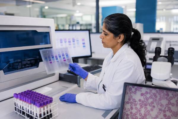
Hemoglobin electrophoresis is a blood test that separates the different forms of hemoglobin inside red blood cells. Hemoglobin is the protein that carries oxygen, and small inherited changes in its structure or production can lead to sickle cell disease, sickle cell trait, thalassemia, or other hemoglobin variants. The test is often ordered when a complete blood count shows unexplained anemia, a low mean corpuscular volume, abnormal red blood cell shapes, a family history of hemoglobin disorders, or a positive newborn or sickle cell screen. Results are usually reported as percentages of hemoglobin A, A2, F, S, C, E, or other variants. The pattern matters more than any single number. A normal adult pattern is very different from a newborn pattern, and recent transfusion, iron deficiency, pregnancy, or some treatments can change the interpretation.
- Hemoglobin electrophoresis measures hemoglobin types, not the total hemoglobin level measured on a CBC.
- Normal adults usually have mostly HbA, small amounts of HbA2, and little or no HbF.
- HbS suggests sickle hemoglobin; the pattern helps separate sickle trait from sickle cell disease.
- High HbA2 often points toward beta-thalassemia trait, especially with low MCV.
- Alpha-thalassemia trait can have a normal electrophoresis result, so genetic testing may be needed.
- No fasting is usually needed, but recent blood transfusion can make results hard to interpret.
Table of Contents
- What the Hemoglobin Electrophoresis Test Measures
- When Hemoglobin Electrophoresis Is Ordered
- How the Test Is Done and How to Prepare
- Normal Hemoglobin Types and Typical Percentages
- Sickle Cell Trait, Sickle Cell Disease, and HbS Results
- Thalassemia Patterns on Hemoglobin Electrophoresis
- Limitations, False Reassurance, and Misleading Results
- What to Do After an Abnormal Result
What the Hemoglobin Electrophoresis Test Measures
Hemoglobin electrophoresis measures which types of hemoglobin are present in the blood and how much of each type is present. It does this by separating hemoglobin proteins based on their electrical charge. Many laboratories now use related methods such as high-performance liquid chromatography, capillary electrophoresis, or isoelectric focusing, but patients may still hear the whole group called “hemoglobin electrophoresis.”
The test does not replace a complete blood count. A CBC tells you whether the hemoglobin level is low, whether red blood cells are small or large, and whether other blood cell lines are affected. Hemoglobin electrophoresis tells you whether the hemoglobin itself has an inherited variant or an abnormal production pattern.
The main hemoglobin types reported may include:
- HbA: the main adult hemoglobin.
- HbA2: a small adult hemoglobin fraction that becomes important when checking for beta-thalassemia trait.
- HbF: fetal hemoglobin, usually high before birth and in newborns, then much lower after infancy.
- HbS: sickle hemoglobin, seen in sickle cell trait and sickle cell disease.
- HbC: a hemoglobin variant that can cause HbC trait, HbC disease, or HbSC disease when inherited with HbS.
- HbE: a hemoglobin variant more common in people with ancestry from Southeast Asia and nearby regions.
- Other variants: less common hemoglobin forms may require confirmatory testing.
A single result can answer several different questions. It may confirm sickle cell trait after a positive screening test, help diagnose sickle cell disease, support a diagnosis of beta-thalassemia trait, identify a hemoglobin variant in a person with microcytosis, or guide family planning when both partners could pass on a hemoglobin disorder.
Hemoglobin electrophoresis is especially useful because many hemoglobin disorders are inherited. A person can feel well and still carry a hemoglobin variant. That matters because two healthy carriers can have a child with a serious hemoglobin disorder, depending on which genes each parent passes on.
When Hemoglobin Electrophoresis Is Ordered
Hemoglobin electrophoresis is commonly ordered when the blood count, family history, ancestry, newborn screening result, or symptoms suggest a hemoglobin disorder. It is not usually part of a routine CBC, but it is often added when the CBC pattern needs a deeper explanation.
A clinician may order the test for:
- Unexplained anemia.
- Low MCV, meaning small red blood cells.
- Mild anemia that does not fit a simple iron deficiency pattern.
- Target cells or other abnormal red blood cell shapes on a blood smear.
- A positive sickle cell screen.
- Family history of sickle cell disease, thalassemia, or a known hemoglobin variant.
- Carrier screening before or during pregnancy.
- Newborn follow-up after an abnormal screening result.
- Evaluation before certain surgeries or high-risk medical situations when sickle cell disease is possible.
- Clarifying whether a person has trait, disease, or a combined condition such as HbS/beta-thalassemia.
The test is often paired with CBC markers such as hemoglobin, hematocrit, MCV, MCH, and RDW. For example, a person with beta-thalassemia trait often has a low MCV with a normal or only mildly low hemoglobin level. A person with iron deficiency can also have low MCV, so the pattern must be interpreted carefully. The article on MCV and RDW anemia patterns can help explain why cell size and size variation matter before electrophoresis results are interpreted.
Screening during pregnancy and family planning
Carrier screening is one of the most important uses of hemoglobin electrophoresis. A carrier may have no symptoms, but if both reproductive partners carry certain hemoglobin changes, a child may inherit a severe condition.
Examples include:
- One parent with sickle cell trait and one parent with sickle cell trait can have a child with sickle cell disease.
- One parent with sickle cell trait and one parent with HbC trait can have a child with HbSC disease.
- One parent with sickle cell trait and one parent with beta-thalassemia trait can have a child with HbS/beta-thalassemia.
- Two parents with beta-thalassemia trait can have a child with beta-thalassemia major.
Testing both partners is important when one person has an abnormal result. The risk is not fully clear until the other partner’s status is known.
Testing after a positive sickle cell screen
A sickle cell screen can detect the presence of HbS, but it does not fully define the pattern. It may not reliably separate sickle cell trait from sickle cell disease or from combined conditions. Hemoglobin electrophoresis or a comparable hemoglobinopathy evaluation is used to confirm the result. A dedicated sickle cell screen is a screening tool; electrophoresis is part of the diagnostic workup.
How the Test Is Done and How to Prepare
Hemoglobin electrophoresis is done on a blood sample, usually drawn from a vein in the arm. In newborns, the first screening sample is often collected from a heel stick, but confirmatory testing may use another blood sample depending on the program and clinical situation.
Most people do not need to fast. Ordinary meals, water, and most medications do not interfere with the test. The blood draw itself takes only a few minutes. Results may take a few days, although timing varies by laboratory and whether confirmatory testing is needed.
Before the test, the clinician or laboratory should know about:
- A blood transfusion within the last 3 to 4 months.
- Known sickle cell disease or thalassemia.
- Hydroxyurea or other treatments that may raise HbF.
- Bone marrow transplant or stem cell transplant history.
- Pregnancy.
- Iron deficiency or recent iron treatment.
- A newborn or infant’s age in weeks or months.
Recent transfusion is one of the most important details. Transfused red blood cells contain the donor’s hemoglobin pattern, which can partially cover or dilute the patient’s own pattern. If the test is not urgent, clinicians may wait until enough transfused cells have cleared, or they may use genetic testing when waiting is not appropriate.
Electrophoresis versus HPLC and genetic testing
Many reports use the phrase “hemoglobin electrophoresis” even when the laboratory used HPLC or capillary electrophoresis. These methods can all identify and measure common hemoglobin types, but they are not identical. Some variants overlap or migrate together on one method, so laboratories may use a second method to confirm an unusual pattern.
Genetic testing looks directly at hemoglobin gene changes. It is especially helpful when electrophoresis is normal but suspicion remains, as can happen with alpha-thalassemia trait. It can also clarify complex results, prenatal risk, rare variants, or discordant findings between the CBC and hemoglobin analysis.
Normal Hemoglobin Types and Typical Percentages
Normal hemoglobin percentages depend strongly on age. Adults should have mostly HbA. Newborns have mostly HbF because fetal hemoglobin carries oxygen efficiently before birth. During the first months of life, HbF falls and HbA rises.
Reference ranges vary by laboratory, method, and age. The table below shows common adult patterns used for general interpretation.
| Hemoglobin type | Typical adult percentage | General meaning |
|---|---|---|
| HbA | About 95% to 98% | Main adult hemoglobin |
| HbA2 | About 2% to 3.5% | Small adult fraction; often higher in beta-thalassemia trait |
| HbF | Usually less than 1% to 2% | Fetal hemoglobin; higher in newborns and in some inherited or treatment-related states |
| HbS, HbC, HbE, other variants | Usually absent | May suggest inherited hemoglobin variant |
A “normal” adult electrophoresis pattern usually means HbA is dominant, HbA2 is within the lab’s adult reference range, HbF is low, and no abnormal variant is detected. That pattern reduces the chance of beta-thalassemia trait, sickle cell trait, HbC trait, and many other common variants. It does not rule out every inherited red blood cell disorder.
The hemoglobin electrophoresis result should be read beside the measured hemoglobin level. The hemoglobin blood test shows whether oxygen-carrying protein is low, normal, or high. Electrophoresis explains the type of hemoglobin present. A person can have abnormal electrophoresis with a normal hemoglobin level, especially in carrier states.
Newborn and infant results
Newborn results look different from adult results. A healthy newborn usually has a large amount of HbF and a smaller but rising amount of HbA. Because HbF is naturally high, newborn screening programs use age-specific interpretation and follow-up testing.
A newborn pattern may show combinations such as FA, FAS, FS, FAC, or FSC. The letters often list the most abundant hemoglobins in order. For example, FAS usually suggests sickle cell trait in a newborn, while FS can suggest sickle cell disease or sickle beta-zero thalassemia and needs prompt confirmatory evaluation.
Sickle Cell Trait, Sickle Cell Disease, and HbS Results
HbS is sickle hemoglobin. Its presence means a person has inherited at least one sickle hemoglobin gene. The percentage pattern helps separate sickle cell trait from sickle cell disease and from combined hemoglobin disorders.
In sickle cell trait, a person usually has both HbA and HbS, with HbA higher than HbS. A common adult pattern is roughly 50% to 60% HbA and 35% to 45% HbS, although exact percentages vary. HbA2 is usually not markedly elevated, and HbF is usually low. Most people with sickle cell trait do not have chronic anemia or typical sickle cell crises, but trait can matter during extreme dehydration, severe low oxygen exposure, high altitude, intense exertion, kidney symptoms such as blood in the urine, and family planning.
In sickle cell disease due to HbSS, HbS is the main hemoglobin. HbA is absent unless the person has recently received a transfusion. HbF may be present in variable amounts. Higher HbF can soften the disease pattern in some people because HbF interferes with sickling, but HbF does not make the diagnosis disappear.
In HbSC disease, both HbS and HbC are present. HbA is absent unless there has been a transfusion. HbSC disease can cause pain episodes, anemia, eye disease, spleen complications, and other sickle-related problems, though the pattern and severity can differ from HbSS.
In HbS/beta-thalassemia, the result depends on whether the beta-thalassemia gene produces no beta chains or reduced beta chains. HbS/beta-zero thalassemia can look similar to HbSS because HbA is absent. HbS/beta-plus thalassemia usually has some HbA, increased HbA2, and HbS. This is one reason a simple “positive HbS” result is not enough.
| Pattern | Typical electrophoresis clue | General interpretation |
|---|---|---|
| Sickle cell trait | HbA and HbS both present, with HbA usually higher | Carrier state; important for counseling and certain risk situations |
| HbSS sickle cell disease | Mostly HbS, no HbA unless transfused | Sickle cell disease requiring ongoing medical care |
| HbSC disease | HbS and HbC present, no HbA unless transfused | Compound hemoglobin disorder with sickle-related complications |
| HbS/beta-zero thalassemia | HbS present, HbA absent, HbA2 often increased | Often clinically similar to HbSS |
| HbS/beta-plus thalassemia | HbS present, some HbA present, HbA2 often increased | Severity varies; needs hematology interpretation |
Symptoms and electrophoresis do not always match perfectly. Some people with sickle cell disease have fewer symptoms in childhood and still need preventive care. Others have serious complications early. Anyone with a result suggesting sickle cell disease needs timely follow-up, vaccination planning, infection precautions, pain management guidance, and hematology care.
Thalassemia Patterns on Hemoglobin Electrophoresis
Thalassemia is different from many hemoglobin variants. In thalassemia, the body has trouble making enough alpha or beta globin chains, which are building blocks of hemoglobin. The hemoglobin may be structurally normal, but production is imbalanced. This often causes small red blood cells, reflected by a low MCV.
Beta-thalassemia trait is one of the clearest electrophoresis patterns. It often shows increased HbA2, commonly above the adult reference range, with normal or mildly increased HbF. The CBC often shows low MCV and low MCH, sometimes with a normal or high red blood cell count despite mild anemia. That combination can look different from iron deficiency, where RDW is often higher and ferritin may be low.
A person with a low MCV should not automatically be told they have iron deficiency. Iron studies help separate iron deficiency from thalassemia trait, and both can occur together. The pattern of low MCV with high RDW often supports iron deficiency, while thalassemia trait may show low MCV with a relatively preserved or high RBC count. Still, real-life patterns overlap.
Beta-thalassemia trait
Beta-thalassemia trait usually causes a mild, lifelong microcytosis. The person may feel well and may not need treatment. The result matters because it can be mistaken for iron deficiency and because it can be passed to children.
Common clues include:
- Low MCV, often more striking than the degree of anemia.
- Normal or slightly low hemoglobin.
- Normal or high RBC count.
- HbA2 above the lab’s reference range.
- HbF normal or mildly increased.
Iron deficiency can lower HbA2 and hide beta-thalassemia trait. If ferritin is low, clinicians may treat iron deficiency first and repeat testing later. The iron panel test is often used with electrophoresis when microcytosis is present.
Beta-thalassemia major and intermedia
Beta-thalassemia major usually appears in infancy or early childhood with severe anemia, poor growth, jaundice, enlarged spleen, and need for regular transfusions. Electrophoresis often shows very high HbF, increased HbA2, and absent or very low HbA.
Beta-thalassemia intermedia is less severe than major but more significant than trait. Hemoglobin levels, symptoms, transfusion needs, iron overload risk, and spleen size vary widely. Laboratory percentages help support the diagnosis, but clinical severity guides management.
Alpha-thalassemia trait
Alpha-thalassemia trait can be easy to miss because adult hemoglobin electrophoresis may look normal. HbA2 is usually not high in the same way it is in beta-thalassemia trait. A person may have low MCV, mild or no anemia, normal iron studies, and a normal electrophoresis result.
This is one of the most common reasons a “normal electrophoresis” does not end the evaluation. If the CBC strongly suggests thalassemia and iron deficiency has been excluded, genetic testing for alpha-globin gene deletions may be appropriate, especially when pregnancy or family planning is involved.
Limitations, False Reassurance, and Misleading Results
Hemoglobin electrophoresis is powerful, but it is not perfect. The result can be misleading when the timing, clinical context, or related blood tests are ignored.
A recent transfusion can make the patient’s hemoglobin pattern look more normal because donor red blood cells contain donor hemoglobin. This can reduce the apparent percentage of HbS or add HbA to someone who would otherwise have none. Always tell the clinician if a transfusion occurred within the past few months.
Iron deficiency can complicate thalassemia interpretation. In beta-thalassemia trait, HbA2 is often elevated. Iron deficiency may lower HbA2 into the normal range, making beta-thalassemia trait harder to detect. When microcytosis is present, ferritin and transferrin saturation often need to be reviewed before calling the result normal.
Age also changes interpretation. A newborn’s high HbF is normal. An adult’s high HbF may reflect an inherited persistence of fetal hemoglobin, thalassemia, sickle cell disease, bone marrow stress, pregnancy-related shifts, or medication effect. The same percentage can mean different things at different ages.
Some variants overlap on one method. For example, one technique may place two hemoglobins in a similar location. A laboratory may report a “variant” rather than a final identity until a second method or genetic test confirms it.
The test also does not explain every cause of anemia. Low hemoglobin can come from iron deficiency, chronic inflammation, kidney disease, bleeding, B12 deficiency, folate deficiency, hemolysis, bone marrow disorders, or mixed causes. The hemoglobin and ferritin pattern may be more useful when the main question is iron deficiency, while electrophoresis is more useful when an inherited hemoglobin disorder is suspected.
Warning signs deserve faster care than routine lab follow-up. Severe anemia symptoms, chest pain, shortness of breath, fainting, severe pain episodes, fever in a child with sickle cell disease, stroke-like symptoms, or dark urine with sudden weakness should be treated urgently.
What to Do After an Abnormal Result
The next step depends on the pattern. Some results only need explanation and family counseling. Others need hematology referral, newborn follow-up, genetic testing, or treatment planning.
For a result suggesting sickle cell trait, many people need education rather than daily treatment. The result should be documented, shared with healthcare professionals when relevant, and considered during family planning. People with trait should understand risk situations such as severe dehydration, extreme exertion, high altitude, and unexplained blood in the urine.
For a result suggesting sickle cell disease, follow-up should be prompt. Care may include hematology referral, infection prevention, vaccination review, penicillin prophylaxis in young children when appropriate, stroke screening in childhood, pain plans, kidney monitoring, eye exams, transfusion planning, and discussion of disease-modifying therapies. The exact plan depends on age, genotype, symptoms, and local guidelines.
For beta-thalassemia trait, iron should not be taken long-term unless iron deficiency is proven. The most useful next steps are usually iron studies if not already done, explanation of carrier status, partner testing when pregnancy is possible, and avoidance of repeated unnecessary iron treatment.
For suspected alpha-thalassemia trait, normal electrophoresis may not be enough. If MCV remains low and iron studies are normal, genetic testing may be the most direct way to confirm carrier status. This is especially important when both partners may carry alpha-thalassemia deletions, because certain combinations can cause serious fetal disease.
For unclear or rare variants, the report may recommend confirmatory testing. That can include repeat electrophoresis, HPLC, capillary electrophoresis, DNA testing, review of parental results, or a hematology consultation. Do not assume a rare variant is harmless or dangerous based on the name alone; many variants require expert interpretation.
A practical follow-up plan often looks like this:
- Review the CBC, especially hemoglobin, MCV, MCH, RDW, and RBC count.
- Review iron studies if red blood cells are small.
- Check whether there was a transfusion in the last 3 to 4 months.
- Compare the result with age-specific reference ranges.
- Test the reproductive partner if carrier status could affect pregnancy risk.
- Ask whether genetic testing is needed when electrophoresis and CBC results do not match.
- See a hematologist for sickle cell disease, thalassemia intermedia or major, complex patterns, or symptomatic anemia.
The result should answer a specific clinical question: carrier status, cause of microcytosis, confirmation after screening, newborn diagnosis, or explanation of anemia. When that question remains unanswered, further testing is reasonable.
References
- Significant haemoglobinopathies: A guideline for screening and diagnosis 2023 (Guideline)
- Hemoglobinopathies in Pregnancy 2022 (Practice Advisory)
- Society for Maternal-Fetal Medicine Consult Series #68: Sickle cell disease in pregnancy 2024 (Guideline)
- Hemoglobin Electrophoresis in Sickle Cell Disease: A Primer for the Clinician – Hematology.org 2021 (Review)
- Beta Thalassemia 2023 (Review)
- Sickle Cell Trait 2024 (Review)
Disclaimer
Hemoglobin electrophoresis results should be interpreted with age, CBC findings, iron studies, transfusion history, symptoms, and family history. Abnormal results can affect pregnancy planning and family members, so genetic counseling or hematology review may be appropriate. Seek urgent medical care for severe anemia symptoms, chest pain, stroke-like symptoms, fever in a child with sickle cell disease, or a severe pain episode.