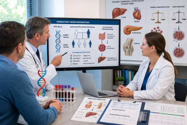
A hereditary hemochromatosis genetic test looks for variants that can disrupt the body’s regulation of iron absorption. Most first-line tests examine the HFE gene, especially the common C282Y and H63D variants. The result can clarify inherited susceptibility, support a diagnosis in someone with abnormal iron studies, and guide testing of adult relatives. It cannot show by itself whether excess iron has accumulated or whether the liver, heart, pancreas, joints, or endocrine organs have been injured. That distinction matters because many people with an HFE genotype associated with risk never develop clinically important iron overload. Conversely, high ferritin may arise from inflammation, fatty liver disease, alcohol use, infection, malignancy, or metabolic illness rather than hereditary hemochromatosis. Accurate interpretation combines genotype with transferrin saturation, ferritin, blood counts, liver tests, symptoms, family history, and—when indicated—magnetic resonance imaging or fibrosis assessment.
- C282Y homozygosity is the HFE genotype most strongly associated with classic adult hemochromatosis.
- Genotype indicates susceptibility; transferrin saturation and ferritin show the current iron phenotype.
- C282Y/H63D compound heterozygosity usually has much lower penetrance than C282Y homozygosity.
- H63D alone rarely explains substantial iron overload without another cause.
- Early recognition and therapeutic phlebotomy can prevent many irreversible complications.
Table of Contents
- How HFE-Related Hemochromatosis Develops
- C282Y, H63D, and Other HFE Results
- Who Should Consider HFE Testing
- How the Genetic Test Is Performed
- Interpreting Positive, Negative, and Uncertain Results
- Confirming Iron Overload and Organ Involvement
- Treatment, Monitoring, and Lifestyle
- Inheritance, Family Testing, and Long-Term Outlook
How HFE-Related Hemochromatosis Develops
Iron is essential for hemoglobin, enzymes, energy metabolism, and many cellular processes. The body has no regulated pathway for excreting large amounts of excess iron, so absorption from the intestine must be tightly controlled. The hormone hepcidin, produced mainly by the liver, is central to that control. Hepcidin limits the activity of ferroportin, the protein that releases iron from intestinal cells and macrophages into the bloodstream.
The HFE protein participates in the signaling system that tells the liver how much hepcidin to make. Pathogenic HFE variation can reduce appropriate hepcidin signaling. Ferroportin then remains more active than it should, intestinal iron absorption increases, and iron is released into plasma faster than the body requires. Transferrin becomes increasingly saturated. When binding capacity is exceeded, reactive non-transferrin-bound iron can enter tissues and promote oxidative injury.
Classic HFE-related hemochromatosis usually progresses slowly over decades. Iron tends to accumulate first in the liver, although the pancreas, heart, pituitary, skin, and joints may also be affected. Potential manifestations include fatigue, arthralgia—particularly involving the second and third knuckles—liver enzyme abnormalities, fibrosis or cirrhosis, diabetes, hypogonadism, skin pigmentation, and cardiomyopathy or arrhythmia. Many affected people are now identified before these complications develop.
Penetrance is incomplete. Not everyone with a high-risk HFE genotype develops biochemical iron overload, and only a subset develops organ damage. Sex assigned at birth, menstruation, pregnancy, blood donation, diet, alcohol intake, body size, metabolic liver disease, inflammation, and additional genetic factors modify expression. Iron accumulation has historically been more frequent and severe in men and in postmenopausal women because regular blood loss reduces iron stores.
The term “hereditary hemochromatosis” covers more than HFE. Rare disorders involving HJV, HAMP, TFR2, SLC40A1, and other genes can cause iron overload with different ages of onset and biochemical patterns. Juvenile forms may cause severe endocrine and cardiac disease at a young age. A negative common-variant HFE test therefore does not exclude every inherited iron disorder.
Secondary iron overload is also distinct. Repeated transfusions, ineffective red-cell production, chronic hemolytic disorders, excessive iron administration, and some liver diseases can increase iron independently of HFE. Treatment and family implications differ, so the cause must be established rather than inferred from ferritin alone.
C282Y, H63D, and Other HFE Results
The HFE variants commonly called C282Y and H63D are protein-level names. C282Y replaces cysteine with tyrosine at amino acid 282; H63D replaces histidine with aspartic acid at amino acid 63. Reports may also list DNA-level nomenclature and reference sequence identifiers. The laboratory should state whether one or two copies were detected.
C282Y homozygosity means a person inherited C282Y from both biological parents. This is the genotype most strongly associated with typical HFE-related hemochromatosis in populations of Northern European ancestry. It supports the diagnosis when transferrin saturation is persistently elevated and iron stores are increased. It still does not prove current tissue injury. Some homozygotes maintain normal ferritin, while others develop progressive overload.
C282Y heterozygosity means one C282Y copy and one HFE copy without that variant. Most carriers do not develop HFE-related iron overload solely because of carrier status. Mild changes in iron indices can occur, but marked ferritin elevation or organ iron should prompt evaluation for another genetic, hepatic, inflammatory, metabolic, hematologic, or environmental cause.
C282Y/H63D compound heterozygosity means C282Y is present on one HFE copy and H63D on the other. This genotype can be associated with modestly higher transferrin saturation or ferritin, but the likelihood of clinically important overload is far lower than with C282Y homozygosity. Current European guidance emphasizes management according to the demonstrated phenotype, not the genotype label alone. Substantial overload should trigger a search for cofactors such as alcohol-related liver injury, metabolic dysfunction-associated steatotic liver disease, viral hepatitis, iron supplementation, or another iron gene.
H63D homozygosity means two H63D copies. H63D is common and is generally considered a low-penetrance susceptibility variant rather than a sufficient cause of classic hemochromatosis. Most homozygotes do not develop clinically significant iron loading. Elevated ferritin in this setting should not automatically be attributed to H63D.
H63D heterozygosity is common carrier status and usually has little clinical consequence. It does not justify phlebotomy in the absence of documented iron overload. Reporting language should avoid implying that a single H63D allele is a disease diagnosis.
A rarer HFE variant, S65C, may be included on some panels. Its effect is generally modest, and interpretation depends on the second allele and the biochemical phenotype. Full HFE sequencing or deletion/duplication analysis may detect uncommon pathogenic variants, but routine first-line testing often focuses on C282Y because it accounts for most classic HFE-related disease in the relevant ancestry groups.
Population frequency is important. C282Y is most prevalent in people with Northern European ancestry and is much less common in many other populations. Testing strategies that assume all iron overload is HFE-related can miss non-HFE disorders and can perform poorly when ancestry differs from the populations in which classic HFE disease was characterized.
Who Should Consider HFE Testing
HFE testing is most useful when there is evidence that iron regulation may be abnormal. Persistent elevation of transferrin saturation—often the earliest biochemical signal—combined with elevated ferritin is a common reason to test. A single abnormal value is usually repeated and interpreted in context because acute illness, laboratory variation, recent iron intake, and liver injury can affect results.
Testing may be appropriate for adults with otherwise unexplained iron overload, characteristic liver findings, suggestive arthropathy, early endocrine complications, cardiomyopathy with iron evidence, or a first-degree relative with confirmed HFE-related hemochromatosis. It can also clarify the cause when imaging shows increased liver iron.
A ferritin test by itself is not a specific screening test for hereditary hemochromatosis. Ferritin is both an iron-storage protein and an acute-phase reactant. It may rise with obesity, metabolic liver disease, alcohol use, infection, inflammation, cancer, kidney disease, or cellular injury. Transferrin saturation helps distinguish increased circulating iron availability from many non-iron causes of hyperferritinemia, although no single threshold is perfect.
General population genetic screening remains debated because penetrance is incomplete and prevalence varies widely by ancestry. Some health systems use biochemical screening first, while others incorporate opportunistic genotyping. The potential benefit of identifying treatable overload must be balanced against unnecessary labeling of people whose genotype will never produce disease.
Testing children for typical adult-onset HFE-related hemochromatosis is usually deferred because serious iron accumulation is uncommon before adulthood and there is rarely an immediate childhood intervention. Adult relatives can make their own informed decision. Earlier specialist assessment is warranted when severe childhood or adolescent iron overload suggests a juvenile or non-HFE form.
Before testing, the clinician should define the question. In a person with abnormal iron studies, the goal may be diagnostic clarification. In a relative, the goal may be predictive testing and a plan for biochemical surveillance. In a person with severe early disease or a non-European ancestry pattern, a broader hemochromatosis panel may be more informative than C282Y/H63D testing alone.
Genetic counseling is particularly useful when the phenotype and genotype do not match, when broader sequencing is considered, when testing is offered to relatives, or when reproductive questions arise. Counseling can address incomplete penetrance, autosomal recessive inheritance, privacy, and the difference between susceptibility and established overload.
How the Genetic Test Is Performed
HFE testing usually requires a blood sample, although saliva or a cheek swab may be accepted by some laboratories. DNA is extracted and analyzed for specific variants. Targeted genotyping can efficiently detect C282Y and H63D; some assays also include S65C. Sequencing examines more of the coding region and splice boundaries, while deletion/duplication analysis looks for larger changes not detected by routine sequencing.
The report should identify the gene, transcript, DNA and protein nomenclature, zygosity, method, limitations, and classification. For common HFE genotypes, the report may provide an interpretive statement about associated risk. That statement must be read alongside iron studies rather than as a stand-alone diagnosis.
Preanalytic preparation is minimal for DNA testing. Iron studies may have separate laboratory instructions, and a clinician may repeat fasting morning transferrin saturation when an initial result is borderline or unexpected. The genetic result remains stable over life, but ferritin and transferrin saturation can change substantially with age, treatment, blood loss, inflammation, and liver health.
Targeted testing has limitations. It can identify the common variants it was designed to detect but will not evaluate rare HFE variants, non-HFE genes, transfusional overload, or other causes of high ferritin. A “negative HFE test” may mean only that C282Y and H63D were absent. The exact assay scope should be checked before concluding that hereditary disease has been excluded.
Broader multigene testing is considered when iron overload is convincing but C282Y homozygosity is absent, particularly with young onset, severe loading, endocrine or cardiac involvement, consanguinity, or a family pattern suggesting another inheritance mechanism. The panel may include HJV, HAMP, TFR2, SLC40A1, and genes associated with rare iron disorders. Broader testing also increases the chance of uncertain findings, so phenotype-guided interpretation is essential.
Clinical laboratories generally classify rare variants as pathogenic, likely pathogenic, uncertain significance, likely benign, or benign. C282Y and H63D are often reported with genotype-specific risk language rather than treated like rare fully penetrant variants. A common allele’s presence should not be exaggerated by generic software that labels every sequence difference a “mutation.”
Interpreting Positive, Negative, and Uncertain Results
A positive C282Y/C282Y result with elevated transferrin saturation and ferritin strongly supports HFE-related hemochromatosis. The next task is to quantify iron burden and assess the liver and other organs. Management is based on the biochemical and clinical phenotype. A person with normal ferritin may need periodic monitoring rather than immediate iron removal.
A C282Y/C282Y result with normal iron studies indicates genetic susceptibility without current biochemical expression. It should prompt a plan for repeat testing at an interval chosen for age, sex, family history, and clinical context. It does not mean that organ damage is silently present, nor does it guarantee future disease.
A C282Y/H63D result should be interpreted cautiously. Mildly abnormal iron indices may be related, but clinically important overload must be demonstrated. If ferritin is high while transferrin saturation is normal or only minimally elevated, metabolic liver disease, alcohol, inflammation, and other explanations may be more likely. Phlebotomy should not be prescribed solely because the genotype sounds “double positive.”
An H63D/H63D or single-allele result usually does not establish HFE-related hemochromatosis. Marked iron overload requires another explanation. The genotype may be one modifier among several, but it should not end the diagnostic investigation.
A negative targeted HFE result lowers the likelihood of classic C282Y-associated disease. It does not exclude non-HFE hereditary hemochromatosis, a rare HFE variant outside the assay, or secondary iron overload. The strength of the phenotype determines whether broader testing, MRI, hematology review, hepatology review, or evaluation for transfusional and liver causes is appropriate.
A variant of uncertain significance is not diagnostic. It should not trigger therapeutic phlebotomy or predictive testing of healthy relatives as though causality were proven. Reclassification may depend on population frequency, functional data, segregation in the family, and whether the person’s biochemical pattern matches the gene’s known mechanism.
An unexpected result from direct-to-consumer testing should be confirmed clinically before it is used for care. Raw-data interpretation may miscall variants, omit phase information needed to determine whether two findings are on opposite chromosomes, or use disease claims that do not reflect current penetrance evidence.
The phrase “carrier” also requires context. HFE-related hemochromatosis is usually inherited in an autosomal recessive manner, so a person with one C282Y allele is often called a carrier. Carrier status has reproductive implications but usually does not produce classic iron-loading disease by itself. A carrier with high ferritin still deserves medical evaluation—the abnormal laboratory result should not be dismissed, but neither should it automatically be blamed on HFE.
Confirming Iron Overload and Organ Involvement
Transferrin saturation is calculated from serum iron and total iron-binding capacity or transferrin. Persistent elevation suggests that circulating transferrin is carrying an unusually high proportion of iron. Ferritin estimates storage iron but is influenced by inflammation and tissue injury. Trends are often more informative than one measurement.
When HFE-related hemochromatosis is suspected, assessment commonly includes a complete blood count, liver enzymes, creatinine, fasting glucose or hemoglobin A1c, and evaluation for alcohol use, viral hepatitis, and metabolic liver disease. Symptoms and examination may direct cardiac, endocrine, or joint evaluation.
Ferritin concentration helps stratify liver risk. Very high ferritin, abnormal liver enzymes, thrombocytopenia, hepatomegaly, or other evidence of chronic liver disease may justify fibrosis assessment. Noninvasive elastography can estimate stiffness. Magnetic resonance imaging can quantify liver iron and, with appropriate sequences, assess cardiac iron when clinically indicated. MRI is particularly useful when the genotype is not C282Y homozygosity and proof of tissue iron is needed.
Liver biopsy is no longer required for most straightforward cases. It may be used when the cause of liver disease remains uncertain, noninvasive fibrosis tests conflict, or histology would change management. Biopsy can stage fibrosis and measure iron distribution, but it is invasive and subject to sampling variation.
Organ assessment should be proportional. Cardiac testing may include electrocardiography and echocardiography when there are palpitations, heart failure symptoms, severe loading, or a juvenile phenotype. Endocrine evaluation may address diabetes, hypogonadism, thyroid or pituitary concerns. Joint symptoms may persist even after iron depletion, particularly metacarpophalangeal arthropathy.
A central safety principle is that iron overload and ferritin elevation are not interchangeable. Someone can have high ferritin without excess tissue iron, and unnecessary repeated phlebotomy can cause anemia, fatigue, venous access problems, and loss of trust. Conversely, a genetically susceptible person with rising transferrin saturation and ferritin should not be reassured solely because symptoms are absent.
People with cirrhosis require management of chronic liver disease and surveillance for hepatocellular carcinoma according to specialist guidance. A high-risk genotype without cirrhosis does not automatically require liver-cancer surveillance. The indication is driven by established advanced fibrosis or cirrhosis, not by HFE status alone.
Treatment, Monitoring, and Lifestyle
Therapeutic phlebotomy is the standard treatment for most people with HFE-related iron overload who can safely undergo blood removal. Each session removes iron contained in red blood cells. The body then draws on stored iron to make new hemoglobin, gradually reducing excess stores.
Treatment usually has an induction phase followed by maintenance. During induction, blood may be removed weekly or every one to two weeks, depending on hemoglobin, symptoms, venous access, cardiovascular status, and the degree of overload. Ferritin is monitored to guide progress. The target range varies somewhat by guideline and clinical setting, but the goal is iron depletion without causing anemia or iron deficiency.
During maintenance, phlebotomy is performed less often—sometimes several times per year—to keep ferritin within the agreed range. Frequency differs widely because iron reaccumulation varies. Hemoglobin should be checked so that treatment is delayed or adjusted when anemia develops. Transferrin saturation may remain elevated even after ferritin is controlled and should not be used in isolation to drive excessive blood removal.
Early treatment can prevent or improve fatigue, liver injury, skin pigmentation, and some cardiac or endocrine effects. Established cirrhosis, diabetes, hypogonadism, or arthropathy may not fully reverse. This is why identifying biochemical overload before irreversible injury is valuable.
Iron chelation is not first-line for uncomplicated HFE-related disease but may be considered when phlebotomy is unsafe or impossible, such as in significant anemia or poor venous access. Specialist supervision is required because chelators have important renal, hepatic, hematologic, auditory, ocular, and gastrointestinal risks depending on the agent.
People with confirmed iron overload should avoid iron supplements unless specifically prescribed. High-dose vitamin C supplements can increase iron absorption and mobilization and are generally avoided; ordinary food sources are usually acceptable. Raw or undercooked shellfish carries a particular risk of severe infection from iron-loving Vibrio species and should be avoided. Alcohol should be limited, especially when liver iron or liver disease is present.
A highly restrictive low-iron diet is rarely a substitute for phlebotomy. Balanced nutrition is preferable. Red meat and iron-fortified products can be consumed in moderation according to individual guidance, while unnecessary supplements should be reviewed. Tea or coffee with meals can reduce non-heme iron absorption, but dietary strategies have a relatively small effect compared with removing blood.
Blood donation may be possible for some medically stable people after appropriate evaluation, depending on local blood-service rules. Therapeutic schedules and routine donation requirements are not identical. Patients should not conceal the diagnosis or use unsupervised donation as a replacement for monitoring.
Inheritance, Family Testing, and Long-Term Outlook
HFE-related hemochromatosis is usually autosomal recessive. A person with C282Y homozygosity inherited one C282Y allele from each biological parent. Each child will inherit at least one C282Y allele; whether a child can inherit two depends on the other biological parent’s genotype. Siblings have a higher chance of sharing the same genotype than more distant relatives.
Adult first-degree relatives of a person with confirmed HFE-related hemochromatosis should be offered an informed approach to testing. This may combine HFE genotyping with transferrin saturation and ferritin. Testing only the genotype can identify susceptibility but may miss the opportunity to detect current overload; testing only ferritin may miss an early biochemical pattern. The exact strategy depends on local guidance and the family’s known result.
When one biological parent has C282Y homozygosity, testing the other parent can sometimes clarify children’s risk without immediately testing minors. If the other parent has no relevant HFE variant, the children are expected to be carriers rather than C282Y homozygotes. Reproductive counseling can explain residual uncertainty and the limited predictive value of genotype for severity.
Relatives who carry one allele usually do not need hemochromatosis treatment. They should receive a copy of the result and understand that iron studies are ordered for clinical reasons, not because carrier status itself proves disease. Relatives with C282Y homozygosity need baseline iron studies and an individualized monitoring plan even if they feel well.
The prognosis is excellent when HFE-related overload is detected before cirrhosis, diabetes, or cardiomyopathy and is treated effectively. Life expectancy can approach that of the general population. Prognosis is less favorable after advanced liver disease develops, which makes fibrosis assessment and ongoing hepatology care important.
The genetic result should remain in the medical record because it does not change, while the phenotype should be updated over time. A concise summary can include genotype, peak transferrin saturation and ferritin, liver iron or fibrosis findings, treatment history, maintenance target, and which relatives have been informed. This prevents future clinicians from confusing an isolated genotype with active disease or overlooking a documented iron-loading phenotype.
A well-interpreted HFE test can end a diagnostic search, direct simple preventive treatment, and identify relatives before injury occurs. Its value comes from disciplined integration: genotype establishes inherited context, iron studies establish biochemical expression, and organ assessment establishes clinical consequences.
References
- HFE-Related Hemochromatosis — 2024 GeneReviews chapter.
- Diagnosis and management of hereditary hemochromatosis — 2024 clinical review.
- Hereditary haemochromatosis: A review — 2024 review.
- Hemochromatosis — 2024 clinical reference.
- TFR2-Related Hemochromatosis — 2023 GeneReviews chapter.
- EASL Clinical Practice Guidelines on haemochromatosis — 2022 clinical guideline.
Disclaimer
This article is for general education and does not diagnose iron overload or replace individualized medical care. HFE results must be interpreted with transferrin saturation, ferritin, blood counts, liver health, symptoms, and family history. Do not begin iron removal, stop prescribed supplements, or change treatment without guidance from a qualified clinician.