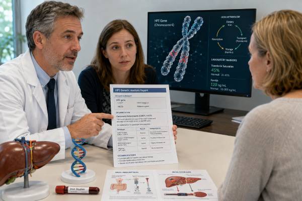
An HFE genetic test looks for inherited variants associated with the most common form of adult hereditary hemochromatosis. The key result is usually C282Y, now written p.Cys282Tyr. H63D, or p.His63Asp, is also commonly reported but has much lower clinical impact and should not be treated as a diagnosis by itself. Genetics must be interpreted with iron studies. Transferrin saturation reflects how much circulating transferrin is loaded with iron, while ferritin is influenced by iron stores, inflammation, liver injury, alcohol use, metabolic disease, and other conditions. Two C282Y copies create the strongest common HFE predisposition, yet many people with that genotype never develop clinically important iron overload. One C282Y copy, one H63D copy, or two H63D copies usually requires another explanation when ferritin is markedly elevated. The test is therefore best used to explain a biochemical phenotype or to evaluate relatives—not as a stand-alone verdict based on DNA alone.
- C282Y homozygosity is the common HFE genotype most strongly associated with iron-loading hemochromatosis.
- Genotype does not equal disease; transferrin saturation, ferritin, and evidence of tissue iron determine clinical significance.
- C282Y/H63D compound heterozygosity has low penetrance and usually needs cofactors before major overload develops.
- H63D alone is generally not considered a sufficient explanation for hemochromatosis.
- A high ferritin is common and can reflect inflammation, fatty liver, alcohol, infection, or metabolic disease rather than excess iron.
- First-degree relatives of a person with confirmed HFE hemochromatosis can benefit from targeted testing and iron studies.
Table of Contents
- What HFE testing can and cannot show
- Reading C282Y and H63D genotypes
- Iron studies come first
- Confirming iron overload and liver risk
- When another gene or cause is likely
- Family testing and inheritance
- Treatment, monitoring, and daily life
- How to review your result
What HFE testing can and cannot show
HFE helps regulate hepcidin, the hormone that controls intestinal iron absorption and iron release from storage cells. When hepcidin signaling is inappropriately low, the body absorbs more iron than it needs. Because there is no normal pathway for actively excreting large amounts of iron, excess can accumulate over years.
HFE testing is most useful in three settings:
A person with abnormal iron studies: Persistently elevated transferrin saturation, with or without elevated ferritin, can prompt testing for C282Y. The result helps distinguish common HFE-related hemochromatosis from other causes.
A first-degree relative of an affected person: Adult siblings, parents, and children can be evaluated before organ damage develops. Testing may combine HFE genotyping with transferrin saturation and ferritin.
Clarification of an incidental result: C282Y or H63D may be reported on a broad panel, exome, or consumer test. Clinical confirmation and iron studies are needed before the label “hemochromatosis” is applied.
The test cannot show how much iron is stored in the liver, whether fibrosis is present, or whether symptoms are caused by iron. It also cannot predict with certainty who will progress. Penetrance depends on age, sex-related physiology, menstrual and pregnancy-related iron loss, blood donation, alcohol, obesity, metabolic disease, viral hepatitis, diet, and other genetic factors.
Symptoms such as fatigue, joint pain, abdominal discomfort, reduced libido, diabetes, skin darkening, arrhythmia, or liver disease are not specific. They deserve evaluation, but a common HFE genotype should not be used to attribute every symptom to iron overload.
Many laboratories test only C282Y and H63D, sometimes S65C. That may be appropriate for common HFE hemochromatosis, but it is not full-gene sequencing and does not evaluate non-HFE iron disorders. Read the methodology before describing the test as “negative.”
A broad genetic variant result guide can help distinguish a high-impact pathogenic genotype from a common risk-modifying allele.
Reading C282Y and H63D genotypes
The report may use older shorthand or current Human Genome Variation Society names:
- C282Y = HFE c.845G>A, p.Cys282Tyr;
- H63D = HFE c.187C>G, p.His63Asp;
- S65C = HFE c.193A>T, p.Ser65Cys.
C282Y homozygous
Two C282Y copies are the common genotype most strongly associated with HFE-related iron loading. A person may have:
- a genetic predisposition with normal iron studies;
- biochemical hemochromatosis with elevated transferrin saturation and ferritin but no proven organ injury;
- clinical hemochromatosis with iron overload causing liver, endocrine, cardiac, joint, or other complications.
These are not interchangeable. Many C282Y homozygotes never develop severe disease. The genotype justifies periodic assessment, but treatment is based on iron loading and clinical context.
C282Y/H63D compound heterozygous
One C282Y copy and one H63D copy are on opposite HFE chromosomes in a compound heterozygote. This genotype can be associated with mildly abnormal iron measures, but clinically important iron overload is uncommon. Marked ferritin elevation should prompt evaluation for alcohol-related liver injury, metabolic dysfunction-associated steatotic liver disease, inflammation, hepatitis, iron supplementation, transfusions, or another genetic cause.
The genotype may contribute when strong cofactors are present. It should not be used as the sole proof of hereditary iron overload.
C282Y heterozygous
One C282Y copy generally indicates carrier status. Carriers may have slightly altered iron measures but usually do not develop classic HFE hemochromatosis. If a carrier has substantial iron overload, clinicians should look beyond the single result.
H63D homozygous or heterozygous
One or two H63D copies commonly appear in the population. Current European guidance emphasizes that H63D should not be treated as a disease-causing genotype on its own. Some people have mild shifts in transferrin saturation or ferritin, but another cause is usually needed to explain significant loading.
No common variants detected
A negative C282Y/H63D test makes common HFE hemochromatosis less likely. It does not exclude rare HFE variants, juvenile hemochromatosis, ferroportin disease, aceruloplasminemia, transfusional iron overload, chronic liver disease, or other disorders.
S65C or a VUS
S65C generally has limited penetrance and is not equivalent to C282Y homozygosity. A VUS should not lead to phlebotomy or family labeling without phenotype and expert review.
The central rule is simple: interpret the genotype against repeated iron studies and objective evidence of overload.
Iron studies come first
Transferrin saturation and ferritin answer different questions.
Transferrin saturation (TSAT) is calculated from serum iron and transferrin or total iron-binding capacity. A persistently elevated TSAT is a hallmark of hepcidin-deficient iron loading and often appears before ferritin rises. Thresholds vary by guideline and laboratory; values around or above 45% commonly trigger further evaluation.
TSAT can fluctuate with meals, time of day, acute illness, laboratory variation, and recent supplements. A single borderline result should often be repeated under standardized conditions rather than treated as diagnostic.
Ferritin is an iron-storage protein and an acute-phase reactant. A high ferritin can reflect:
- actual iron overload;
- inflammation or infection;
- fatty liver or other liver injury;
- alcohol use;
- obesity and metabolic syndrome;
- malignancy;
- kidney disease;
- recent strenuous illness or tissue damage.
A normal ferritin with C282Y homozygosity generally indicates no substantial iron accumulation at that time. A high ferritin with normal or low TSAT often points away from classic HFE hemochromatosis, although exceptions require clinical judgment.
The complete assessment may include a blood count, liver enzymes, C-reactive protein, glucose or A1c, lipid profile, hepatitis testing, alcohol history, medication and supplement review, and family history. Menstrual history, prior pregnancy, blood donation, and transfusions can change the phenotype.
Serum iron alone is not a reliable screening test. It varies widely and should be interpreted through TSAT. Ferritin alone is also insufficient because it is nonspecific.
People sometimes begin blood donation or stop eating iron-containing foods after seeing a high ferritin. That can obscure the diagnosis or create anemia. Testing should be completed before self-treatment when possible.
A useful report review asks whether both TSAT and ferritin were measured on more than one occasion, whether inflammation was present, and whether the pattern fits the genotype. DNA establishes predisposition; biochemical tests establish expression.
Confirming iron overload and liver risk
When C282Y homozygosity accompanies elevated TSAT and ferritin, the diagnosis is often straightforward. In other genotypes, guidelines place greater emphasis on demonstrating tissue iron before calling the condition hemochromatosis.
Magnetic resonance imaging: Validated MRI methods can estimate liver iron concentration without biopsy. MRI can also show whether iron distribution favors hepatocytes or the reticuloendothelial system, which may suggest different mechanisms.
Liver fibrosis assessment: Transient elastography, blood-based fibrosis scores, imaging, and clinical findings help estimate scarring. Ferritin level is one risk marker but does not replace fibrosis assessment.
Liver biopsy: Biopsy is used less often for diagnosis because genetics and MRI provide noninvasive information. It may still be considered when the cause is uncertain, another liver disease is suspected, or precise fibrosis staging would change management.
Organ evaluation depends on the degree and duration of loading. It may include glucose testing, cardiac assessment for symptoms, hormone evaluation, and joint review. The liver is especially important because cirrhosis changes long-term cancer surveillance needs.
A markedly elevated ferritin—particularly around or above 1,000 micrograms per liter—raises concern for advanced liver disease or another serious inflammatory or malignant process, but it is not specific. Prompt evaluation is appropriate rather than assuming the number simply reflects HFE status.
People with cirrhosis from hemochromatosis remain at increased risk for hepatocellular carcinoma even after iron depletion and generally need liver cancer surveillance according to specialist guidance. Those without advanced fibrosis do not automatically need the same surveillance.
The terms “iron overload,” “hemochromatosis,” and “hyperferritinemia” should not be used as synonyms. Hyperferritinemia is a lab finding. Iron overload is excess body iron. Hemochromatosis is a genetic disorder of inappropriate iron absorption with a compatible phenotype.
When another gene or cause is likely
A person with substantial iron overload but no C282Y homozygosity may need a different diagnostic path. The next step depends on age, TSAT, MRI distribution, anemia, family history, transfusion history, and liver disease.
Non-HFE genetic causes include variants in:
- HJV and HAMP, associated with juvenile, often severe hepcidin-deficient hemochromatosis;
- TFR2, associated with another recessive hepcidin-deficient form;
- SLC40A1, associated with ferroportin disease, which may be dominant and can produce different TSAT patterns;
- CP, associated with aceruloplasminemia and neurologic or metabolic findings;
- other rare iron-regulation genes.
Early-onset overload, cardiac or endocrine disease in youth, a strong family history without C282Y homozygosity, or MRI-confirmed loading may support a broader panel. Indiscriminate sequencing in every person with a mildly high ferritin is less useful because most such elevations are acquired or metabolic.
Secondary iron overload can result from repeated transfusions, ineffective red-cell production, excessive iron therapy, chronic hemolytic conditions, or some liver diseases. The body’s iron distribution and TSAT pattern can differ from HFE hemochromatosis.
Metabolic hyperferritinemia is common. Fatty liver, insulin resistance, central adiposity, high triglycerides, and alcohol can raise ferritin, sometimes with modest hepatic iron. Treatment focuses on the underlying metabolic and liver conditions rather than automatically using an HFE phlebotomy schedule.
A high ferritin with anemia, low platelets, fever, weight loss, or systemic illness needs broader medical assessment. Genetic testing should not distract from malignancy, infection, inflammatory disease, or hematologic disorders.
If an older test examined only C282Y, H63D, and S65C, its scope may still be adequate for common HFE disease but not for a convincing non-HFE phenotype. The decision to expand testing belongs with a liver, hematology, or genetics specialist.
Age at presentation can be a valuable clue. Classic HFE-related loading is usually recognized in adulthood after years of increased absorption. Severe iron overload in a child, teenager, or young adult—especially with heart failure, rhythm disturbance, delayed puberty, or endocrine dysfunction—raises concern for juvenile forms and warrants urgent specialist assessment. Conversely, an older adult with only a modest ferritin elevation, normal TSAT, fatty liver, and metabolic syndrome is more likely to have a common acquired explanation than a rare high-penetrance iron gene.
Iron distribution also helps. Hepcidin-deficient disorders often produce high TSAT and hepatocyte-predominant liver iron. Some ferroportin variants can cause iron retention in macrophages with normal or only mildly elevated TSAT, and phlebotomy may be less well tolerated because anemia can develop. Aceruloplasminemia can combine high ferritin with low serum iron or TSAT, diabetes, retinal changes, and neurologic disease. These patterns are not diagnostic by themselves, but they prevent the reflex assumption that every inherited iron disorder behaves like C282Y homozygosity.
Family testing and inheritance
HFE-related hemochromatosis is inherited in an autosomal recessive pattern, but counseling is complicated by low and variable penetrance.
A person with two C282Y copies inherited one from each parent. Their siblings have the classic recessive probabilities if both parents are carriers: 25% chance of two copies, 50% chance of one copy, and 25% chance of neither. These probabilities describe genotype, not the chance of clinical disease.
Adult first-degree relatives of a person with confirmed HFE hemochromatosis can be offered HFE testing and iron studies. Testing both the family variant and the phenotype is useful because a C282Y homozygote with normal iron needs monitoring, while a relative with abnormal iron but a different genotype may need another explanation.
Children of a C282Y homozygote inherit at least one C282Y copy. Whether they can inherit two depends on the other parent. Routine testing of young children is usually not urgent because classic HFE iron loading is an adult-onset condition. Testing is commonly considered when the child reaches adulthood or when results would change near-term care.
A C282Y carrier generally does not need treatment or ongoing specialist surveillance solely because of carrier status. The person may share the result with adult relatives planning testing. Partner testing is usually a reproductive-information choice rather than a medical necessity because even C282Y homozygous children may never develop disease and effective monitoring exists.
H63D should not drive broad cascade testing in the same way as a clearly affected C282Y-homozygous family. Testing relatives for H63D alone can create unnecessary labeling without a meaningful prevention benefit.
An autosomal recessive inheritance guide explains the Mendelian probabilities, but HFE counseling must add the distinction between inheriting a genotype and developing iron-overload disease.
When contacting relatives, share the actual laboratory report. “Hemochromatosis runs in the family” is not enough to know whether the family has common HFE disease, a non-HFE disorder, or only unexplained high ferritin.
Treatment, monitoring, and daily life
For confirmed iron-loading HFE hemochromatosis, therapeutic phlebotomy is the standard treatment. Removing blood forces the body to use stored iron to make new red cells. Treatment usually has an iron-depletion phase followed by maintenance, with hemoglobin and ferritin monitored to avoid anemia.
Phlebotomy is not indicated solely because a person carries H63D or one C282Y copy. It is also not a treatment for every elevated ferritin. Starting without confirmed iron loading can cause iron deficiency while leaving the real cause untreated.
The target ferritin and schedule vary by guideline, tolerance, age, comorbidities, and treatment phase. TSAT may remain elevated even after ferritin reaches target, so clinicians avoid chasing every TSAT result with excessive phlebotomy.
Lifestyle advice is usually practical rather than extreme:
- avoid iron supplements unless specifically prescribed;
- avoid high-dose vitamin C supplements during active iron overload because they can increase iron absorption and mobilization;
- limit alcohol, especially when liver injury or fibrosis is present;
- avoid raw or undercooked shellfish because iron-overloaded people are more vulnerable to severe Vibrio vulnificus infection;
- follow a balanced diet rather than trying to eliminate all iron;
- manage weight, diabetes, blood pressure, and fatty liver risk.
Tea, coffee, calcium, and other dietary factors affect absorption, but diet alone cannot remove established excess iron. Phlebotomy should not be replaced by restrictive eating.
Blood collected during therapeutic phlebotomy may be eligible for donation in some systems if standard donor criteria are met. Policies differ by region and service.
Joint symptoms, especially in the second and third knuckles, may not fully reverse after iron depletion. Advanced cirrhosis, endocrine damage, or heart injury can also persist, which is why diagnosis before organ damage is valuable.
People with a predisposing genotype but normal ferritin typically need periodic iron studies rather than treatment. The interval is individualized. Monitoring can be less intensive in older people who have repeatedly normal results and no cofactors, but this decision should be documented.
How to review your result
Use the following sequence:
- Identify the exact genotype. Is it C282Y/C282Y, C282Y/H63D, one C282Y, H63D/H63D, or no common variant?
- Confirm the test scope. Was it a two- or three-variant assay, full HFE analysis, or a broader iron panel?
- Review repeated TSAT and ferritin. Do they show a hepcidin-deficient pattern or isolated hyperferritinemia?
- Look for cofactors. Alcohol, fatty liver, inflammation, supplements, transfusions, hepatitis, and metabolic disease matter.
- Assess tissue and organ risk. Is MRI, fibrosis testing, or specialist evaluation needed?
- Decide whether phlebotomy is indicated. The answer should be based on iron loading, not genotype alone.
- Identify relatives who benefit. Prioritize adult first-degree relatives of a person with confirmed disease.
- Plan follow-up. Record the monitoring interval, target ferritin if treated, and re-evaluation triggers.
Common errors are calling C282Y/H63D “double hemochromatosis,” treating H63D as a high-penetrance mutation, and assuming high ferritin always means too much iron. Another is dismissing a C282Y homozygote with normal ferritin as a false positive. The genotype is real, but the phenotype may not yet be expressed and may never become clinically important.
Seek prompt care for jaundice, confusion, vomiting blood, black stools, severe abdominal swelling, chest pain, fainting, new arrhythmia, or symptoms of serious infection after raw shellfish exposure. Routine HFE results are not emergencies, but complications of liver or cardiac disease can be.
The clearest diagnosis combines three layers: a credible genetic predisposition, a compatible biochemical pattern, and evidence about organ iron or injury. Keeping those layers separate prevents both underdiagnosis and overtreatment.
References
- HFE-Related Hemochromatosis 2024 (Review)
- EASL Clinical Practice Guidelines on haemochromatosis 2022 (Guideline)
- HFE genotypes, haemochromatosis diagnosis and clinical penetrance 2024
- Diagnosis and management of hereditary hemochromatosis 2024 (Review)
- Diagnosis and Treatment of Hemochromatosis 2025 (Review)
- Hemochromatosis (HFE) 3 Variants 2025 (Test Fact Sheet)
Disclaimer
This article is for education and does not replace liver, hematology, genetics, or primary care evaluation. HFE results must be interpreted with repeated transferrin saturation, ferritin, liver status, symptoms, and other causes of hyperferritinemia. Do not begin phlebotomy, blood donation, supplements, or major dietary restriction solely from a genotype without clinician guidance.