
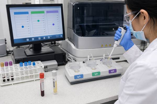
An immunoglobulin panel measures the main antibody classes circulating in blood: IgG, IgA, and IgM. The panel can show whether antibody concentrations are broadly low, one class is selectively reduced, or one or more classes are elevated. It is often ordered for recurrent or unusual infections, unexplained inflammation, liver disease, abnormal total protein, suspected immune deficiency, or monitoring of a known disorder. The test measures quantity, not antibody quality. A person can have immunoglobulin levels within range but respond poorly to vaccines, while another person can have a low value without clinically important infection risk. High results can reflect a broad polyclonal immune response or a monoclonal protein produced by one abnormal cell clone. Interpretation therefore depends on age-specific reference ranges, infection history, medicines, protein loss, blood counts, organ function, electrophoresis, and functional antibody tests. The pattern across all three classes is usually more informative than any single number.
- IgG provides most long-term circulating antibody protection, IgA supports mucosal defense, and IgM acts early in immune responses.
- Children require age-specific ranges because immunoglobulin concentrations mature over time.
- Low levels should be confirmed and assessed for secondary causes before a primary immune disorder is diagnosed.
- High immunoglobulins may be polyclonal or monoclonal, which require different follow-up.
- Normal quantities do not guarantee normal vaccine responses or complete immune function.
Table of Contents
- What the three-part panel measures
- Normal ranges, units, and age effects
- Patterns of low IgG, IgA, and IgM
- Patterns of high immunoglobulins
- Why quantity is not the same as antibody function
- Secondary causes, medicines, and protein loss
- Follow-up tests and diagnostic pathways
- Treatment, monitoring, and when to seek care
What the three-part panel measures
Immunoglobulins are antibodies produced by plasma cells, the mature descendants of B lymphocytes. Each class has a distinct heavy chain and characteristic role. The standard quantitative panel measures total serum IgG, IgA, and IgM. It does not usually include IgE, IgD, IgG subclasses, or antibodies against particular microbes.
| Class | Main distribution and role | Examples of questions raised by an abnormal result |
|---|---|---|
| IgG | Largest circulating antibody pool; long-term systemic protection, neutralization, opsonization, and placental transfer | Broad antibody deficiency, chronic immune stimulation, monoclonal protein, or treatment effect |
| IgA | Major antibody at respiratory, gastrointestinal, and genitourinary surfaces; also present in serum | Selective IgA deficiency, mucosal immune problems, liver disease, or polyclonal elevation |
| IgM | Large pentameric antibody produced early in primary responses and by natural-antibody B cells | Early immune stimulation, selective deficiency, class-switch defect, or IgM monoclonal gammopathy |
IgG commonly accounts for most immunoglobulin mass in serum. IgA is the second largest component, while IgM contributes a smaller fraction despite its large molecular size. The panel reports concentrations, not the number of B cells or plasma cells.
Clinicians order it when the history suggests antibody dysfunction. Repeated bacterial sinusitis, otitis, bronchitis, or pneumonia are common reasons. Other indications include chronic diarrhea, unusual infections, poor growth, autoimmune disease, enlarged lymph nodes or spleen, unexplained lung damage, a family history of immune deficiency, or pre-treatment assessment before B-cell-targeted therapy.
The same panel can investigate high total protein, a widened globulin gap, chronic liver abnormalities, inflammation, or suspected plasma-cell disease. In that setting, the question is not whether the immune system is deficient but why antibody production is increased.
The panel is also used to monitor immunoglobulin replacement, chemotherapy, transplantation, immunosuppressive drugs, chronic lymphocytic leukemia, multiple myeloma, and other conditions that change antibody concentrations. Timing relative to an infusion or medication dose matters.
A panel cannot map the entire immune system. It does not assess T-cell function, neutrophil killing, complement activity, spleen function, or barrier defenses. Even within humoral immunity, it cannot show whether antibodies recognize relevant vaccine antigens. It is an entry point rather than a complete immune evaluation.
Normal ranges, units, and age effects
Reference intervals depend on age, laboratory method, calibration, and population. Results may be reported in milligrams per deciliter (mg/dL), milligrams per liter (mg/L), or grams per liter (g/L). One g/L equals 100 mg/dL. The interval printed on the individual report is the correct comparator.
One major laboratory lists adult intervals around 767 to 1,590 mg/dL for IgG, 61 to 356 mg/dL for IgA, and 37 to 286 mg/dL for IgM. These are examples, not universal standards. Other laboratories use different lower and upper limits and may stratify adults by sex.
Age is particularly important in pediatrics. Maternal IgG crosses the placenta, so a newborn begins life with passively acquired IgG that then declines. The infant’s own production rises gradually, creating a normal physiologic low point during early infancy. Maternal IgA and IgM do not cross the placenta in meaningful amounts, and the infant produces these classes over time.
Because each class matures on a different schedule, an adult range should never be applied to a young child. A value flagged by a general patient portal may need reclassification using the laboratory’s pediatric interval and the child’s exact age.
Reference intervals describe a population distribution. A result a few units below or above the limit can occur in a healthy person. Clinicians assess how far the result lies outside range, whether more than one class is affected, and whether it persists.
Preanalytic and clinical context can modify the number:
- dehydration can concentrate proteins;
- fluid overload can dilute them;
- acute infection can temporarily increase immunoglobulins;
- recent immunoglobulin infusion can raise IgG;
- plasma exchange can lower circulating proteins;
- monoclonal antibody drugs may be detected as immunoglobulin;
- pregnancy and age can shift concentrations; and
- different laboratories may produce nonidentical values.
A surprising result is often repeated when the person is clinically stable. Comparing trends requires attention to units and laboratory method. A change from 7.5 g/L to 750 mg/dL is no change at all; it is the same concentration expressed differently.
The severity of reduction is clinically relevant, but no isolated cutoff defines infection risk for every patient. Someone with moderately low IgG and repeated pneumonia may need more evaluation than someone with a lower stable level and no infections. The phenotype anchors interpretation.
Patterns of low IgG, IgA, and IgM
The combination of low classes narrows the differential diagnosis. Before assigning a primary immune disorder, clinicians confirm the result and exclude medicines, protein loss, malignancy, and other acquired causes.
Low IgG with low IgA and/or IgM suggests broad hypogammaglobulinemia. Possibilities include common variable immunodeficiency, an inborn error of immunity, medication-induced B-cell dysfunction, hematologic malignancy, protein loss, or prolonged immunosuppression. Common variable immunodeficiency is not diagnosed from the panel alone; it requires a compatible age and clinical picture, repeatedly low IgG, impaired antibody function or other supporting immune findings, and exclusion of secondary causes.
Low IgG with normal IgA and IgM may occur in transient states, secondary deficiency, an evolving antibody disorder, or selected primary conditions. A total IgG result should be interpreted with albumin, urine protein, medications, and vaccine responses. An IgG blood test page may explain the class in more depth, but the surrounding pattern remains essential.
Low IgA with normal IgG and IgM can fit selective IgA deficiency when the reduction is marked, persistent, and measured at an appropriate age after other causes are excluded. Many people are asymptomatic. Others have respiratory or gastrointestinal infections, allergies, autoimmunity, or celiac disease. Partial IgA reduction is common and does not automatically carry the same implications.
Low IgM with normal IgG and IgA may be incidental or fit selective IgM deficiency after repeat confirmation. The condition is heterogeneous: some people remain well, while others have recurrent infections, allergy, or autoimmunity. Functional antibody testing helps determine significance.
Low IgG and IgA with normal or high IgM raises concern for a class-switch recombination defect in the appropriate child or young person. Historically called hyper-IgM syndromes, these rare conditions can cause severe bacterial and opportunistic infections and require specialist evaluation.
Low levels of all three classes can occur in profound B-cell deficiency, severe combined immune disorders, anti-CD20 treatment, leukemia or lymphoma, major protein loss, and other acquired states. Lymphocyte subsets help distinguish absent B cells from B cells that are present but functioning poorly.
A person receiving intravenous or subcutaneous immunoglobulin cannot have baseline IgG production assessed directly from a routine trough value because the measured IgG includes donor antibodies. The treatment goal is individualized around infection control and pharmacokinetics, not simply achieving the center of a population interval.
Low immunoglobulins may coexist with autoimmunity, granulomatous inflammation, enteropathy, or lymphoproliferation. Recurrent infection is important but not the only presentation of antibody disorders.
Patterns of high immunoglobulins
Elevated immunoglobulins are first separated into polyclonal and monoclonal patterns. A quantitative panel cannot make that distinction by itself.
A polyclonal increase reflects activity from many plasma-cell clones. More than one class may be high, or one class may predominate. Common causes include:
- acute or chronic infection;
- autoimmune and connective-tissue disease;
- chronic liver disease;
- chronic inflammatory disorders;
- immune dysregulation;
- some malignancies; and
- recovery after immune suppression.
Liver disease is a frequent cause of broad hypergammaglobulinemia. Patterns can offer clues: IgA often rises in cirrhosis, IgG may be prominent in autoimmune hepatitis, and IgM can be elevated in primary biliary cholangitis. These associations are not diagnostic and must be combined with liver enzymes, autoantibodies, imaging, and sometimes biopsy.
Persistent polyclonal elevation can also point toward chronic infection, sarcoidosis, Sjögren disease, systemic lupus, inflammatory bowel disease, or rarer inflammatory syndromes. C-reactive protein, blood counts, liver testing, targeted autoantibodies, infection studies, and clinical examination guide the workup.
A monoclonal increase is produced by one cell clone. It may occur with monoclonal gammopathy of undetermined significance, multiple myeloma, Waldenström macroglobulinemia, lymphoma, amyloidosis, or another clonal disorder. Total IgG, IgA, or IgM may be high, normal, or even low if the monoclonal protein is small and normal immunoglobulins are suppressed.
Serum protein electrophoresis and immunofixation identify a restricted protein. Serum free light chains add sensitivity for light-chain disorders. A sharp M spike differs biologically from a broad inflammatory rise even if both produce the same total IgG concentration.
Very high polyclonal or monoclonal immunoglobulins can occasionally contribute to hyperviscosity. Risk depends on antibody class, concentration, molecular behavior, and other proteins. IgM causes viscosity problems at lower mass concentrations than IgG because it is a large pentamer. Symptoms, not the panel alone, determine urgency.
One high class does not prove that the corresponding arm of immunity is strong. A monoclonal immunoglobulin may have little protective function, and the clone can suppress diverse normal antibodies. High total protein can coexist with increased infection risk.
Why quantity is not the same as antibody function
Quantitative immunoglobulins answer “how much?” Functional antibody testing asks “does the person make useful antibodies to a defined antigen?” Both are needed in some immune evaluations.
A person can have normal IgG, IgA, and IgM but respond inadequately to polysaccharide antigens. This pattern may support specific antibody deficiency when recurrent infections and carefully timed vaccine titers agree. Conversely, mildly low IgG may be clinically unimportant if vaccine responses are robust and infections are ordinary.
Functional assessment often uses antibodies to previous protein vaccines, such as tetanus, and serotype-specific antibodies to pneumococcus. When baseline titers are insufficient or unclear, clinicians may vaccinate and repeat titers after a defined interval. Interpretation accounts for age, prior vaccines, vaccine type, baseline values, number of serotypes tested, and laboratory method.
The panel also cannot measure mucosal IgA directly. Serum IgA correlates imperfectly with secretory IgA at body surfaces. A normal serum value does not guarantee that every mucosal defense is intact, and low serum IgA does not predict exactly how often a person will become ill.
IgG subclasses can be measured in selected cases, but isolated subclass reductions are common and can fluctuate. They become meaningful when persistent, age-adjusted, and supported by a characteristic infection history and impaired antibody function. Routine subclass testing after every normal panel is not necessary.
The quality of an infection history matters. Viral colds in a child attending school or daycare are expected. More concerning features include multiple radiographically confirmed pneumonias, invasive bacterial infection, infections at unusual sites, poor response to appropriate antibiotics, chronic sinus disease with structural damage, bronchiectasis, or opportunistic organisms.
Immune function also depends on exposure and anatomy. Smoking, aspiration, cystic fibrosis, primary ciliary dyskinesia, airway obstruction, allergies, and structural sinus disease can cause recurrent infections despite normal antibodies. Functional evaluation should not stop after finding a borderline immunoglobulin result.
Secondary causes, medicines, and protein loss
Acquired hypogammaglobulinemia is more common than many rare primary antibody disorders. Identifying a secondary cause may change treatment and prevent an incorrect lifelong diagnosis.
Medicines associated with low immunoglobulins include B-cell-depleting antibodies, some anticonvulsants, corticosteroids, immunosuppressants, chemotherapy, and selected targeted cancer drugs. The effect varies by agent, dose, duration, previous therapies, and baseline immune health. The prescriber should review risks; patients should not stop essential treatment on their own.
Hematologic diseases can reduce normal antibody production. Chronic lymphocytic leukemia, lymphoma, multiple myeloma, and other marrow disorders may cause low uninvolved immunoglobulins even when a monoclonal protein is present. Blood counts, smear, electrophoresis, free light chains, and clinical findings direct evaluation.
Protein loss lowers immunoglobulins without a primary failure of production. Kidney disease can spill protein into urine, while protein-losing enteropathy loses it through the gut. Low albumin, edema, urine protein, chronic diarrhea, and gastrointestinal testing help identify these mechanisms. Major burns and some skin disorders can also cause loss.
Malnutrition and severe systemic illness may reduce protein synthesis or reflect complex immune suppression. HIV and other chronic infections can cause varied patterns, including high polyclonal immunoglobulins despite functional immune compromise.
After stem-cell or organ transplantation, the pattern depends on conditioning, immunosuppression, graft function, infections, and immune reconstitution. Timing is central. A low result soon after B-cell-depleting therapy has a different meaning from a progressive decline years later.
Secondary hypergammaglobulinemia likewise requires cause-based assessment. Persistent antigen stimulation from liver disease, autoimmune inflammation, or infection may raise several classes. Treating the underlying condition often reduces the concentrations; directly targeting the number is rarely the goal.
Repeating the panel before immunosuppressive or B-cell-directed therapy can establish a baseline. That baseline is valuable if recurrent infections develop later, because it distinguishes preexisting deficiency from a treatment-emergent change.
Follow-up tests and diagnostic pathways
Follow-up is tailored to the direction and pattern of abnormality. There is no single universal “immune panel” that answers every question.
For low immunoglobulins, common next steps include:
- repeat IgG, IgA, and IgM when clinically stable;
- complete blood count with differential;
- lymphocyte subsets for T cells, B cells, and natural killer cells;
- albumin, total protein, kidney and liver tests, and urinalysis;
- serum and urine protein studies when a clonal disorder is possible;
- HIV testing or other infection studies when appropriate;
- vaccine-specific antibody titers;
- medication and treatment review; and
- imaging or pulmonary testing if recurrent infection has caused structural damage.
An immunologist may analyze B-cell maturation subsets, assess complement, review vaccine records, and consider genetic testing. Genetic testing is most useful when the phenotype suggests an inborn error, there is a family history, or results would change treatment and counseling.
For high values, SPEP and immunofixation distinguish broad from restricted production. A broad pattern prompts investigation of liver disease, inflammation, infection, and autoimmunity. A monoclonal result often leads to hematology evaluation, free light chains, urine studies, and risk-based marrow or imaging tests.
A panel can be normal even when another immune pathway is defective. Severe or unusual infections may justify neutrophil, complement, T-cell, spleen, or genetic evaluation despite normal immunoglobulins. A primary immunodeficiency blood test panel is best understood as a staged evaluation rather than a single fixed bundle.
Results should be linked to objective outcomes. For a patient with recurrent pneumonia, the key questions include organism, imaging, airway damage, and response to prevention. For someone with a globulin gap, the key issue is polyclonal inflammation versus monoclonality. The same three numbers lead to different pathways.
Borderline abnormalities often require observation, not immediate invasive testing. Persistent, substantial, or progressive changes and those accompanied by organ findings deserve greater urgency.
Treatment, monitoring, and when to seek care
Treatment is not prescribed solely to normalize IgG, IgA, or IgM. It targets the diagnosis and its clinical consequences.
For confirmed clinically significant antibody deficiency, management may include prompt treatment of infections, vaccination planning, prophylactic antibiotics, airway-clearance measures, and immunoglobulin replacement. Standard replacement products contain predominantly IgG. They do not replace IgA or IgM directly, but supplied IgG can reduce bacterial infections in disorders with impaired antibody production.
The dose and route of intravenous or subcutaneous immunoglobulin are individualized. Clinicians consider infection burden, trough or steady-state IgG, body size, protein loss, lung disease, adverse effects, and patient preference. A laboratory target alone is not sufficient.
Selective IgA deficiency usually does not require immunoglobulin replacement. Care focuses on infections, allergies, autoimmune complications, and transfusion awareness when there has been a prior reaction. Selective low IgM is managed according to infection and vaccine-response findings.
Polyclonal high levels often improve when the underlying infection, inflammatory disease, or liver condition is controlled. A monoclonal protein is observed or treated according to the clonal diagnosis and evidence of organ damage.
Seek timely medical review for recurrent pneumonia, infections requiring intravenous antibiotics, persistent diarrhea and weight loss, unusual organisms, new enlarged lymph nodes, unexplained anemia, kidney dysfunction, bone pain, neuropathy, or a steadily changing protein level.
Urgent care is appropriate for breathing difficulty, signs of sepsis, meningitis symptoms, severe dehydration, acute kidney failure, confusion, sudden visual changes, major bleeding, or other possible hyperviscosity symptoms. People receiving immunosuppressive therapy may become seriously ill without a high fever.
A useful panel interpretation ends with a plan: whether to repeat the values, test antibody function, search for protein loss, characterize a high protein, refer to immunology or hematology, or simply observe. The numbers are valuable when they organize that next decision rather than being treated as diagnoses in isolation.
References
- Immunoglobulins (IgG, IgA, and IgM), Serum 2026 (Laboratory Test Catalog)
- Practical guidance for the diagnosis and management of secondary hypogammaglobulinemia: A Work Group Report of the AAAAI Primary Immunodeficiency and Altered Immune Response Committees 2022 (Guidance)
- Hypergammaglobulinemia (Polyclonal Gammopathy) 2023 (Clinical Review)
- Etiological study of polyclonal hypergammaglobulinemia in a French cohort and proposal of a diagnostic aid algorithm 2024
- Common Variable Immunodeficiency 2025 (Clinical Review)
- Human inborn errors of immunity: 2024 update on the classification from the International Union of Immunological Societies Expert Committee 2025 (Classification)
Disclaimer
This article provides general educational information and cannot diagnose immune deficiency, infection, liver disease, or a plasma-cell disorder. Reference ranges and testing pathways vary, so a qualified clinician should interpret the panel with age, symptoms, medicines, and related results. Seek urgent care for severe infection, breathing difficulty, neurologic or visual symptoms, major bleeding, or acute organ dysfunction.





