
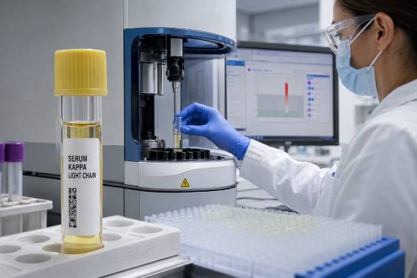
A kappa light chain test measures one component of immunoglobulins, the proteins made by plasma cells to form antibodies. Most clinical orders measure free kappa light chains circulating unattached to a heavy chain and report free lambda light chains plus a kappa/lambda ratio at the same time. A high kappa value is not automatically evidence of multiple myeloma. Kidney impairment and broad immune activation often raise both kappa and lambda, sometimes substantially, while preserving a relatively balanced ratio. A high kappa concentration with a disproportionately high ratio is more suggestive of a kappa-producing cell clone, but still requires confirmation and clinical correlation. The result is interpreted with kidney function, assay-specific ranges, serum protein electrophoresis, immunofixation, blood counts, calcium, urine protein, symptoms, and sometimes bone marrow or tissue biopsy. This article explains the protein’s biology, common reasons for elevation, and how clinicians separate reduced clearance or inflammation from monoclonal gammopathy.
- Kappa and lambda values should be interpreted together, not as isolated numbers.
- Reduced kidney filtration commonly raises free kappa light chains.
- A normal or kidney-appropriate ratio often favors a polyclonal or clearance-related increase.
- A skewed ratio can indicate clonality but is not a stand-alone cancer diagnosis.
- Rapidly rising involved light chains with kidney injury require prompt assessment.
Table of Contents
- Where kappa light chains come from
- Serum free, total, and urine light-chain tests
- Reference ranges and assay variation
- High kappa with a balanced ratio
- High kappa with a kappa-skewed ratio
- Conditions linked to monoclonal kappa production
- Diagnostic workup and result combinations
- Monitoring, treatment, and urgent warning signs
Where kappa light chains come from
A complete immunoglobulin contains two heavy chains and two light chains. Every individual antibody uses either kappa or lambda light chains, never one of each. Normal plasma cells include many clones, some making kappa antibodies and others making lambda antibodies, so both types are present in blood.
During antibody assembly, plasma cells produce a small excess of light chains. The unpaired molecules are released as free light chains. They are much smaller than intact immunoglobulins and circulate for only a short time before the kidneys filter, reabsorb, and metabolize them. This rapid turnover lets serum free light-chain concentrations change quickly when production or kidney clearance changes.
Kappa free light chains usually circulate as single molecules, whereas lambda chains are more often dimers. Kappa is cleared somewhat faster by normal kidneys. These biological differences help explain why the expected kappa/lambda ratio is not exactly 1.0 and why declining kidney function changes the distribution.
Normal immunity creates a polyclonal mixture. When infection or inflammation activates many B-cell clones, production of both kappa and lambda can increase. A plasma-cell or B-cell clone, by contrast, usually produces one restricted light-chain type. A kappa-producing clone raises kappa disproportionately and suppresses or fails to raise lambda to the same degree.
The free light-chain test does not show what antigen an antibody recognizes. It is not an allergy test, an infection-specific antibody test, or a direct measure of immune protection. Its principal uses are detecting, classifying, and monitoring monoclonal gammopathies and interpreting abnormal protein or kidney findings.
Kappa can also be measured as part of intact immunoglobulins or as total light chains in urine, but those tests answer different questions. The wording on the report—“free kappa,” “total kappa,” “urine kappa,” or “immunofixation”—must be confirmed before interpreting the value.
Serum free, total, and urine light-chain tests
The most common test is a serum free light-chain assay. It uses antibodies that recognize exposed regions of kappa and lambda chains when they are not attached to heavy chains. The report generally includes:
- free kappa concentration;
- free lambda concentration;
- kappa/lambda ratio; and sometimes
- the difference between involved and uninvolved free light chains.
The “involved” light chain is the type produced by a suspected or known clone. In a kappa disorder, kappa is involved and lambda is uninvolved. The difference, often abbreviated dFLC, is calculated by subtracting the uninvolved value from the involved value. It is used in selected diagnostic and response criteria, particularly for light-chain amyloidosis and oligosecretory disease.
Serum total light-chain tests measure free plus antibody-bound light chains. Because most light chains are part of intact immunoglobulins, total values are less sensitive to a small free-light-chain clone. They should not be substituted for a free-light-chain assay without understanding the method.
Urine electrophoresis and immunofixation can detect monoclonal free light chains that pass into urine, historically called Bence Jones proteins. The amount in urine depends on production, kidney handling, tubular damage, and collection quality. A 24-hour collection may be requested for quantification or formal response assessment.
Serum immunofixation identifies intact monoclonal proteins and some free-light-chain bands. Combining serum electrophoresis, immunofixation, and free light chains improves detection because no single method identifies every clone.
The assay is ordered for suspected multiple myeloma, light-chain myeloma, monoclonal gammopathy, AL amyloidosis, light-chain deposition disease, unexplained kidney injury, neuropathy, cardiomyopathy, anemia, high calcium, bone lesions, or a globulin abnormality. It is also used to monitor a known light-chain-secreting disorder.
No fasting is generally needed. The blood draw itself is routine. Interpretation becomes less routine when kidney function is reduced, an inflammatory illness is active, or results from different assay platforms are compared. Recent dialysis timing, plasma exchange, and major fluid shifts should also be recorded because they can alter concentrations.
Reference ranges and assay variation
Free light-chain reference intervals are method-specific. Historically quoted examples for one assay are approximately 3.3 to 19.4 mg/L for free kappa, 5.7 to 26.3 mg/L for free lambda, and 0.26 to 1.65 for the ratio. These numbers should not be copied onto a result generated by another platform.
Assays use different antibodies, calibrators, and reaction systems. They can produce meaningfully different kappa and lambda concentrations from the same specimen. In 2026, some major laboratories updated reference values after multi-institutional work confirmed platform-dependent variation. The report’s own interval and method are therefore essential.
Age and kidney function also influence results. Free light chains rise with age largely because estimated filtration declines and chronic inflammation becomes more common. Using a single narrow range for all adults can label healthy older people or patients with stable chronic kidney disease as abnormal.
Kidney-adjusted ratio intervals have been proposed. A frequently used broader range in chronic kidney disease has been about 0.37 to 3.1, although newer studies support estimated-glomerular-filtration-rate-specific intervals and different values for dialysis populations. The laboratory or specialist should apply a validated approach rather than a universal internet cutoff.
The absolute kappa value can be many times the standard upper limit in severe kidney failure without a kappa plasma-cell clone. The ratio, trend, lambda concentration, dialysis status, and clinical findings determine whether that elevation is expected.
A report may use mg/L or mg/dL. One mg/dL equals 10 mg/L, so 2.0 mg/dL equals 20 mg/L. Unit errors can make a result appear tenfold different. Serial monitoring should ideally use the same laboratory, platform, and units.
A result close to a boundary is less decisive than a markedly abnormal pattern. Analytical variation, infection, hydration, and temporary kidney changes can move values around a cutoff. Unexpected borderline results are often repeated after reversible factors have stabilized.
Free light-chain assays can also show nonlinearity or antigen excess at extreme concentrations. Laboratories dilute samples and apply internal checks, but no method is free from interference. If a result conflicts sharply with immunofixation, kidney findings, or a prior trend, direct discussion with the laboratory can identify a platform change, dilution issue, or specimen problem. Biotin is not a universal interference for these assays, and patients should not assume that stopping supplements will correct an unexplained value without laboratory advice.
The ratio is unitless because it divides one concentration by the other. It should be interpreted only when both values were measured by the same assay in the same sample. A ratio that is technically calculable may still be unstable when one result is near the assay’s lower measuring limit. In that setting, the absolute concentrations and confirmatory protein studies deserve greater weight.
High kappa with a balanced ratio
When free kappa and free lambda are both high and their ratio remains within the laboratory’s standard or kidney-adjusted interval, the pattern usually reflects reduced clearance or polyclonal immune activation rather than one dominant clone.
Chronic kidney disease is the most common reason. The kidneys normally filter free light chains and metabolize them in proximal tubules. As glomerular filtration falls, both types accumulate. Kappa may rise proportionally more because its normal clearance is faster, which shifts the ratio upward without necessarily crossing an appropriate renal interval.
Acute kidney injury can produce a rapid increase. The clinical priority is determining why kidney function changed. Dehydration, infection, medication toxicity, obstruction, glomerular disease, and light-chain injury are among the possibilities. A balanced ratio lowers suspicion for a monoclonal process but does not completely eliminate it.
Polyclonal immune stimulation increases light-chain production. Causes include:
- acute or chronic infection;
- autoimmune and connective-tissue disease;
- chronic liver disease;
- inflammatory disorders;
- chronic lung disease;
- some cancers without a light-chain-secreting clone; and
- immune recovery after treatment.
In these settings, quantitative immunoglobulins may also be broadly elevated and SPEP may show a diffuse gamma-region increase. Inflammatory markers and disease-specific testing help identify the cause.
A balanced pattern can coexist with a small monoclonal protein that is below the assay’s ability to distort the ratio. This is why a normal ratio does not negate a clear M protein on electrophoresis or immunofixation. Likewise, biclonal production of opposite light chains can occasionally produce an apparently balanced result.
Kidney impairment plus inflammation can create very high absolute values. The correct response is not to diagnose myeloma from the kappa concentration alone. Clinicians review creatinine, estimated GFR, prior trends, lambda, ratio, electrophoresis, immunofixation, blood counts, calcium, symptoms, and urine findings.
If kidney function improves and the elevation was clearance-related, both light chains often decline. Persistent disproportionate kappa elevation after renal recovery deserves further evaluation.
High kappa with a kappa-skewed ratio
A kappa concentration above range with a high kappa/lambda ratio suggests that kappa production exceeds lambda production. The farther the ratio departs from an assay- and kidney-appropriate interval, the stronger the evidence for a kappa-restricted clone. It is still not a diagnosis by itself.
Clonal plasma cells may secrete:
- free kappa alone;
- an intact immunoglobulin such as IgG-kappa plus excess free kappa;
- more than one kappa-containing protein; or
- very small quantities detectable only by sensitive methods.
The uninvolved lambda value matters. A high ratio can arise because kappa is increased, lambda is suppressed, or both. Suppression of normal immunoglobulin production can occur as a clone expands. Comparing absolute values avoids overinterpreting a ratio driven by an unusually low denominator.
The magnitude has different uses. A mildly high ratio may be caused by kidney disease, inflammation, assay variation, or a small monoclonal gammopathy. A very high ratio with a substantially elevated involved kappa level is more concerning. In the appropriate setting, an involved-to-uninvolved ratio of at least 100, with the involved free light chain at least 100 mg/L, is one validated myeloma-defining biomarker—but only when other diagnostic requirements are met and the finding is attributable to a clonal plasma-cell process.
That biomarker does not mean every person with kappa above 100 mg/L has myeloma. Severe kidney disease can raise absolute kappa beyond that level while the ratio remains relatively balanced. Both components of the criterion, clonal evidence, and full diagnostic evaluation are necessary.
A skewed ratio may appear before an M spike is visible because free light chains can be the only secreted product. This occurs in light-chain MGUS, light-chain myeloma, and some amyloid or deposition disorders. Serum and urine immunofixation help confirm restriction.
New population and kidney-function studies have shown that older reference definitions can overdiagnose light-chain MGUS. Updated intervals reduce false positives, especially in older adults and those with reduced filtration. An abnormal portal flag should therefore be interpreted by a clinician familiar with the assay and renal context.
Conditions linked to monoclonal kappa production
A confirmed kappa-restricted protein can occur across a spectrum. The diagnosis depends on clone size, type, symptoms, and organ effects.
Light-chain monoclonal gammopathy of undetermined significance involves an abnormal free light-chain ratio, increased involved light chain, no intact heavy-chain monoclonal protein, limited marrow clonal cells, and no attributable myeloma-defining damage. It is monitored rather than treated, but current research is refining reference intervals to avoid labeling physiologic variation as disease.
Non-IgM MGUS can produce an IgG-kappa or IgA-kappa protein with excess free kappa. It has no attributable end-organ damage and a lower cell burden than smoldering or active myeloma.
Smoldering multiple myeloma has a higher monoclonal protein or marrow plasma-cell burden but no myeloma-defining event. Free light chains contribute to risk assessment and may identify a biomarker that reclassifies disease as active.
Multiple myeloma can secrete intact kappa immunoglobulin, free kappa alone, or both. Clinical injury may include anemia, bone lesions, hypercalcemia, kidney impairment, and increased infection risk. Free kappa can contribute directly to cast nephropathy when production is very high.
AL amyloidosis results when a monoclonal light chain misfolds and deposits in tissues. Lambda clones are more common overall, but kappa amyloidosis occurs. The clone can be small and the M protein faint. Symptoms may involve the heart, kidneys, nerves, liver, gastrointestinal tract, or soft tissues.
Light-chain deposition disease often involves kappa and deposits nonfibrillar light chains, frequently in kidneys. Proteinuria, blood in urine, hypertension, and declining kidney function may occur. Kidney biopsy defines the lesion and proves that the monoclonal protein is causing damage.
Other B-cell lymphomas, chronic lymphocytic leukemia, solitary plasmacytoma, monoclonal gammopathy of renal significance, and rare heavy-chain or deposition syndromes can also produce kappa restriction.
The word “monoclonal” does not equal “malignant.” MGUS is a precursor state, while monoclonal gammopathy of clinical significance describes a small clone whose protein is already harming an organ. The latter may require clone-directed treatment despite not meeting myeloma criteria.
Diagnostic workup and result combinations
Evaluation begins with the complete report, not just the kappa flag. The clinician records kappa, lambda, ratio, assay platform, units, kidney function, and prior values. Timing relative to acute illness, dialysis, treatment, or contrast exposure can matter.
Common companion tests include:
- complete blood count;
- creatinine, estimated GFR, calcium, albumin, and total protein;
- SPEP and serum immunofixation;
- quantitative IgG, IgA, and IgM;
- urine protein, urine electrophoresis, and urine immunofixation;
- cardiac biomarkers when amyloidosis is suspected;
- imaging for bone lesions or plasmacytoma; and
- bone marrow aspiration and biopsy when indicated.
Some useful pattern combinations are:
- Kappa high, lambda high, ratio appropriate: often kidney impairment or polyclonal inflammation.
- Kappa high, lambda normal or low, ratio high: possible kappa clone.
- Kappa normal, ratio high because lambda is very low: requires cautious review; suppression, assay variation, or a small clone may explain it.
- Kappa high, ratio normal, immunofixation positive: an intact monoclonal protein or small clone can still be present.
- Kappa high, ratio high, kidney function rapidly worsening: urgent evaluation for light-chain-related kidney injury is needed.
- Kappa falling during treatment: may indicate response if it is the established involved marker and laboratory conditions are comparable.
A hematologist may perform marrow flow cytometry, cytogenetics, fluorescence in situ hybridization, and molecular studies. A nephrologist may recommend kidney biopsy when proteinuria or declining function could represent monoclonal gammopathy of renal significance. Tissue biopsy with definitive protein typing is required for amyloidosis.
The kappa level does not determine whether a biopsy is necessary. A modest clone can cause amyloid or renal injury, while a large clearance-related increase may not require marrow testing. Organ findings set the urgency.
Serial values should be compared on the same assay. Switching platforms can create apparent changes that are analytical rather than biological. If a laboratory changes its method or reference range, the report and clinical team should document the transition.
Monitoring, treatment, and urgent warning signs
There is no treatment for a high kappa number in isolation. Management targets reduced kidney clearance, inflammation, a plasma-cell clone, lymphoma, amyloidosis, or a deposition disorder.
For stable chronic kidney disease with a balanced ratio and no monoclonal evidence, clinicians monitor kidney function and the underlying renal condition. Repeating free light chains may be unnecessary unless the clinical picture changes.
For MGUS, follow-up is risk-based. It may include kappa, lambda, ratio, SPEP, blood counts, creatinine, calcium, and symptom review. A rising involved kappa or increasingly skewed ratio prompts reassessment but does not independently prove progression.
During treatment of light-chain myeloma or amyloidosis, the involved kappa and dFLC can change quickly and serve as response markers. Interpretation accounts for kidney function, because worsening filtration can raise both values and mimic biochemical progression. Deep response may be assessed with immunofixation, marrow minimal residual disease testing, and organ-specific outcomes.
Acute light-chain cast nephropathy is time-sensitive. Very high monoclonal free light-chain production can obstruct and injure renal tubules. Rapid clone-directed therapy and supportive kidney care are central; decisions about dialysis or extracorporeal removal are specialist matters.
Seek urgent assessment for sharply reduced urine output, sudden swelling, severe dehydration, confusion, weakness, persistent vomiting, or rapidly worsening kidney test results. New severe back pain with weakness, numbness, or bladder dysfunction may indicate spinal cord compression and is an emergency.
Other prompt-review symptoms include persistent bone pain, unexplained fracture, marked fatigue, recurrent infections, weight loss, neuropathy, foamy urine, leg swelling, shortness of breath, easy bruising, or unexplained heart thickening or failure.
The practical interpretation is comparative: Is kappa elevated because both light chains are accumulating, or is kappa separating from lambda because one clone is expanding? The ratio, kidney function, protein studies, and organ findings answer that question far more reliably than the kappa concentration by itself.
References
- Immunoglobulin Free Light Chains, Serum 2026 (Laboratory Test Catalog)
- Free Light Chains 2024
- New free light chain range decreases LC-MGUS prevalence 2025
- Reference interval of free light chains ratio in patients with end-stage renal disease on chronic hemodialysis 2024
- New Definition of Light Chain Monoclonal Gammopathy of Undetermined Significance 2025
- IMWG Criteria for the Diagnosis of Multiple Myeloma 2026 (Diagnostic Criteria)
Disclaimer
This article provides general education and cannot determine whether an individual kappa elevation is caused by kidney disease, inflammation, or a monoclonal disorder. Assay methods and reference ranges vary, and results require interpretation with lambda, the ratio, kidney function, and clinical findings. Seek urgent care for acute kidney symptoms, neurologic deficits, severe bone pain with weakness, or other rapidly worsening symptoms.





