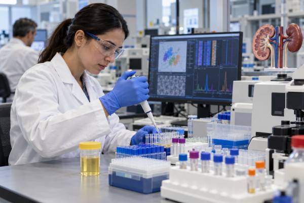
Urine biomarkers such as kidney injury molecule-1 (KIM-1), aquaporin-1 (AQP1), and perilipin-2 (PLIN2) are being studied as noninvasive ways to detect renal cell carcinoma, the most common type of kidney cancer. Research is promising, particularly for AQP1 and PLIN2 in clear cell and papillary renal cell carcinoma, but there is an important practical limitation: there is currently no standard, clinically implemented urine panel using KIM-1, AQP1, and PLIN2 for routine kidney cancer screening or diagnosis. These markers remain investigational. KIM-1 can rise with kidney injury from many noncancer causes, which limits cancer specificity. AQP1 and PLIN2 have shown strong results in some early studies, yet later evidence has been inconsistent and external validation remains limited. Today, kidney masses are still evaluated mainly with imaging, clinical assessment, and sometimes renal mass biopsy. Urine biomarker research may eventually help distinguish benign from malignant masses or identify cancer earlier, but a urine result should not replace established diagnostic care.
- KIM-1 is a kidney-injury protein that can be elevated in renal cell carcinoma, but it is not cancer-specific and may rise with acute or chronic kidney damage.
- AQP1 and PLIN2 have shown the most consistent promise for clear cell and papillary renal cell carcinoma, although results vary across studies and methods.
- No accepted clinical reference range or cutoff exists for a combined KIM-1/AQP1/PLIN2 kidney cancer panel. Research thresholds should not be used as personal diagnostic ranges.
- These urine markers cannot confirm kidney cancer, determine tumor grade, or replace imaging and pathology.
- A normal experimental biomarker result cannot rule out all renal cell carcinoma subtypes, especially because marker expression differs by tumor biology.
Table of Contents
- What the KIM-1, AQP1, and PLIN2 Panel Is
- KIM-1 and Kidney Cancer
- Aquaporin-1 and Perilipin-2
- What Research Shows About Diagnostic Performance
- Limitations and Causes of Misleading Results
- Current Role in Kidney Cancer Diagnosis
- Future Uses and Practical Next Steps
What the KIM-1, AQP1, and PLIN2 Panel Is
A KIM-1/AQP1/PLIN2 urine “panel” is best understood as a research concept, not a standardized commercial diagnostic test. Investigators have measured these proteins individually or in combinations to see whether their urinary concentrations can distinguish renal cell carcinoma (RCC) from healthy controls, benign renal masses, or other kidney conditions.
The three markers reflect different biology:
- KIM-1, also called HAVCR1, is a transmembrane protein strongly induced in injured proximal tubular cells. Its extracellular portion can be shed and detected in urine or blood.
- Aquaporin-1 (AQP1) is a water-channel protein expressed in parts of the kidney, including proximal tubules. It can be overexpressed in clear cell and papillary RCC and released into urine.
- Perilipin-2 (PLIN2), formerly called adipose differentiation-related protein or ADFP, coats intracellular lipid droplets. Clear cell RCC is characteristically rich in lipid and glycogen, making PLIN2 biologically plausible as a marker.
Combining markers is attractive because cancer is heterogeneous. A single protein may be absent in one tumor or elevated for a noncancer reason. A multi-marker pattern could theoretically improve discrimination. However, combining three promising molecules does not automatically create a clinically validated panel. The markers must be measured with standardized assays, tested prospectively in the intended population, validated independently, and shown to improve real clinical decisions.
That validation step remains incomplete. A 2025 systematic review of urinary biomarkers for RCC identified more than 100 candidate markers and multiple panels, but concluded that external validation was severely lacking and that none of the reviewed urinary diagnostic biomarkers had been implemented in routine clinical practice.
This is why an experimental result should be interpreted very differently from an established laboratory test with standardized reference intervals.
KIM-1 and Kidney Cancer
KIM-1 is especially interesting because many renal cancers arise from tubular epithelial cells. It is overexpressed in several proximal-tubule-derived RCC subtypes, including many clear cell and papillary tumors. Its shed ectodomain can be measured in urine, creating a potential noninvasive signal of tumor-related kidney biology.
Small clinical studies found that urinary KIM-1 could be higher before kidney tumor removal and fall afterward in patients whose tumors expressed KIM-1. More recent meta-analytic work has supported an association between liquid-based KIM-1 measurements and RCC, although study methods and specimen types vary.
The major limitation is specificity. KIM-1 is widely used in kidney research as a marker of tubular injury. Acute kidney injury, chronic kidney disease, toxic kidney damage, ischemia, and other renal conditions can increase KIM-1 without cancer. That makes a high urine KIM-1 result fundamentally different from a tumor-specific mutation.
A high KIM-1 value therefore cannot answer, “Do I have kidney cancer?” It may reflect injury, tumor biology, or both. Likewise, a low value does not exclude RCC because not all renal tumors express KIM-1 strongly. Chromophobe RCC, for example, arises from different tubular biology and has shown less consistent KIM-1 expression in earlier studies.
Researchers often normalize urinary KIM-1 to urine creatinine to reduce the effect of urine dilution. Results may be expressed as a ratio such as ng of KIM-1 per mg of creatinine. Those numbers are assay- and study-specific. There is no universal kidney-cancer cutoff that patients should apply to a routine KIM-1 measurement.
KIM-1 also illustrates why a marker that is biologically compelling can still be difficult to use diagnostically. Sensitivity for kidney injury can work against cancer specificity. Future panels may need to combine KIM-1 with more tumor-selective proteins, DNA changes, or imaging features rather than rely on KIM-1 alone.
Aquaporin-1 and Perilipin-2
AQP1 and PLIN2 have generated some of the strongest early urine-protein data in RCC research. Multiple studies from the same research program found substantially higher urinary concentrations in patients with clear cell or papillary RCC than in controls, with concentrations decreasing after tumor removal.
In one study of 61 patients with clear cell or papillary RCC and 43 matched controls, median urinary AQP1 was about 35-fold higher and PLIN2 about 9-fold higher in the cancer group. The reported sensitivity and specificity were both 100% for AQP1, while PLIN2 had 92% sensitivity and 100% specificity. Concentrations correlated with tumor size and some aspects of stage but not tumor grade.
A later prospective screening-oriented study examined urine from hundreds of people undergoing abdominal CT. AQP1 and PLIN2 again showed high diagnostic performance, with areas under the receiver-operating-characteristic curve above 0.90 in that cohort. These results made the pair look especially promising for detecting clear cell and papillary tumors.
However, later evidence has not been uniformly positive. The 2025 systematic review found that AQP1 results were contradictory across studies. Some investigators reproduced strong differences, while others found little or no useful discrimination. PLIN2 appeared more consistent, but it too had been studied by relatively few independent groups.
Subtype biology is another important limitation. AQP1 and PLIN2 are not equally elevated across every kidney tumor. Earlier work suggests they perform better for clear cell and papillary RCC than for chromophobe RCC. A test designed around these proteins could therefore miss a biologically different malignancy even if it performs well for the most common subtype.
Because of these issues, there is no validated “normal AQP1” or “normal PLIN2” kidney-cancer range for routine patient care. Researchers have used different measurement platforms, normalization methods, and cutoffs. A number from one study cannot safely be transferred to another laboratory.
What Research Shows About Diagnostic Performance
The broad RCC urine-biomarker literature is much larger than these three proteins. Investigators have studied metabolites, microRNAs, DNA methylation, extracellular vesicles, proteins, and volatile organic compounds. That breadth is encouraging, but it also shows that the field has not converged on one clinically proven assay.
The most comprehensive recent systematic review included 46 studies, identifying 105 individual urinary biomarkers and 29 multi-biomarker panels. AQP1 and PLIN2 were among the proteins considered promising, but only a small number of candidate markers had been evaluated in more than one study. The authors emphasized that validation is the major bottleneck.
A few lessons emerge from the evidence:
- Early case-control results can look better than real-world performance. Comparing known cancer patients with healthy volunteers creates a clearer biological contrast than testing people with indeterminate renal masses, kidney disease, cysts, or competing diagnoses.
- Independent replication matters. A marker repeatedly tested by the same group is not equivalent to confirmation across independent laboratories and populations.
- Assay method matters. Western blot, ELISA, mass spectrometry, and other platforms can yield different absolute values and cutoffs.
- RCC is not one disease. Clear cell, papillary, chromophobe, and rarer renal tumors have different molecular profiles.
- Clinical utility is harder than diagnostic accuracy. Even a good marker must show that it changes management—for example, safely avoiding biopsy or surgery for a benign mass without missing aggressive cancer.
This last point is crucial. A high AUC or impressive sensitivity in a research paper does not automatically mean a test is ready for population screening. Screening also requires evidence about false positives, downstream imaging and procedures, cost, overdiagnosis, and whether earlier detection improves outcomes.
Researchers have explored using AQP1 and PLIN2 alongside renal mass biopsy to characterize small renal masses. A 2021 study proposed an algorithm in which elevated markers could help identify clear cell or papillary RCC while biopsy would still be needed for marker-negative masses, including possible chromophobe tumors. The work was promising but exploratory rather than a standard-of-care protocol.
Limitations and Causes of Misleading Results
The largest risk in interpreting an experimental kidney cancer urine marker is treating a research signal as if it were a validated diagnosis. Both false-positive and false-negative results are possible.
KIM-1 false positives are particularly plausible because many forms of tubular kidney injury can raise the marker. Reduced kidney function, recent ischemic injury, inflammation, nephrotoxic medication exposure, or other renal disease can complicate interpretation.
AQP1 and PLIN2 variability can arise from tumor subtype, tumor size, specimen handling, assay technique, and normalization. A small tumor may shed less marker than a large tumor. A chromophobe tumor may not produce the same pattern as clear cell RCC. Differences in urine concentration can also affect raw measurements if results are not appropriately normalized.
Other limitations include:
- No standardized clinical assay: laboratories may use research-developed techniques rather than a harmonized commercial platform.
- No agreed decision thresholds: proposed cutoffs vary by study and cannot be treated as universal reference ranges.
- Limited prospective validation: many studies are relatively small or use selected patient groups.
- Uncertain performance in screening: the test characteristics seen in known-cancer cohorts may not hold in people without symptoms or known masses.
- No direct staging ability: urinary protein concentration cannot reliably establish whether a tumor has invaded beyond the kidney or spread to lymph nodes or distant organs.
- No definitive grading: tumor grade requires pathologic examination of tissue.
Kidney cancer itself often causes no urinary abnormality, especially when a tumor is small. A normal routine urinalysis or absence of blood in the urine does not exclude a renal mass. Experimental protein markers do not change that principle.
A person who sees KIM-1, AQP1, or PLIN2 on a research or specialty report should ask exactly which assay was used, what population the reference range came from, whether the result is clinically validated, and what decision the ordering clinician intends the result to influence.
Current Role in Kidney Cancer Diagnosis
At present, KIM-1, AQP1, and PLIN2 do not replace the established evaluation of a suspected kidney tumor. Most renal masses are first found on ultrasound, CT, or MRI, often incidentally during imaging performed for another reason.
When a renal mass is identified, clinicians assess factors such as size, enhancement pattern, growth over time, presence of fat, complexity of cystic features, kidney function, symptoms, age, comorbidities, and risk of spread. Contrast-enhanced CT or MRI usually provides much more anatomic information than a urine protein measurement.
A renal mass biopsy may be recommended when tissue information is likely to change management. It can help distinguish benign from malignant lesions or identify tumor subtype before ablation, systemic therapy, or selected surveillance decisions. Biopsy has limitations, including nondiagnostic samples, but it provides information that a urine biomarker cannot currently supply.
Management options for a localized small renal mass can include active surveillance, partial nephrectomy, radical nephrectomy, or thermal ablation depending on the clinical situation. An experimental biomarker should not be used by itself to choose among these treatments.
This is different from established urine markers in some bladder or prostate diagnostic pathways. The fact that a marker can be measured in urine does not mean it has reached the same level of clinical evidence. For RCC, recent systematic reviews still describe urinary biomarkers as promising candidates requiring further validation.
Research laboratories may also report biomarker concentrations in different ways, such as absolute concentration, concentration adjusted to urine creatinine, or a value generated by an experimental model. Those formats are not interchangeable. A threshold from one published assay should not be applied to another laboratory method unless that method has been analytically and clinically validated against the same cutoff. This is one reason a single universal “normal range” cannot yet be given for a KIM-1/AQP1/PLIN2 kidney-cancer panel.
Anyone with visible blood in the urine, persistent flank pain, an unexplained renal mass, or concerning imaging should follow the recommended diagnostic pathway rather than seeking reassurance from an investigational urine test.
Future Uses and Practical Next Steps
Urine biomarkers could eventually fill several important gaps in kidney cancer care. A reliable panel might help screen selected high-risk populations, distinguish benign from malignant renal masses, determine which small masses need biopsy, identify recurrence after treatment, or reduce unnecessary imaging. A multi-marker approach may ultimately outperform any single protein.
KIM-1, AQP1, and PLIN2 could also be combined with newer molecular signals such as DNA methylation patterns, microRNAs, extracellular-vesicle cargo, or metabolomic signatures. Machine-learning models may integrate these data with CT or MRI characteristics. The goal would be a clinically interpretable probability of cancer, not simply three separate protein concentrations.
Before such a panel becomes routine, it needs several layers of evidence: standardized analytical methods, reproducibility across laboratories, independent prospective validation, subtype-specific performance, prespecified cutoffs, and studies showing that using the test actually improves patient outcomes or reduces unnecessary procedures without increasing missed cancers.
If you are offered one of these markers today, ask:
- Is this test being used in a research study or as routine clinical care?
- Is the assay validated for kidney cancer, kidney injury, or another purpose?
- What does the laboratory consider a valid reference interval, and was that interval developed for cancer diagnosis?
- Could chronic kidney disease or recent kidney injury influence my result?
- What imaging or biopsy decision will this result change?
- What happens if the biomarker result conflicts with CT or MRI findings?
For someone with a kidney mass, the most useful next step is usually a discussion with a urologist or kidney-cancer specialist about imaging features and the need for surveillance, biopsy, or treatment. Experimental urine markers can contribute to research, but they should not be allowed to create false reassurance or unnecessary alarm.
References
- Renal cell carcinoma detection: a systematic review in diagnostic urinary biomarkers 2025 (Systematic Review)
- Liquid-based kidney injury molecule-1 as a diagnostic and prognostic indicator in renal cell carcinoma: A systematic review and meta-analysis 2025 (Systematic Review)
- Biomarkers in Renal Cell Carcinoma: A Systematic Review and Immunohistochemical Validation Study 2025 (Systematic Review)
- Epidemiology and Prevention of Renal Cell Carcinoma 2022 (Review)
- Urinary aquaporin 1 and perilipin 2: Can these novel markers accurately characterize small renal masses and help guide patient management? 2019
- Urinary concentrations of aquaporin-1 and perilipin-2 in patients with renal cell carcinoma correlate with tumor size and stage but not grade 2014
Disclaimer
This article describes research on kidney cancer urine biomarkers and is not a substitute for medical diagnosis or treatment. KIM-1, AQP1, and PLIN2 are not a validated stand-alone urine panel for routine kidney cancer diagnosis, and research values should not be interpreted without specialist guidance. A suspicious renal mass or concerning urinary symptoms require standard clinical evaluation with appropriate imaging and, when indicated, tissue diagnosis.