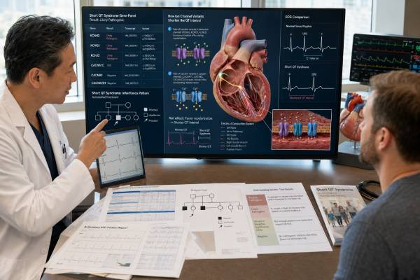
Short QT syndrome is a very rare inherited electrical disorder in which the heart’s recovery time between beats is abnormally brief, creating vulnerability to atrial and ventricular arrhythmias. Genetic testing can support the diagnosis, clarify the molecular mechanism in selected families, and make targeted testing possible for relatives. It cannot diagnose the syndrome from DNA alone. A short corrected QT interval may occur because of normal variation, measurement error, high calcium, high potassium, acidosis, fever, medications, or other clinical conditions. Even among people with convincing short QT syndrome, no disease-causing variant is found in many cases. The strongest gene evidence is concentrated in a small group—particularly KCNH2, with additional support for KCNQ1, KCNJ2, and SLC4A3—while several historically marketed genes have weaker or disputed associations. Because an incorrect label can lead to unnecessary restrictions or invasive treatment, testing should follow expert electrocardiographic evaluation and be interpreted by an inherited-arrhythmia team.
- A low QTc value is a diagnostic clue, not proof of short QT syndrome by itself.
- Serial high-quality ECGs and exclusion of acquired causes come before genetic interpretation.
- KCNH2 has the strongest established gene-disease relationship; other genes require evidence-aware review.
- A pathogenic result can enable targeted family testing but does not precisely predict sudden-death risk.
- A variant of uncertain significance is not a diagnosis and should not drive an ICD decision.
- A negative panel does not exclude short QT syndrome when the clinical phenotype is convincing.
Table of Contents
- A short QT is a clue, not the syndrome
- Building a clinical diagnosis
- Channel genes and the strength of evidence
- Choosing and performing genetic testing
- Reading the result correctly
- Risk, treatment, and daily life
- Family testing and unresolved cases
A short QT is a clue, not the syndrome
The QT interval on an electrocardiogram begins with ventricular electrical activation and ends when ventricular repolarization is complete. Because the interval changes with heart rate, clinicians usually interpret a corrected value, or QTc. Short QT syndrome, abbreviated SQTS, involves pathologically accelerated repolarization and shortened refractory periods. That electrical environment can permit rapidly re-entering rhythms, including atrial fibrillation, ventricular tachycardia, and ventricular fibrillation.
The diagnosis is not synonymous with “QTc below the laboratory range.” QT correction formulas behave differently at high and low heart rates, automated measurements can be wrong, and the end of the T wave may be difficult to identify. Bazett correction, which is widely displayed by ECG machines, can distort QTc at heart-rate extremes. A specialist may measure the interval manually in several leads, compare formulas, and repeat the ECG at a more stable rate. The relationship between QT and heart rate—sometimes called rate adaptation—can also provide useful context.
Published criteria use very short QTc thresholds and allow somewhat less extreme shortening when additional evidence is present, such as a pathogenic variant, a family history of SQTS, or survival from otherwise unexplained ventricular fibrillation. Exact thresholds should be applied by a clinician rather than used as a self-diagnosis tool. Sex, age, heart rate, tracing quality, T-wave morphology, symptoms, and family history all affect interpretation. Tall, narrow, or peaked T waves and a short or nearly absent ST segment may support the phenotype but are not specific.
Acquired QT shortening must be considered. Hypercalcemia, hyperkalemia, acidosis, hyperthermia, digoxin effect, and selected drugs can reduce the QT interval. Some metabolic or systemic disorders produce a short QT-like tracing without primary SQTS. The ECG should be reassessed after a reversible abnormality is corrected whenever possible. Structural heart disease, ischemia, toxic exposure, and other channelopathies may also change repolarization or produce ventricular arrhythmia through another mechanism.
Symptoms range from none to palpitations, fainting, seizures caused by transient cerebral hypoperfusion, atrial fibrillation, cardiac arrest, or sudden death. Events can occur at rest, during sleep, or with activity. An asymptomatic person may be evaluated because of a family member’s diagnosis or an unexpectedly short QTc. Conversely, fainting in a person with a short QT interval may have a non-arrhythmic cause. Establishing that the electrical phenotype and clinical event belong together is central to avoiding both missed disease and overdiagnosis.
SQTS should also be distinguished from long QT syndrome. Some of the same genes can be involved in both conditions, but the direction and mechanism of channel dysfunction differ. A gene name alone therefore cannot tell whether a variant lengthens or shortens repolarization.
Building a clinical diagnosis
Evaluation starts with the original ECG, not merely the QTc printed on a report. An electrophysiologist reviews tracing speed and calibration, rhythm, QRS duration, T-wave termination, U waves, and whether bundle-branch block or pacing makes the QT difficult to interpret. Several resting ECGs are preferable to a single outlier. Holter monitoring or exercise testing may show how the QT adapts across different heart rates, although neither test has a single pattern that independently proves SQTS.
A detailed history asks about sudden loss of consciousness, circumstances surrounding episodes, documented atrial or ventricular arrhythmias, resuscitated cardiac arrest, unexplained drowning, single-vehicle crashes, nocturnal death, and seizure diagnoses that did not respond as expected to neurologic treatment. Medical records are essential whenever available. The family pedigree should cover at least three generations and include ages at events, ECG findings, autopsy information, and whether relatives had structural heart disease.
The examination and testing look for alternatives. Electrolytes, calcium, kidney function, acid-base status, medication and supplement exposure, and acute illness are reviewed. Echocardiography is commonly performed to exclude significant structural disease. Cardiac magnetic resonance imaging may be useful when cardiomyopathy, myocarditis, scar, or another substrate is suspected. In a cardiac-arrest survivor, the workup is broader because coronary disease, cardiomyopathy, Brugada syndrome, catecholaminergic polymorphic ventricular tachycardia, early repolarization syndrome, toxic causes, and other diagnoses may be more likely than SQTS.
Provocative electrophysiology testing is not a universal diagnostic requirement. Short ventricular refractory periods may be observed, but inducibility of arrhythmia has imperfect reproducibility and does not replace the clinical picture. Likewise, an implantable loop recorder can document the rhythm during recurrent unexplained symptoms but does not establish the genetic cause.
Clinical certainty matters before sequencing. SQTS is so rare that a person with a borderline QTc and no supportive history has a low pretest probability. In that setting, a rare DNA change—especially in a weakly associated gene—is more likely to confuse than clarify. By contrast, a survivor of unexplained ventricular fibrillation with repeatedly extreme QT shortening and similarly affected relatives has a much stronger indication for genetic counseling and testing.
Children require age-aware ECG interpretation. Infants normally have faster heart rates, and QT measurement can be challenging. Sudden infant death in a family can raise concern, but it is not specific for SQTS. Pediatric electrophysiology review, comparison with age-appropriate norms, and careful exclusion of metabolic causes are important before assigning a lifelong diagnosis.
Channel genes and the strength of evidence
The heart’s action potential depends on coordinated movement of potassium, sodium, calcium, chloride, and bicarbonate across cell membranes. Disease-causing SQTS variants generally increase outward repolarizing current or reduce inward current, shortening the action potential. The functional direction is crucial. A loss-of-function variant and a gain-of-function variant in the same channel gene may lead to different syndromes.
KCNH2 encodes the hERG potassium channel that carries the rapid delayed rectifier current. Gain-of-function variants can accelerate repolarization and cause the best-established molecular subtype of SQTS. Other KCNH2 variants reduce channel function and cause long QT syndrome type 2. This contrast demonstrates why “a KCNH2 variant” is not a complete interpretation; the exact variant, functional evidence, segregation, and phenotype must agree.
KCNQ1 encodes a potassium-channel subunit involved in the slow delayed rectifier current. Certain gain-of-function variants have been reported in SQTS, sometimes with atrial fibrillation or striking QT shortening at slow heart rates. Many pathogenic KCNQ1 variants instead reduce current and cause long QT syndrome type 1. KCNJ2 encodes the inward-rectifier potassium channel Kir2.1. A small number of gain-of-function variants have convincing or moderate evidence for SQTS, while other KCNJ2 mechanisms cause Andersen–Tawil syndrome.
SLC4A3 encodes a chloride-bicarbonate exchanger. Human genetic and experimental evidence supports an association with SQTS, but far fewer families are known than for common inherited arrhythmia disorders. Laboratories may include this gene on a focused evidence-based panel. Because the published variant count is small, classification often depends heavily on family segregation, rarity, functional data, and whether the ECG phenotype is definite.
Older literature and some commercial panels list calcium-channel genes such as CACNA1C, CACNB2, and CACNA2D1, as well as SCN5A and several additional genes. Some reported individuals had Brugada features, carnitine-transport deficiency, developmental findings, or simply a shorter-than-average QT rather than definite isolated SQTS. Contemporary gene-validity reviews have therefore narrowed the set of genes considered clinically actionable for classic SQTS. A broad arrhythmia panel may still be appropriate when the phenotype overlaps another syndrome, but the report must distinguish established SQTS genes from disputed or phenotype-like associations.
Genetic architecture is not fully solved. Even in clinically persuasive cases, testing often finds no causal variant. This may reflect undiscovered genes, noncoding or structural changes, mosaicism, technical limitations, or a more complex genetic mechanism. Low yield is not a reason to avoid testing when the result could help a family; it is a reason to set realistic expectations and resist overinterpreting weak findings.
Choosing and performing genetic testing
Testing is most informative in a person with the clearest clinical phenotype. In a family with several affected members, this is usually the individual with repeatedly marked QT shortening, documented ventricular arrhythmia, or the most complete records. Starting with an unaffected relative creates a common interpretive trap: a negative result cannot exclude a familial condition when no family variant has been identified.
A focused SQTS panel should emphasize genes with validated disease associations and use a laboratory experienced in cardiac channelopathies. The test description should specify which genes and exons are analyzed, whether deletion and duplication changes are assessed, what coverage limitations exist, and how variants are classified. Copy-number variants are not the predominant known mechanism for SQTS, but understanding assay scope is still important. Broader arrhythmia or cardiomyopathy panels may be chosen when the differential diagnosis remains open.
Pretest counseling covers the possible outcomes—pathogenic or likely pathogenic, uncertain, benign or likely benign, and negative—as well as implications for relatives. It should address privacy, insurance rules in the relevant country, potential incidental findings from broad sequencing, and whether the patient consents to recontact if a classification changes. Genetic testing does not forecast the exact age of an event and cannot convert a low-quality ECG into a definite diagnosis.
Blood or saliva is generally sufficient. The ordering clinician should provide the laboratory with QT measurements, heart rates, ECG morphology, symptoms, arrhythmia documentation, family history, and suspected overlapping diagnoses. Phenotypic detail helps analysts judge whether a variant’s known mechanism fits the case. A laboratory interpreting KCNH2, for example, needs to know that the phenotype is short QT rather than assuming any previously reported pathogenic KCNH2 variant is relevant.
Postmortem testing may be considered after sudden unexplained death when an adequate DNA sample exists. The best approach coordinates molecular autopsy with expert cardiac pathology and clinical evaluation of first-degree relatives. A variant found after death should not be treated as causal solely because it is rare. The decedent’s circumstances, autopsy, available ECGs, family segregation, and gene validity remain necessary.
Consumer raw-data files are not an adequate diagnostic test. They may omit most rare variants, miscall technically difficult positions, use outdated annotations, or report research associations without clinical validation. Any potentially important finding requires confirmation in an accredited clinical laboratory and interpretation in the context of a specialist evaluation.
Reading the result correctly
A pathogenic or likely pathogenic variant in a well-established SQTS gene can provide molecular confirmation when the ECG and clinical phenotype fit. It can identify the channel mechanism, support targeted testing of relatives, and end an otherwise uncertain family search. Classification labels are not interchangeable with clinical severity. A likely pathogenic variant is considered sufficiently convincing for clinical use, but neither it nor a pathogenic label predicts whether a carrier will experience atrial fibrillation, fainting, or cardiac arrest.
The report should be checked for the exact nucleotide and protein change, transcript, inheritance, zygosity, evidence summary, and the gene’s mechanism. Functional data must be interpreted carefully: an experiment showing altered current does not automatically prove human disease if the direction, magnitude, model, or phenotype is inconsistent. Population frequency is especially important for an extremely rare syndrome; a variant common in healthy populations is unlikely to explain highly penetrant SQTS.
A variant of uncertain significance has insufficient or conflicting evidence. It must not be used to diagnose SQTS, clear a relative from surveillance, prescribe quinidine, restrict sports, or justify an implantable cardioverter-defibrillator. Testing relatives solely to learn whether they carry a VUS is generally not predictive testing. Carefully designed segregation studies can help the laboratory gather evidence, but the genetics team should define which relatives are informative and how results will be used.
A negative test does not rule out SQTS. It means that the laboratory did not identify a reportable causal variant with the method and knowledge available. If the clinical diagnosis is strong, management and family ECG screening continue according to phenotype. If the diagnosis was weak, a negative panel should prompt reconsideration of measurement, acquired causes, and alternative arrhythmia syndromes rather than repeated indiscriminate sequencing.
A benign or likely benign variant is not the cause of the syndrome and should not be used for family decisions. Occasionally a report includes a pathogenic variant for another condition discovered because a broad panel was ordered. The clinician must determine whether that finding explains the patient, represents carrier status, creates an unrelated health implication, or is an incidental result covered by consent.
Variant classifications can change. The SQTS evidence base is unusually small, and recent reanalyses have downgraded or questioned many historically reported variants while strengthening a limited subset. Patients should retain their full report and ask how the laboratory handles amended classifications. Re-evaluation is particularly important when an old report used terms such as “mutation,” “possibly disease-causing,” or “risk variant” without modern criteria.
Risk, treatment, and daily life
Risk assessment relies most heavily on the person’s clinical history. Previous cardiac arrest or documented sustained ventricular tachycardia or fibrillation is a major warning sign. Recurrent arrhythmic syncope and a family history of sudden death may add concern, but risk prediction in asymptomatic individuals is imprecise because patient numbers are small and long-term data are limited. QTc length and genotype alone have not produced a reliable calculator that can tell an individual carrier whether or when an event will occur.
An implantable cardioverter-defibrillator, or ICD, is the main protection for survivors of cardiac arrest and selected patients with documented malignant ventricular arrhythmia. It can terminate ventricular fibrillation but does not prevent every arrhythmia and carries important burdens, including infection, lead failure, inappropriate shocks, psychological effects, and repeated procedures over a lifetime. T-wave oversensing may be a particular programming concern in SQTS because tall T waves can be mistaken for ventricular events. Device decisions belong in an expert electrophysiology setting.
Quinidine or hydroquinidine has the strongest pharmacologic experience in SQTS and can prolong repolarization in some molecular subtypes, especially certain KCNH2-mediated disease. It may be considered when an ICD is not possible, is declined, or as adjunctive therapy in selected patients. Response is not guaranteed across all genotypes. QT prolongation, gastrointestinal effects, drug interactions, and proarrhythmia require supervised initiation and monitoring. Other antiarrhythmic drugs have less consistent evidence and should not be substituted casually.
Atrial fibrillation may be an early manifestation and can produce palpitations or embolic risk. Rhythm and anticoagulation decisions are individualized rather than based solely on the SQTS diagnosis. Fever, electrolyte disturbance, dehydration, and new medications should be handled with awareness of the person’s arrhythmia history. Unlike long QT syndrome, the central medication issue is not simply avoiding all QT-prolonging drugs; treatment choices require electrophysiologic judgment because deliberately extending an abnormally short action potential may sometimes be therapeutic.
Exercise advice should be personalized. Evidence does not support assuming that every event is exertional or banning all activity for every genotype-positive person. The team considers symptoms, prior arrhythmia, ICD status, medication response, QT phenotype, and the safety plan available during sport. Competitive athletes need shared decision-making with clinicians experienced in inherited arrhythmias, including emergency planning and access to an automated external defibrillator where appropriate.
People with SQTS and their close family members should know how to respond to collapse. Cardiopulmonary resuscitation training and ready access to defibrillation can be valuable. Seizure-like episodes, unexplained fainting, sustained palpitations, or gasping collapse require urgent assessment. Genetic testing is not an emergency test and should never delay resuscitation, rhythm documentation, or treatment of reversible causes.
Family testing and unresolved cases
Most established SQTS families show autosomal dominant inheritance. A person with a causal variant usually has a 50% chance of passing it to each child. That probability does not mean half of the person’s children will necessarily have symptoms, nor does it predict severity. Penetrance and expression vary, and the available family data for many individual variants are sparse. A variant may also be de novo, meaning it arose in the affected person rather than being inherited.
When a pathogenic or likely pathogenic familial variant is known, targeted testing can identify relatives who carry it. Carriers need a baseline electrophysiology evaluation and follow-up plan even if they feel well. Relatives who test negative for the established family variant can generally be released from variant-based surveillance, provided the variant convincingly explains the family phenotype and they have no independent clinical abnormality.
When no causal variant is found, first-degree relatives are evaluated clinically rather than reassured by the proband’s negative panel. Screening usually includes history and ECG, with additional testing guided by findings and the family event. Because QTc can vary and borderline measurements are difficult, one normal tracing may not settle every case. The specialist chooses intervals according to age, family history, and diagnostic confidence.
Testing children is often appropriate when a familial pathogenic variant is known because SQTS can present in childhood and surveillance may change care. Counseling should be age appropriate, and parents should understand that a positive result indicates susceptibility rather than an inevitable event. Reproductive counseling can discuss prenatal diagnosis or preimplantation genetic testing when a clearly causal familial variant is established; a VUS is not an appropriate foundation for those procedures.
If the result is unresolved, the team may review original ECGs, update electrolyte and medication information, test a more clearly affected relative, request laboratory reanalysis, or use a broader method when another syndrome is plausible. Repeating a large panel without addressing the original uncertainty often adds more VUS findings. New evidence should be sought in a structured way, not by treating every rare internet-listed variant as causal.
Periodic reinterpretation is especially relevant in SQTS because gene validity has changed substantially. The useful question is not merely whether a gene once appeared in a case report, but whether multiple lines of human and experimental evidence now support a specific variant, mechanism, and phenotype. Until that standard is met, clinical care should follow documented rhythm risk and family history rather than speculative DNA findings.
References
- 2022 ESC Guidelines for the management of patients with ventricular arrhythmias and the prevention of sudden cardiac death. European Society of Cardiology guideline, 2022.
- Evaluation of gene validity for CPVT and short QT syndrome in sudden arrhythmic death. ClinGen evidence evaluation, 2022.
- Interpreting the actionable clinical role of rare variants associated with short QT syndrome. Variant reanalysis study, 2024.
- JCS/JHRS 2022 Guideline on Diagnosis and Risk Assessment of Arrhythmia. Clinical guideline, English publication 2024.
- The Uncommon Phenomenon of Short QT Syndrome. Clinical review, 2025.
- A novel variant in KCNQ1 associated with short QT syndrome. Case and functional report, 2021.
Disclaimer
This article provides general education and is not a diagnosis or individualized treatment plan. QT measurement, genetic classification, medication decisions, sports advice, and ICD assessment should be handled by qualified inherited-arrhythmia professionals using the complete clinical record. Call emergency services for collapse, sustained palpitations with severe symptoms, chest pain, or unexplained loss of consciousness.