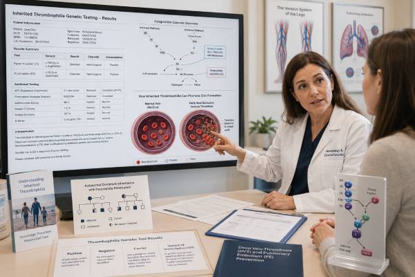
A thrombophilia genetic test looks for inherited changes that increase susceptibility to venous blood clots, most commonly factor V Leiden in F5 and prothrombin G20210A in F2. These variants can raise the chance of deep vein thrombosis or pulmonary embolism, but they do not mean that a clot is inevitable, and they usually do not explain heart attacks or most strokes. Testing is most useful when the result is likely to change a defined decision—such as whether anticoagulation is stopped in a selected clinical scenario, how estrogen exposure is approached, or whether relatives should be assessed for a high-risk familial disorder. Routine testing after every clot often adds cost and anxiety without changing treatment. A complete thrombophilia evaluation may also involve functional assays for antithrombin, protein C, and protein S and antibody tests for antiphospholipid syndrome; these are not interchangeable with DNA testing and can be distorted by acute thrombosis, pregnancy, liver disease, or anticoagulant medication.
- Factor V Leiden and prothrombin G20210A mainly increase venous, not arterial, thrombosis risk.
- Most heterozygous carriers never develop a clot.
- Testing should begin with the clinical decision it is intended to change.
- Protein C, protein S, antithrombin, and antiphospholipid testing require attention to timing and medication interference.
- A positive result alone usually does not justify lifelong anticoagulation.
- MTHFR polymorphisms are not part of an evidence-based inherited thrombophilia panel.
Table of Contents
- What a thrombophilia workup contains
- Factor V Leiden and APC resistance
- Prothrombin G20210A and combined risk
- When testing can change care
- Timing, functional tests, and interference
- Turning results into decisions
- Family testing and residual risk
What a thrombophilia workup contains
Thrombophilia means an increased tendency toward thrombosis. In clinical hematology, the term usually concerns venous thromboembolism, or VTE: deep vein thrombosis in a limb and pulmonary embolism when clot travels to the lungs. Clots in cerebral, splanchnic, retinal, or other unusual veins may also prompt specialized evaluation. The term does not imply that all thrombosis is inherited or that every laboratory abnormality has the same strength of effect.
The two common DNA tests are specific-variant assays. Factor V Leiden is a particular F5 change, historically called G1691A and now commonly reported as c.1601G>A, p.Arg534Gln using current numbering. Prothrombin G20210A is a noncoding F2 change, c.*97G>A, that increases prothrombin expression. A laboratory may test only these positions because they account for the established common variants; sequencing the entire F5 or F2 gene is not normally needed for a typical thrombophilia screen.
Other hereditary thrombophilias involve antithrombin, protein C, or protein S deficiency. Initial evaluation usually relies on activity and antigen assays rather than broad sequencing. Genetic testing of SERPINC1, PROC, or PROS1 may be useful in selected families after a true deficiency is established and acquired causes are excluded. These deficiencies are less common than factor V Leiden but can confer greater thrombosis risk, so family context and accurate laboratory confirmation are important.
Antiphospholipid syndrome, or APS, is acquired rather than inherited. Testing includes lupus anticoagulant, anticardiolipin antibodies, and anti-beta-2 glycoprotein I antibodies, with persistence confirmed at the required interval when diagnostic criteria are being applied. APS can affect both veins and arteries and may alter anticoagulant selection. A negative F5/F2 genetic test says nothing about APS.
Additional disorders enter the differential in specific circumstances. Myeloproliferative neoplasms and JAK2 variants are considered in splanchnic or cerebral venous thrombosis, abnormal blood counts, or related clues. Paroxysmal nocturnal hemoglobinuria is assessed by flow cytometry, not a standard germline panel. Cancer, surgery, immobility, pregnancy, estrogen, obesity, infection, inflammatory disease, nephrotic syndrome, and central venous catheters are acquired or circumstantial risk factors that may matter more than a common inherited variant.
The popular label “full thrombophilia panel” can therefore be misleading. It combines tests with different biology, timing requirements, and clinical uses. An evidence-based workup names the question first, then selects only the tests capable of answering it. Adding MTHFR testing, broad coagulation-gene sequencing, or unvalidated polymorphism panels does not improve routine VTE care.
Factor V Leiden and APC resistance
Factor V participates in the coagulation cascade by helping generate thrombin. Activated protein C, with protein S as a cofactor, normally switches off activated factor V and limits clot propagation. Factor V Leiden changes a key cleavage site, making factor V relatively resistant to inactivation. The result is activated protein C resistance and a longer-lasting procoagulant signal.
The variant is most common in people with European ancestry and is uncommon in many East Asian, sub-Saharan African, and Indigenous populations. Prevalence is not a substitute for testing, but ancestry affects pretest probability and the chance that a rare thrombosis in a given population has another explanation. Laboratories should report the exact variant rather than a vague statement that the “factor V gene is positive.”
A person may be heterozygous, with one altered copy, or homozygous, with two. Heterozygosity produces a moderate relative increase in first-VTE risk; homozygosity generally carries substantially greater risk. Absolute risk still depends on age and exposures. Many heterozygotes never clot, while a carrier exposed to major surgery, prolonged immobility, estrogen, pregnancy, or several additional risk factors may cross a clinically important threshold.
Factor V Leiden is primarily a risk factor for venous thrombosis. Its relationship to myocardial infarction, common ischemic stroke, and pregnancy complications is weak, inconsistent, or not sufficient to support routine testing in most settings. Recurrent miscarriage or placental complications alone should not automatically trigger an inherited-thrombophilia panel. Obstetric history may justify evaluation for APS or another cause depending on the pattern.
An activated protein C resistance assay can detect the phenotype, but DNA testing identifies factor V Leiden directly and is not altered by anticoagulants or an acute clot. Functional resistance testing may be affected by other factors and can detect non-Leiden causes in some methods. The appropriate first test depends on the laboratory algorithm and clinical question.
A positive factor V Leiden result is not a measurement of current clot burden. It cannot diagnose a DVT or pulmonary embolism, determine whether chest pain is a PE, or show whether anticoagulation is working. Suspected acute VTE requires clinical assessment, D-dimer in appropriate low-risk settings, and diagnostic imaging—not a genetic report.
Prothrombin G20210A and combined risk
Prothrombin, or coagulation factor II, is converted to thrombin during clot formation. The F2 G20210A variant lies in the 3′ untranslated region rather than changing the amino-acid sequence. It is associated with increased prothrombin levels and a greater tendency to form venous clots. Like factor V Leiden, it is seen most often in people of European ancestry, with geographic variation.
Most carriers are heterozygous. Their relative risk of a first VTE is increased, but many remain asymptomatic throughout life. Homozygosity is rare and is believed to confer higher risk, although estimates are less precise because few people have been studied. The report should distinguish one copy from two and should not use “positive” as though all genotypes were equivalent.
A person can inherit both factor V Leiden and prothrombin G20210A. Combined heterozygosity generally creates more risk than either variant alone, particularly when acquired triggers are present. It still does not guarantee thrombosis. Conversely, a person with neither variant can develop VTE because these tests capture only two contributors within a much larger risk system.
The F2 variant is not the same as a prothrombin time test. Prothrombin time and INR assess parts of clotting function and are used to monitor vitamin K antagonist therapy; they do not reveal whether G20210A is present. A carrier can have a normal routine coagulation screen. Likewise, an elevated D-dimer reflects fibrin turnover and is not a genetic test.
The variant is mainly associated with venous events. Testing after isolated arterial thrombosis, coronary disease, or routine stroke is usually low yield unless the clinical setting raises a different, specific concern. Cerebral venous sinus thrombosis is a venous event and should not be confused with a typical arterial ischemic stroke.
The inherited label can overstate certainty. Factor V Leiden and prothrombin G20210A are susceptibility variants whose clinical expression depends on context. They are unlike highly penetrant single-gene disorders in which a pathogenic variant nearly defines a disease. Communicating this distinction prevents a healthy carrier from believing that the body is continuously forming dangerous clots.
When testing can change care
The best reason to order thrombophilia testing is that a possible result would alter a decision. Before drawing blood, the clinician should specify the branch point: Would a positive result change planned anticoagulation duration? Would it affect whether a person uses estrogen-containing contraception? Would it guide prophylaxis during pregnancy or postpartum? Is there a known high-risk deficiency in the family for which targeted testing could change prevention?
Testing is often unnecessary after VTE provoked by major surgery because the trigger is clear and treatment duration is generally based on the event, not a common inherited variant. It is also often unhelpful after unprovoked VTE when the patient will receive extended or indefinite anticoagulation regardless of the result. In both cases, a panel may produce information without changing the endpoint.
Guidelines identify selected scenarios in which testing can be considered, but recommendations are conditional and depend on the management strategy used in that health system. Examples may include VTE associated with pregnancy, postpartum, combined oral contraceptives, or a nonsurgical transient factor when clinicians would otherwise stop anticoagulation and a thrombophilia result could change that plan. Unusual-site thrombosis requires specialist judgment because evidence varies by site and acquired disorders may be more important.
A strong family history changes the question. If a first-degree relative has documented antithrombin, protein C, or protein S deficiency, selective testing may identify relatives who need advice around surgery, pregnancy, or estrogen. Routine screening of all relatives for factor V Leiden or F2 G20210A has a smaller management impact and is not universally recommended. Knowing the exact familial finding is more useful than ordering a generic panel.
Testing in asymptomatic people before estrogen exposure is not automatically indicated. Personal VTE history, first-degree family history, type of familial thrombophilia, age, smoking, obesity, and available non-estrogen options all matter. In many situations, a person with a strong clotting family history may reasonably avoid estrogen even if genetic testing is negative, because the family’s causal factor may be unknown.
Children with typical provoked venous catheters rarely benefit from broad inherited testing. Neonatal thrombosis, purpura fulminans, recurrent unprovoked VTE, or a known severe familial deficiency requires pediatric hematology expertise. Adult prevalence and risk estimates should not simply be applied to infants and children.
Testing should not be ordered to reduce anxiety without a plan for both possible outcomes. A negative panel can create false reassurance, while a common heterozygous variant can create disproportionate fear. Pretest counseling is valuable because the choice not to test may be the most evidence-based option.
Timing, functional tests, and interference
F5 and F2 genotypes do not change during life and are not altered by acute thrombosis, pregnancy, or anticoagulant therapy. Their DNA assays can therefore be performed at any time from a technical perspective. Clinical timing still matters: testing during an acute admission may generate results before the long-term decision is clear, and informed counseling can be difficult during a crisis.
Protein C, protein S, and antithrombin assays are different. Acute thrombosis, severe illness, disseminated intravascular coagulation, liver disease, nephrotic syndrome, pregnancy, estrogen, and anticoagulants can lower or otherwise distort results. Warfarin reduces protein C and protein S. Heparin and direct oral anticoagulants can interfere with selected antithrombin or clot-based assays, depending on the platform. Laboratories may use drug-removal methods or alternative assays, but a specialist must interpret them.
An isolated low activity level does not automatically prove hereditary deficiency. Confirmation usually requires repeat testing under appropriate conditions, comparison of activity and antigen, review of acquired causes, and sometimes testing relatives or sequencing the relevant gene. Mildly low protein S is particularly vulnerable to overdiagnosis. Reference ranges vary with age, sex, pregnancy, and laboratory method.
Lupus anticoagulant testing is also affected by anticoagulants and must be planned carefully. Anticardiolipin and anti-beta-2 glycoprotein I immunoassays have different interference patterns. A diagnosis of APS requires the appropriate clinical event and persistent qualifying antibodies, not one weakly positive result during infection. Stopping anticoagulation solely to obtain testing may be unsafe and should never be done without the treating clinician.
Sample and identity errors matter because these results can affect family members. Clinical laboratories use validated methods and confirm unusual findings according to their protocols. Direct-to-consumer raw data or wellness panels should not be used to diagnose thrombophilia; any reported F5 or F2 finding requires confirmation in an accredited laboratory.
The laboratory request should state current medication, recent thrombosis, pregnancy status, liver or kidney disease, and transfusion history. A technically accurate value without this context can still be clinically misleading. When timing is unsuitable, the best action may be to defer the functional component while treating the clot normally.
Turning results into decisions
A heterozygous factor V Leiden or prothrombin G20210A result usually modifies risk rather than dictating treatment. For a person who has already had VTE, the decision to continue anticoagulation weighs whether the event was provoked, recurrence history, clot location, ongoing triggers, bleeding risk, age, preferences, and other illnesses. Common heterozygosity alone is generally not a sufficient reason for lifelong therapy.
Homozygosity, combined F5/F2 heterozygosity, or a confirmed natural-anticoagulant deficiency may increase concern, but management is still individualized. The strength of association with a first clot is not identical to the risk of recurrence after treatment stops. A result should be interpreted using evidence for the exact genotype rather than a generic “thrombophilia positive” label.
For an asymptomatic carrier, continuous anticoagulation is usually not used because bleeding harm can outweigh the absolute prevention benefit. Temporary prophylaxis may be appropriate during major surgery, hospitalization, prolonged immobilization, or another high-risk period. The plan should follow standard risk assessment; carrier status is one factor among many.
Estrogen-containing contraception and hormone therapy raise VTE risk. Factor V Leiden, prothrombin G20210A, a prior VTE, and a strong family history can shift the balance toward progestin-only or nonhormonal alternatives. The safest choice depends on the person’s total risk and therapeutic goals. A negative two-variant test does not erase risk from obesity, smoking, age, family history, or estrogen itself.
Pregnancy and the six-week postpartum period are prothrombotic. Prophylaxis decisions depend on personal VTE history, genotype, family history, other risk factors, and the specific guideline being followed. A healthy heterozygous carrier with no personal VTE does not automatically require injections throughout pregnancy. A prior estrogen-related or pregnancy-related VTE may lead to a different plan. Hematology and maternal-fetal medicine should coordinate antepartum and postpartum strategy.
Thrombophilia results rarely change the initial emergency treatment of DVT or pulmonary embolism. Anticoagulant choice may be affected by APS, kidney function, cancer, pregnancy, body weight, drug interactions, and bleeding risk more than by factor V Leiden or F2 G20210A. Severe chest pain, breathlessness, coughing blood, fainting, or a swollen painful limb requires urgent medical evaluation regardless of genotype.
Family testing and residual risk
Factor V Leiden and prothrombin G20210A are inherited in an autosomal dominant susceptibility pattern. A heterozygous carrier has a 50% chance of passing the variant to each child. Inheriting the variant means increased susceptibility, not certain disease. A homozygous person generally passes one altered copy to every child; the child’s second copy depends on the other parent.
Family testing is most useful when it changes prevention. A relative facing pregnancy, estrogen exposure, or major surgery may benefit from knowing about a documented high-risk antithrombin, protein C, or protein S deficiency. Testing healthy relatives for a common low-to-moderate-risk variant is more debatable because standard VTE precautions may be recommended regardless. The family should discuss the exact finding and intended decision with a clinician rather than treating all thrombophilias as equivalent.
A negative result for factor V Leiden and prothrombin G20210A leaves substantial residual risk. It does not exclude antithrombin, protein C, or protein S deficiency, APS, an unrecognized familial factor, or acquired triggers. It also does not guarantee that estrogen, surgery, or prolonged immobility is safe. Prevention follows the complete personal and family history.
A broad sequencing result may contain a variant of uncertain significance. A VUS should not be used to prescribe anticoagulation, prohibit pregnancy, or label relatives. Coagulation genes contain many rare benign changes, and functional assays plus segregation may be needed before a molecular finding can be considered causal.
Reproductive testing for common susceptibility variants is not generally approached like testing for a severe childhood genetic disorder. Most carriers remain healthy, and risk can often be reduced through awareness of temporary triggers. Genetic counseling may still be helpful for rare severe deficiencies, compound states, or families with neonatal purpura fulminans.
Practical prevention is more valuable than carrying a “thrombophilia card” without context. Family members should know the signs of DVT and pulmonary embolism, remain mobile during long travel when possible, discuss prophylaxis before major procedures, and tell clinicians about personal VTE and confirmed familial findings. The goal of testing is not to predict every clot; it is to make a limited number of decisions more accurate.
References
- American Society of Hematology 2023 guidelines for management of venous thromboembolism: thrombophilia testing. Evidence-based clinical guideline, 2023.
- Thrombophilia testing: A British Society for Haematology guideline. Clinical guideline, 2022.
- Factor V Leiden Thrombophilia. GeneReviews clinical reference, updated 2024.
- Prothrombin Thrombophilia. GeneReviews clinical reference, updated 2021.
- The dos, don’ts, and nuances of thrombophilia testing. American Society of Hematology educational review, 2023.
- Factor V Leiden and Prothrombin Gene Mutation. Thrombosis Canada clinical guide, 2025.
Disclaimer
This article is for general education and does not determine whether an individual needs thrombophilia testing, anticoagulation, hormone changes, or pregnancy prophylaxis. Results must be interpreted by qualified clinicians using the clot history, medication exposure, laboratory timing, bleeding risk, and family context. Seek urgent care for sudden breathlessness, chest pain, coughing blood, fainting, or a new swollen painful limb.