
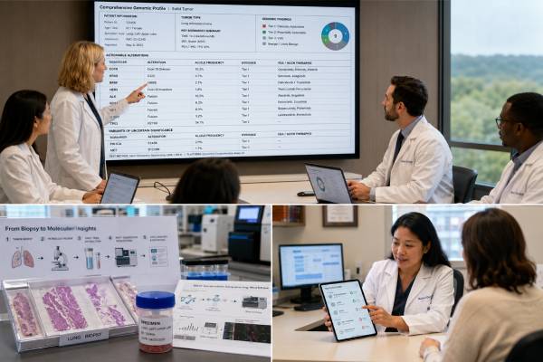
Tumor molecular profiling is an umbrella term for laboratory tests that characterize the biological changes driving a cancer. A profile may combine DNA sequencing, RNA analysis, protein staining, gene-fusion testing, copy-number assessment, and genomic biomarkers such as microsatellite instability or tumor mutational burden. The results can help confirm the tumor type, identify an approved treatment target, reveal resistance, support immunotherapy decisions, or match a patient to a clinical trial.
Profiling is not one standardized test. A focused assay may examine a few established biomarkers, while comprehensive genomic profiling can assess hundreds of genes and several alteration classes. The best approach depends on the cancer, stage, available tissue, treatment urgency, and clinical question. A result is also time- and specimen-specific: one biopsy may not represent every metastasis, and tumors can evolve after therapy. “No actionable alteration” does not mean the tumor lacks molecular changes, and a listed drug is not automatically appropriate. The report must be interpreted with pathology, current guidelines, treatment history, assay limitations, and the patient’s goals.
- Molecular profiling may include DNA, RNA, protein, copy-number, and genomic-signature tests rather than sequencing alone.
- Testing is most useful when a result can change diagnosis, standard therapy, clinical-trial eligibility, or resistance management.
- Evidence for a target depends on the exact alteration, cancer type, disease setting, and available drug.
- Tissue and plasma profiling are complementary, but a negative blood test can miss low-shedding disease.
- A tumor result can suggest inherited cancer risk but usually requires confirmation in a separate normal sample.
Table of Contents
- What Tumor Molecular Profiling Includes
- When Profiling May Help
- Choosing the Specimen and Test
- Common Biomarker Result Types
- Interpreting Actionability and Evidence
- Treatment Targets and Molecular Tumor Boards
- Repeat Profiling and Resistance
- Limitations, Inherited Risk, and Next Steps
What Tumor Molecular Profiling Includes
A tumor profile describes molecular features of cancer cells. The term can refer to one assay or an integrated set of tests. It is broader than next-generation sequencing and may include methods selected by a pathologist for the suspected tumor type.
Common components include:
- Immunohistochemistry, which uses antibodies to detect proteins in tissue. Examples include hormone receptors, HER2, mismatch-repair proteins, PD-L1, and lineage markers.
- In situ hybridization, including fluorescence in situ hybridization, which can detect gene amplification, deletion, or rearrangement in individual cells.
- PCR or digital PCR, which can sensitively detect a known mutation, fusion, or viral sequence.
- DNA next-generation sequencing, which assesses many genes for substitutions, insertions, deletions, copy-number changes, and some rearrangements.
- RNA sequencing, which can identify expressed gene fusions, exon-skipping events, and occasionally expression signatures.
- Methylation profiling, used in selected central nervous system tumors and other diagnostically challenging cancers.
- Genomic signatures, such as microsatellite instability, tumor mutational burden, homologous recombination deficiency, or mutational patterns linked to a repair defect.
These techniques answer different questions. A protein stain may show whether a target is actually expressed. DNA sequencing can identify the underlying alteration. RNA can confirm that a fusion is transcribed. FISH can visualize an amplification or rearrangement. No single method is best for every biomarker.
A focused profile may be appropriate when only a few validated markers affect treatment. For example, an early-stage tumor may require receptor testing and one predictive assay rather than a broad panel. Comprehensive profiling becomes more attractive when many biomarkers are relevant, tissue is scarce, the cancer is rare, or standard options are limited.
The profile can serve four major purposes:
- Diagnostic: defining what the tumor is.
- Predictive: estimating whether a treatment is more or less likely to work.
- Prognostic: describing likely behavior independent of a specific treatment.
- Hereditary signal: identifying a tumor finding that may warrant germline evaluation.
These categories can overlap. Loss of a mismatch-repair protein can help classify a tumor, predict immunotherapy sensitivity, and raise concern for Lynch syndrome. A fusion can define a sarcoma and identify a kinase inhibitor. A molecular alteration can also be diagnostically useful without being targetable.
Tumor profiling generally examines acquired changes. It does not replace a hereditary cancer evaluation. The same gene can carry a somatic driver in one patient and a germline predisposition variant in another. Specimen source and confirmatory testing determine the distinction.
When Profiling May Help
Molecular profiling is most valuable when the result has a realistic path to action. That action may be choosing a standard drug, avoiding an ineffective drug, confirming diagnosis, enrolling in a trial, changing surgery, or recommending germline testing.
It is commonly considered for:
- Advanced or metastatic cancers with multiple established genomic targets.
- Rare tumors for which histology alone is insufficient or matched trials are available.
- Cancer of unknown primary, when molecular and immunophenotypic data may refine classification.
- Tumors with ambiguous morphology or an unexpected clinical course.
- Progression after targeted therapy, when resistance mechanisms could guide the next treatment.
- Selected early-stage cancers in which biomarkers predict adjuvant benefit or recurrence risk.
- Patients who may qualify for a tumor-agnostic therapy based on a fusion, mutation, MSI-high status, or another validated marker.
- Cases in which a tumor feature suggests hereditary cancer risk.
Guidelines increasingly support multigene testing in advanced cancers for which approved targets or clinically meaningful trial options exist. The expected yield varies widely. Lung adenocarcinoma may have several standard driver targets, while some other cancers rarely produce an immediately treatable finding.
The timing should allow the result to influence care. Profiling late in a rapidly declining illness may not benefit the patient because tissue retrieval, sequencing, interpretation, authorization, and drug access take time. Testing earlier in the advanced-disease course can preserve options, but it should not delay urgent proven treatment.
A patient’s treatment goals matter. Someone seeking every reasonable trial option may value broad profiling despite a low chance of an approved match. Another person may prefer to avoid an invasive biopsy when the result is unlikely to alter care. The decision should include expected benefit, risk, cost, and turnaround time.
Not all molecular questions require comprehensive testing. A rapid single-marker assay may be better when one result is urgently needed. Some cancers have validated protein or cytogenetic tests that are more informative than broad sequencing. Profiling should complement, not replace, expert pathology.
The treating team should define whether testing is intended for present care or future options. A profile obtained during curative treatment may reveal findings that matter only if disease recurs. The report should be retained, but its interpretation must be updated later because drug approvals and evidence change.
Patients should also understand that profiling frequently finds variants with uncertain or no established clinical significance. A larger panel increases information, but not necessarily useful information. More genes can lead to more ambiguous findings, incidental hereditary concerns, and out-of-pocket costs.
Choosing the Specimen and Test
The ideal specimen contains enough viable, representative tumor and has been handled in a way that preserves DNA, RNA, and proteins. Formalin-fixed, paraffin-embedded tissue from a recent biopsy or surgery is most common. Cytology specimens and body fluids can also work when the laboratory has validated them.
A pathologist reviews the slides, confirms the diagnosis, estimates tumor percentage, and selects the area for testing. Necrosis, inflammation, fibrosis, and normal cells dilute the tumor signal. Macrodissection can enrich the specimen. Bone specimens exposed to strong acid decalcification may have severely degraded nucleic acid.
When several samples are available, selection considers:
- Recency and whether the specimen was collected before or after relevant therapy.
- Tumor content and tissue quality.
- Primary versus metastatic site.
- Whether the lesion represents current progression.
- Need to preserve tissue for diagnostic stains or future testing.
- Procedure risk if a new biopsy is being considered.
Archival tissue may still contain the original driver, but a new biopsy can better reflect acquired resistance. A metastatic specimen may differ from the primary tumor. No single site captures all heterogeneity.
The test should match the expected biomarkers. A DNA-only panel may be excellent for hotspot mutations but less sensitive for certain fusions. Adding RNA can improve detection of expressed rearrangements and exon-skipping events. A panel that reports tumor mutational burden may need a sufficiently large validated genomic footprint and appropriate germline filtering.
For some cancers, immunohistochemistry or FISH remains essential. HER2 assessment, PD-L1 scoring, mismatch-repair protein evaluation, and specific rearrangements may have therapy-linked methods and cutoffs that cannot be assumed from a general sequencing result. A comprehensive profile can still require companion testing.
Plasma circulating tumor DNA is an alternative when tissue is unsafe, insufficient, or too slow to obtain. It can sample DNA from multiple tumor sites and is useful for resistance testing. However, sensitivity depends on tumor shedding. Low-volume disease, brain-only metastases, and some tumor types may produce a false-negative plasma result.
A combined strategy can be appropriate: tissue for histology, fusions, and broad profiling; plasma for speed or additional heterogeneity. Discordant results should be reviewed rather than averaged. The collection dates, treatment interval, and assay coverage often explain the difference.
Before ordering, verify the laboratory’s accreditation, specimen requirements, gene list, alteration types, limit of detection, reporting policy, and turnaround time. A test name such as “comprehensive” does not guarantee whole-genome coverage or every clinically relevant biomarker.
Common Biomarker Result Types
A molecular report may contain individual alterations and composite biomarkers. Each type requires a different interpretive framework.
Activating mutations can turn on growth pathways. Some are established treatment targets; others are biologically important but not druggable. The exact amino-acid change matters because variants in the same gene can have opposite effects.
Loss-of-function mutations and deletions can disable tumor suppressors or DNA-repair genes. A single altered copy may not prove complete pathway loss. The report may need allele-specific copy-number analysis, loss of heterozygosity, protein staining, or a second event.
Amplifications increase gene copy number and can lead to overexpression. Tumor purity and ploidy affect the estimate. A low-level gain is not equivalent to high-level focal amplification, and some treatments require a validated protein or in situ hybridization result.
Gene fusions and rearrangements join genomic regions. An oncogenic fusion usually retains specific functional domains and is expressed in-frame. A rearrangement of unknown partner or uncertain transcript may need RNA confirmation. Some fusions define the tumor diagnosis even when no therapy is available.
Microsatellite instability and mismatch-repair deficiency indicate impaired repair of repetitive DNA. Results can come from sequencing, PCR, or protein immunohistochemistry. Discordance may require review for sample quality, tumor type, treatment effects, or rare biological mechanisms.
Tumor mutational burden estimates somatic mutations per megabase. It is assay dependent and should be interpreted with the validated threshold, tumor type, treatment indication, and other immune biomarkers. High TMB increases the probability of benefit in some settings but is not a guarantee.
PD-L1 expression is measured by immunohistochemistry, not inferred reliably from a mutation panel. Different drugs and tumor types use different assays and scoring systems, such as tumor proportion score or combined positive score.
Homologous recombination deficiency may be inferred from pathogenic repair-gene variants, genomic scars, or functional methods. The predictive value and approved test differ by cancer. A VUS in a repair gene should not be treated as HRD.
Expression or recurrence signatures estimate biological behavior from patterns of multiple genes. They are usually validated for a specific cancer, stage, and treatment question. They are not interchangeable with broad mutation profiling.
Methylation classes can resolve difficult central nervous system and sarcoma diagnoses. A classifier result needs a confidence score and correlation with morphology and copy-number data.
The report may also show variant allele fraction, read depth, copy number, fusion reads, and quality metrics. These are technical clues, not direct measures of treatment response. Low allele fraction can reflect a subclone, low purity, copy-number context, or sequencing limitations.
Interpreting Actionability and Evidence
“Actionable” is not a uniform category. It can mean that a result changes standard treatment, supports a trial, clarifies diagnosis, predicts resistance, or prompts germline testing. The report should state which meaning applies.
Evidence is strongest when a regulatory approval or high-level guideline links the exact biomarker to a drug in the same cancer, stage, and line of therapy. Evidence becomes progressively less certain when it comes from a different tumor type, a small non-randomized study, a case report, or preclinical models.
| Question | Why it matters |
|---|---|
| Is the exact alteration oncogenic? | Not every variant in a cancer gene activates or disables the pathway. |
| Is evidence from the same cancer type? | Biology and drug response can differ by tissue context. |
| Is the drug approved in this setting? | Stage, prior therapy, and companion-test requirements can restrict use. |
| Are resistance alterations present? | A co-alteration may reduce expected benefit. |
| Was the result analytically reliable? | Low purity or weak fusion evidence may require confirmation. |
| Is treatment accessible? | Insurance, geography, performance status, and trial slots affect feasibility. |
Reports often use evidence tiers, but systems differ among laboratories. A high tier in one scheme may not map exactly to another. The date is important because an emerging target can become standard care, and a once-promising strategy can later prove ineffective.
A VUS is not an actionable mutation. It should not be used alone for off-label therapy, preventive surgery, or family testing. Computational predictions and laboratory functional data can contribute to classification, but clinical action requires adequate evidence.
Tumor-agnostic approvals apply across multiple cancers for a defined biomarker, yet response can still vary by histology. The term does not mean every tumor with the marker has the same sensitivity. Likewise, a drug approved for a gene in one cancer is not automatically effective in all cancers with alterations in that gene.
The report may list therapies associated with resistance. A resistance prediction can be as useful as a sensitivity prediction because it helps avoid ineffective treatment. It must still be specific to the alteration, drug, and disease context.
When evidence is ambiguous, the best interpretation may be “clinical trial preferred” rather than off-label treatment. A trial provides structured eligibility, dosing, safety monitoring, and prospective learning.
Treatment Targets and Molecular Tumor Boards
A treatment target is a molecular dependency that can be inhibited or exploited. Examples include an activated kinase, a defective DNA-repair pathway, hormone-receptor expression, or an immune-evasion marker. Matching is more than finding the same gene name on a drug label.
The oncology team compares the molecular option with established treatments. A matched drug may be appropriate immediately, after first-line therapy, or only after other options fail. Expected response, duration, toxicity, route, interactions, and patient preference all matter.
Co-alterations can modify response. A tumor may have the target but also activate a bypass pathway. The alteration may be present in only part of the tumor. A later resistance mutation may prevent drug binding. These factors explain why a compelling molecular match does not guarantee benefit.
A molecular tumor board can help with rare variants, conflicting assays, unusual cancers, and trial selection. Effective boards typically include oncology, pathology, molecular genetics, bioinformatics, pharmacy, genetic counseling, and relevant disease specialists. They should document the evidence level, recommendation, alternatives, and follow-up.
The board may recommend:
- A standard biomarker-matched treatment.
- Confirmatory testing with another method.
- A clinical trial in the same tumor type.
- A basket trial enrolling by biomarker.
- A new biopsy to assess transformation or resistance.
- Germline testing for a possible hereditary finding.
- No molecularly matched therapy because evidence is insufficient.
“No recommendation” is a valid outcome. It protects patients from treatment based on weak biological analogy. Standard chemotherapy, surgery, radiation, endocrine therapy, immunotherapy, or supportive care may offer greater benefit than an experimental match.
Trial access requires practical review. A report’s trial list can become outdated. Eligibility may depend on prior treatments, organ function, measurable disease, geographic location, and central confirmation. Contacting a trial center is necessary before assuming a match.
Patients should ask what benefit is realistically expected and whether the recommendation changes the current plan. A molecular result can be scientifically interesting without being clinically useful today.
Repeat Profiling and Resistance
Cancer evolves under treatment. Sensitive clones shrink while resistant clones survive and expand. Repeat profiling can identify a new target-dependent mutation, amplification of a bypass pathway, histologic transformation, or loss of the original target.
Reprofiling is most useful when:
- A targeted therapy has stopped working and known resistance mechanisms have treatment implications.
- The cancer recurs after a long interval.
- The original test was limited or lacked RNA fusion analysis.
- New therapies require biomarkers not assessed previously.
- A new metastatic pattern raises concern for transformation or a second primary cancer.
- A trial requires a recent specimen.
A tissue biopsy can reveal both molecular and histologic change. This is important because a cancer can transform into a phenotype with different treatment needs. Plasma cannot show morphology but may detect heterogeneous resistance across several lesions.
A tumor genomic test performed at resistance should be selected for the suspected mechanism. Broad retesting is not always necessary. Sometimes a focused assay for one known resistance mutation is faster and sufficient.
Plasma profiling is useful when biopsy is unsafe or speed is essential. A positive result can be highly informative if the alteration is validated. A negative result may simply mean that too little tumor DNA was present. Tissue should be considered when plasma is negative and another therapy depends on finding the alteration.
Serial testing can produce many small changes that are not actionable. It also adds cost and can consume tissue. Repeating the same broad panel without a clear question is less useful than choosing the right assay at the right clinical transition.
Results from different times should be compared carefully. Variant disappearance in plasma may reflect response or low shedding rather than true eradication. A rising allele fraction can reflect increasing tumor burden, but quantitative interpretation is assay specific.
Limitations, Inherited Risk, and Next Steps
Tumor profiling has technical limits. Low tumor content, degraded DNA or RNA, formalin artifacts, decalcification, incomplete gene coverage, and bioinformatic thresholds can cause false-negative or uncertain results. Some assays do not detect promoter variants, large structural changes, epigenetic silencing, or low-level mosaicism.
It also has biological limits. A sample from one lesion may not represent the entire cancer. The profile can change after therapy. A target may not be essential to every clone. Protein activity and the tumor microenvironment are not fully captured by DNA sequencing.
A possible inherited finding must be handled separately. Tumor-only sequencing cannot reliably distinguish somatic from germline origin. Allele fraction is influenced by purity, copy number, and loss of heterozygosity. Confirmation should use blood, saliva, or another validated normal specimen in a germline laboratory, with genetics involvement.
A confirmed germline result may change cancer surveillance, surgical choices, relatives’ testing, and reproductive planning. A tumor-limited result generally does not carry the same family implications. Some plasma results can also come from clonal hematopoiesis rather than the tumor, particularly variants in common blood-clone genes.
Before making a decision, review:
- What specimen and date were tested?
- Which technologies and biomarkers were included?
- Did the sample meet tumor-content and quality requirements?
- Is the finding diagnostic, predictive, prognostic, resistant, hereditary, or uncertain?
- What is the evidence in this exact cancer and treatment setting?
- Is confirmation required by a companion diagnostic or another method?
- Would a current biopsy or plasma test add useful information?
- Should the case go to a molecular tumor board?
Keep the original report, pathology diagnosis, and any addenda. Laboratories may update classification, but treatment evidence should be checked independently at each decision point. A report generated today may have new relevance later, and a listed therapy may become outdated.
A profile is one component of care. Disease burden, symptoms, organ function, prior toxicity, performance status, personal values, and treatment access can outweigh a molecular option. The best plan integrates the biology with the whole patient.
References
- Recommendations for the use of next-generation sequencing (NGS) for patients with advanced cancer in 2024: a report from the ESMO Precision Medicine Working Group 2024 (Guideline)
- Somatic Genomic Testing in Patients With Metastatic or Advanced Cancer: ASCO Provisional Clinical Opinion 2022 (Guideline)
- Clinical practice recommendations for the use of next-generation sequencing in patients with solid cancer: a joint report from KSMO and KSP 2024 (Guideline)
- Molecular Tumor Boards: A Consensus Statement From the International Association for the Study of Lung Cancer 2025 (Position Statement)
- ESMO Precision Oncology Working Group recommendations on the structure and quality indicators for molecular tumour boards in clinical practice 2025 (Guideline)
- Molecular profiling of solid tumors by next-generation sequencing: an experience from a clinical laboratory 2023 (Study)
Disclaimer
This article is for general education and does not replace medical advice, pathology review, or individualized cancer treatment planning. The meaning of a molecular result depends on the exact alteration, assay, specimen, tumor type, disease setting, prior treatment, and current evidence. Treatment, biopsy, clinical-trial, and germline decisions should be made with qualified oncology, pathology, and genetics professionals.





