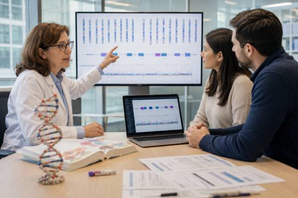
Whole-exome sequencing, usually abbreviated WES or ES, analyzes most protein-coding regions of thousands of genes at the same time. These regions are called exons and make up only a small fraction of the genome, but they contain many known variants that cause Mendelian disorders. WES is often used when a person has a complex or unexplained condition, several diagnoses are possible, or earlier single-gene and panel tests were negative. It can identify a molecular diagnosis, shorten a long diagnostic search, clarify recurrence risk, and sometimes change treatment or surveillance. It can also return uncertain findings, secondary findings unrelated to the original question, or no diagnosis. A negative exome does not exclude genetic disease because some exons are poorly covered and many important repeat, structural, mitochondrial, epigenetic, mosaic, and noncoding changes require other methods. The quality of interpretation depends heavily on detailed clinical information and whether relatives are tested with the patient.
- WES examines most coding exons across thousands of genes, not the entire genome.
- It is most useful for genetically heterogeneous or unexplained disorders.
- Trio testing with both biological parents can improve interpretation.
- Results may be diagnostic, uncertain, negative, or secondary to the testing indication.
- Exome sequencing does not reliably detect every variant type or every exon.
- Reanalysis can provide answers later as gene–disease knowledge improves.
Table of Contents
- What Whole-Exome Sequencing Reads
- Who May Benefit From WES
- How the Test and Analysis Are Performed
- Positive, Uncertain, and Negative Results
- Secondary Findings and Family Information
- Important Limitations of WES
- Reanalysis and Next Diagnostic Steps
What Whole-Exome Sequencing Reads
Genes contain exons that contribute to mature RNA and, for protein-coding genes, specify the amino acid sequence of a protein. The exome is the combined set of these coding regions. Although exons represent roughly one to two percent of human DNA, many currently recognized disease-causing variants lie within them or near exon–intron boundaries.
WES uses next-generation sequencing to read millions of DNA fragments in parallel. Before sequencing, laboratory methods enrich or “capture” exonic regions from the sample. Software aligns the reads to a reference genome and identifies differences. Analysts then filter and interpret variants using the person’s phenotype, inheritance model, population frequency, predicted effect, published cases, functional data, and curated databases.
The word “whole” is relative. Clinical WES does not sequence every exon perfectly and usually does not analyze all noncoding DNA. Capture efficiency varies by gene and region. GC-rich segments, homologous genes, pseudogenes, repetitive sequences, and some first exons may have low coverage. Laboratories set minimum quality thresholds and may fill selected gaps by another method, but practices differ.
WES commonly detects single-nucleotide variants and small insertions or deletions in adequately covered coding regions. Many laboratories also attempt exon-level copy-number analysis, selected mitochondrial variants, or other additions, but these capabilities are not uniform. The methods section of the report determines what was actually assessed.
Exome sequencing can be proband-only, analyzing the affected person; duo, analyzing the person and one relative; or trio, analyzing the person and both biological parents. Larger family analyses may include affected and unaffected siblings or other relatives. Family data can reveal whether a variant is de novo, recessive, X-linked, inherited from an affected parent, or inconsistent with the suspected mechanism.
WES differs from a multigene panel. A panel restricts interpretation to a curated set of genes associated with a particular phenotype and may offer excellent coverage or specialized methods. WES has a wider scope and is useful when the phenotype spans several systems or the responsible gene is uncertain. It can also be reanalyzed later without resequencing if the original data remain adequate.
WES also differs from whole-genome sequencing, which reads coding and noncoding regions more continuously. Genome sequencing may detect some structural and noncoding variants missed by exome sequencing, but neither test detects every clinically relevant change.
Who May Benefit From WES
WES is most likely to help when the medical problem could result from variants in many genes. Common indications include developmental delay, intellectual disability, multiple congenital anomalies, epilepsy, neuromuscular disease, movement disorders, immune dysfunction, kidney disease, hearing or vision loss, skeletal dysplasia, metabolic presentations, and multisystem rare disease.
A child with developmental differences, seizures, unusual growth, and congenital anomalies may fit hundreds of syndromes. Sequentially testing one gene at a time can consume years and tissue while repeatedly returning negative results. WES allows simultaneous consideration of many established and newly described genes.
Adults may also benefit. Some inherited neurologic, cardiac, renal, ophthalmic, connective-tissue, immune, and metabolic disorders present later or were never diagnosed in childhood. Exome testing can be informative when a phenotype is atypical, family history is limited, or the original clinical label is uncertain.
WES may be considered early rather than only after an exhaustive diagnostic odyssey. Evidence-based guidance supports exome or genome sequencing as a first- or second-tier test for many children with congenital anomalies, developmental delay, or intellectual disability. Earlier testing can reduce repeated imaging, invasive procedures, and low-yield sequential assays, although the optimal strategy depends on phenotype and healthcare system.
The test is less suitable when one specific mechanism is strongly suspected and a dedicated assay is better. Examples include:
- A known familial variant suited to targeted testing
- A repeat-expansion disorder requiring repeat sizing
- A methylation or imprinting disorder
- A large chromosomal rearrangement best assessed by chromosome or microarray testing
- A biochemical disorder with a fast, established single-gene or enzyme test
- A condition requiring deep mosaic detection in a particular tissue
WES may follow negative panel testing, but repeating essentially the same genes with similar technology may add little unless exome analysis expands the gene list, evaluates additional variant types, or uses improved interpretation. The prior reports should be reviewed to identify what remains untested.
Clinical information strongly influences yield. Laboratories need specific features, ages of onset, normal and abnormal findings, imaging, pathology, biochemical results, and Human Phenotype Ontology terms where possible. “Developmental delay” alone is less informative than a structured description of motor, language, growth, neurologic, behavioral, and congenital findings.
Testing an affected relative can be more informative than testing only an unaffected family member. In families with several affected people, choosing the relatives most likely to share the genetic cause can help separate disease-related variants from background variation.
WES is not a general health prediction tool. Its principal clinical strength is diagnosing suspected Mendelian disease, not estimating the combined influence of common variants, lifestyle, and environment on ordinary chronic illnesses.
How the Test and Analysis Are Performed
The process begins with pretest counseling and consent. The clinician explains the diagnostic question, possible results, technical limits, family implications, privacy considerations, and options for receiving secondary findings. Parents or guardians consent for children, while older children and adolescents should be involved at a level appropriate to their understanding.
Samples are usually blood or saliva. Blood often provides consistent DNA quality, but another tissue may be needed after stem-cell transplantation or when mosaicism is suspected. Parental samples should be collected at the same time when trio analysis is planned.
The laboratory extracts DNA, fragments it, enriches exonic regions, and sequences them. Quality metrics include average depth, the proportion of target bases above a coverage threshold, contamination checks, and sample relationships. Bioinformatic pipelines call variants and annotate their predicted effect.
Analysis is not a simple search for any rare change. Each person has thousands of coding variants. Filtering may consider:
- Whether the gene is associated with the phenotype
- Dominant, recessive, X-linked, mitochondrial, or de novo inheritance
- Population frequency
- Predicted loss of function, missense effect, or splicing impact
- Segregation with disease in relatives
- Previous clinical reports and expert classifications
- Functional evidence
- Phenotype overlap and age of onset
Trio analysis is powerful because de novo variants can be identified directly and recessive variants can be phased to determine whether they came from different parents. It can also show that a suspicious variant was inherited from a healthy parent, although reduced penetrance and mild parental features must be considered.
Some laboratories use a phenotype-driven exome, reporting only genes plausibly related to the indication. Others analyze a clinical exome or broader set of established disease genes. A full exome dataset may exist even when reporting is restricted. The report should clarify whether analysis was limited and whether broader reanalysis is possible.
Turnaround varies from several weeks to several months. Rapid exome sequencing may be available for critically ill infants or children when a diagnosis could immediately alter intensive care. Rapid analysis prioritizes speed but still requires confirmation, interpretation, and clinical integration.
Candidate variants are reviewed by trained laboratory professionals and classified. Findings likely to explain the condition are typically confirmed when required by laboratory policy and reported with evidence, inheritance, and recommendations. The clinician then compares the molecular result with the patient’s presentation.
WES can identify a molecular diagnosis that changes the name of the condition without changing immediate treatment. That result may still end unnecessary testing, guide anticipatory care, connect the family with specialists, define recurrence risk, or qualify the person for research and trials.
Positive, Uncertain, and Negative Results
A WES report may contain several result categories, and each must be read against the testing indication.
Positive or diagnostic result. A pathogenic or likely pathogenic variant—or an appropriate pair of variants—is identified in a gene that convincingly explains the phenotype. For a dominant disorder, one heterozygous variant may be sufficient. Recessive disease generally requires two pathogenic variants on opposite gene copies. X-linked and mitochondrial mechanisms have different requirements.
A molecular diagnosis can affect treatment, surveillance, anesthesia precautions, medication avoidance, nutritional management, reproductive risk, and testing of relatives. It may also reveal that some symptoms have a separate cause. Not every positive result explains every feature.
Probable or partial diagnosis. The laboratory may find a strong candidate that explains much but not all of the presentation, or a pathogenic variant in a gene with incomplete phenotype overlap. Further clinical evaluation, parental testing, RNA studies, or another assay may be recommended. Some reports use terms such as “likely diagnostic,” while others remain within formal classification categories.
Variant of uncertain significance. A VUS is a change whose role cannot be established. It is not a confirmed diagnosis and should not by itself drive major treatment or reproductive decisions. Family studies can help when they are designed to test a specific hypothesis. Broad exome testing often produces more VUS findings than narrow testing, particularly for people from populations underrepresented in reference databases.
Carrier finding. A person may have one pathogenic variant for an autosomal recessive condition that does not explain their symptoms. Whether carrier findings are routinely reported depends on consent and laboratory policy. Carrier status can be relevant to reproduction but is not generally the diagnosis in a recessive condition.
Negative or nondiagnostic result. No reportable variant adequately explains the condition. This does not exclude a genetic cause. The causal variant may be outside covered regions, a structural or repeat change, mosaic, mitochondrial, epigenetic, or in a gene not yet linked to disease. The phenotype may also have a nongenetic or multifactorial cause.
Unexpected dual diagnosis. WES can show that two different genetic conditions together explain a complex presentation. This is especially important when one diagnosis leaves major features unresolved. A blended phenotype may resemble a single new syndrome until both causes are recognized.
Inconclusive technical result. Poor sample quality, contamination, low coverage, or inability to establish family relationships can limit interpretation. A technical failure should not be described as a negative genetic evaluation.
The report should list exact variant notation, zygosity, classification, inheritance, evidence, relevant transcript, and limitations. A result summary without the full report is inadequate for family testing or future reinterpretation.
Secondary Findings and Family Information
Because WES surveys thousands of genes, it can identify medically important variants unrelated to the original diagnostic question. These are called secondary findings when the laboratory intentionally analyzes a defined set of genes for actionable conditions.
Professional guidance recommends reporting pathogenic or likely pathogenic variants in selected gene–disease pairs where early surveillance or prevention may reduce serious harm. The list is updated as evidence changes. It includes inherited cancer and cardiovascular conditions and other actionable disorders. A secondary finding does not mean the person currently has the disease; it indicates a risk that requires confirmation and condition-specific evaluation.
Consent policies vary by country, laboratory, age, and healthcare system. Some programs allow patients to opt out of secondary findings; others have different rules for children. The discussion should occur before testing so that families understand the scope. A broad request to “tell me everything” is not realistic because most exome variation lacks established health meaning.
Secondary findings differ from incidental findings, which are discovered unexpectedly during analysis rather than deliberately sought. Laboratories may also uncover carrier status, pharmacogenetic variants, or other categories depending on policy.
Family relationships are part of exome analysis. Trio data can reveal that a stated parent is not biologically related, that parents are closely related, or that samples were switched. Laboratories should have a policy for handling unexpected relationship information. Consent should explain that inheritance analysis can expose such findings even when paternity is not the purpose of testing.
A diagnosis in one person can provide information about relatives. A de novo dominant variant may imply a low but nonzero recurrence risk for siblings because of possible parental germline mosaicism. An inherited dominant finding may indicate that a parent and siblings need evaluation. Recessive results may identify both parents as carriers and affect future pregnancies.
Relatives should usually receive targeted testing for the established familial variant rather than full WES. Targeted testing gives a direct answer, costs less, and avoids unrelated uncertain findings. The exact report must be shared so the laboratory tests the correct variant.
Secondary and diagnostic findings can have emotional, insurance, employment, and reproductive implications. Legal protections vary and may not cover life, disability, or long-term-care insurance. Jurisdiction-specific advice may be appropriate before testing.
Important Limitations of WES
WES is broad, but its blind spots are clinically important.
Noncoding variants. Most deep intronic, promoter, enhancer, and intergenic regions are not captured or routinely interpreted. These variants can alter splicing or gene regulation and cause disease.
Uneven exon coverage. Some exons are poorly captured because of high GC content, repeats, homologous sequences, or technical design. A pathogenic variant can be missed in an uncovered region. Coverage summaries should be reviewed for genes of particular concern.
Structural variants. Exon-level deletions and duplications may be inferred from read depth, but sensitivity varies. Balanced translocations, inversions, complex rearrangements, mobile-element insertions, and some copy-number changes are better detected by other methods.
Repeat expansions. Large trinucleotide and other repeat expansions often require dedicated assays. Standard WES is not a reliable replacement for testing conditions such as Huntington disease, fragile X syndrome, myotonic dystrophy, or many hereditary ataxias.
Methylation and imprinting disorders. WES reads DNA sequence, not the methylation pattern required to diagnose many imprinting conditions. It may also miss uniparental disomy unless the analysis is specifically designed and parental data are available.
Mosaicism. A variant present in a small fraction of cells can fall below the assay’s detection threshold. Blood may not contain the variant even when affected skin, brain, or another tissue does.
Mitochondrial DNA. Some laboratories include mitochondrial analysis, but coverage, heteroplasmy thresholds, deletion detection, and tissue relevance differ. A negative blood exome does not exclude a mitochondrial disorder.
Pseudogenes and homologous regions. Genes with highly similar copies can be difficult to map accurately. Specialized long-range PCR or other methods may be required.
Variant interpretation. Even a technically detected change may remain uncertain. Databases contain ancestry gaps, published evidence can conflict, and gene–disease associations continue to evolve. An exome can be negative because science cannot yet interpret the causal variant.
Phenotype mismatch or multiple causes. Incomplete clinical information can lead analysts to filter out the right gene. Conversely, a plausible variant may be overemphasized when the phenotype does not fit. Environmental, infectious, immune, vascular, and multifactorial conditions can mimic genetic disease.
These limitations explain why WES should be selected as part of a diagnostic strategy. Chromosomal microarray, repeat testing, biochemical assays, RNA sequencing, methylation studies, genome sequencing, and tissue-specific analysis remain important tools.
Reanalysis and Next Diagnostic Steps
A nondiagnostic exome is not necessarily the end of the evaluation. Reanalysis can identify diagnoses later because new disease genes are discovered, variant databases expand, classifications change, and the patient’s phenotype becomes more specific.
Reanalysis usually reuses the original sequence data and applies an updated pipeline, gene list, phenotype, and evidence base. Studies in previously negative cohorts show that this can produce additional diagnoses, although yield varies by population, time interval, and original analysis quality.
There is no single ideal reanalysis schedule. Many centers consider review after one to three years, sooner when a new clinical feature appears, another relative is diagnosed, or a relevant gene discovery occurs. Laboratory policies differ: some perform periodic review automatically, while others require a clinician’s request or a new order.
Before requesting reanalysis, update the phenotype. Include new symptoms, normal findings that exclude alternatives, imaging, pathology, biochemical results, and family changes. Add newly available relatives when their samples could clarify inheritance.
The next step after negative WES should address the remaining hypothesis. Options include:
- Genome sequencing for noncoding and structural variants
- Dedicated repeat-expansion analysis
- Chromosomal microarray or karyotype
- Methylation or imprinting testing
- Mitochondrial genome analysis in an appropriate tissue
- RNA sequencing to evaluate expression or splicing
- Long-read sequencing for complex or repetitive regions
- Biochemical, enzyme, metabolomic, or proteomic testing
- Testing another affected relative
- Evaluation for nongenetic causes
A genetics professional can review whether the original data were true WES, a restricted clinical exome, or a virtual panel. If analysis was narrow, expanding the interpretation may be more useful than generating new sequence.
For a positive result, next steps include confirming the clinical diagnosis, arranging specialist care, developing surveillance, identifying treatments or trials, and offering targeted family testing. For a VUS, management remains based on phenotype while the variant is monitored for reclassification.
Keep the complete report and ask who will communicate updates. Laboratories and clinics may lose contact over time. The data can remain valuable for years, but only if the family knows where the test was performed and how to request review.
WES is best understood as both a test and a renewable dataset. It can provide an immediate diagnosis, an incomplete clue, or a negative snapshot that becomes informative as genomic medicine advances.
References
- ACMG Guideline: Exome and Genome Sequencing for Pediatric Congenital Anomalies or Intellectual Disability
- American College of Medical Genetics and Genomics Practice Guidelines and Secondary Findings Updates
- Beyond the Exome: What Is Next in Diagnostic Testing for Mendelian Conditions?
- Reanalysis of Whole-Exome Sequencing Data in Standard Patient Care
- Genome Sequencing for Diagnosing Rare Diseases
- Diagnostic Efficacy and Clinical Utility of Whole-Exome Sequencing in Rare Disease
Disclaimer
This article is for general education and does not replace genetic counseling, laboratory consultation, or individualized medical evaluation. WES capabilities, reportable findings, secondary-finding policies, and limitations vary by laboratory and jurisdiction. Treatment, reproductive, and family-testing decisions should be based on the full report and review by qualified healthcare professionals.