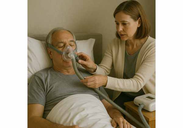
Central sleep apnea (CSA) is a sleep-related breathing disorder characterized by pauses in respiration due to lack of respiratory effort. Unlike obstructive sleep apnea, where airway collapse is the culprit, CSA arises from disrupted communication between the brainstem and respiratory muscles. These recurrent pauses fragment sleep architecture, leading to daytime somnolence, impaired cognition, and cardiovascular complications. CSA can occur in isolation or alongside conditions such as heart failure, stroke, or opioid use, and may manifest as Cheyne-Stokes respiration in advanced cases. Early recognition and targeted treatment—from positive airway pressure devices to pharmacological agents—are essential to restore restful sleep and reduce long-term health risks.
Table of Contents
- Understanding Brainstem-Centered Breathing Pauses
- Spotting Central Apnea Symptoms
- Identifying Contributors and Prevention Methods
- Tools for Diagnostic Assessment
- Management and Therapeutic Options
- Common Questions Around Central Apnea
Understanding Brainstem-Centered Breathing Pauses
At its core, central sleep apnea involves a breakdown in the automatic control of breathing. In healthy sleepers, the brainstem constantly monitors blood levels of carbon dioxide and oxygen, sending rhythmic signals to the diaphragm and intercostal muscles to maintain steady respiration. In CSA, this feedback loop falters: either the brainstem’s chemoreceptors fail to detect rising carbon dioxide, or the neural pathways to respiratory muscles misfire, resulting in transient—or prolonged—pauses in breathing.
There are several recognized subtypes:
- Primary Central Sleep Apnea: No clear underlying cause; thought to involve idiopathic dysfunction of respiratory centers.
- Cheyne-Stokes Respiration (CSR): Cyclic crescendo–decrescendo breathing often seen in heart failure or stroke, where delayed feedback loops cause oscillations in ventilation.
- High-Altitude Periodic Breathing: At high elevations, reduced atmospheric pressure triggers unstable respiratory drive, leading to alternate hyperventilation and pauses.
- Drug- or Substance-Induced CSA: Opioids and certain sedatives depress respiratory drive, increasing apnea events.
- Cheyne-Stokes in Congestive Heart Failure: A hallmark of advanced heart disease, where low cardiac output and circulation delays exacerbate breathing oscillations.
Visualize the respiratory system as an orchestra. In obstructive sleep apnea, the wind instruments (airway) collapse, preventing sound. In central sleep apnea, the conductor (brainstem) loses the score, halting the music altogether. The intermittent silences that follow disrupt the symphony of restorative sleep, particularly the deep (N3) and rapid-eye-movement (REM) stages crucial for memory consolidation and cardiovascular health.
Physiologically, each apnea episode triggers brief arousals—micro-awakenings—to reinstate breathing. Although sleepers often aren’t conscious of these arousals, the cumulative effect is fragmented sleep, reduced sleep efficiency, and elevated sympathetic nervous system activity. Over nights and months, chronic arousals elevate blood pressure, increase arrhythmia risk, and impair glucose metabolism, making CSA a significant contributor to cardiovascular morbidity.
Understanding CSA’s pathophysiology clarifies why treatments aim not only to open the airway but to restore stable central drive—ensuring that signals from the brainstem to respiratory muscles flow smoothly throughout the night.
Spotting Central Apnea Symptoms
Recognizing central sleep apnea requires attention to both nocturnal clues and daytime consequences. While loud snoring is more characteristic of obstructive apnea, CSA may produce subtler sounds—quiet breathing or periodic gasps—yet still fragment sleep architecture profoundly.
Nighttime Indicators
- Intermittent Breathing Silences: Partners may notice cycles of normal breathing followed by silent pauses and then sudden deep breaths or gasps.
- Restless Sleep: Frequent tossing, turning, and repositioning as micro-arousals restore breathing.
- Unusual Snoring Patterns: Softer snores punctuated by whispers or gasps rather than the constant rasp typical of OSA.
Daytime Signs
- Excessive Daytime Sleepiness (EDS): Struggling to stay awake during meetings, while driving, or in social settings, despite adequate time in bed.
- Morning Headaches: Fluctuations in blood oxygen and carbon dioxide can trigger vasodilation and headaches upon waking.
- Cognitive Fog: Difficulty concentrating, memory lapses, reduced problem-solving ability, and slowed reaction times.
- Mood Changes: Irritability, mood swings, and depressive symptoms linked to chronic sleep disruption.
Cardiovascular and Metabolic Effects
- Hypertension: Repeated arousals activate the sympathetic nervous system, raising daytime blood pressure.
- Arrhythmias: Bradycardia–tachycardia cycles during apneas can provoke atrial fibrillation or other rhythm disturbances.
- Glucose Intolerance: Disrupted sleep architecture impairs insulin sensitivity, increasing diabetes risk.
Real-Life Vignette
Consider James, a 68-year-old man with well-controlled hypertension who began waking gasping for air two to three times per night. He assumed it was age-related and didn’t mention it until his daytime fatigue became unbearable. His partner reported episodes of stopped breathing followed by sudden “snort-like” inhalations. A sleep study revealed 25 central apneas per hour, prompting targeted treatment that dramatically improved his energy and blood pressure control.
Because central and obstructive sleep apnea can coexist, a comprehensive evaluation—unbiased by snoring volume—is essential. Patients and partners should note breathing pauses, gasping, and daytime deficits to guide clinicians toward the correct subtype and optimize therapy.
Identifying Contributors and Prevention Methods
Central sleep apnea often emerges from underlying physiological or pharmacological factors. Identifying these contributors is key to prevention and tailored management.
Major Risk Factors
- Heart Failure and Cardiovascular Disease: Low cardiac output and circulation delays in heart failure patients promote Cheyne-Stokes respiration cycles.
- Neurological Disorders: Stroke, traumatic brain injury, or degenerative diseases (e.g., Parkinson’s) can impair brainstem respiratory centers.
- Chronic Kidney Disease: Uremia and fluid shifts may disrupt respiratory control.
- Opioid and Sedative Use: Medications like morphine, methadone, and benzodiazepines depress central respiratory drive, increasing apnea risk.
- High Altitude Exposure: At elevations above 2,500 meters, reduced oxygen tension triggers unstable breathing patterns.
- Advanced Age: Aging brainstem neurons show reduced chemoreceptor sensitivity, weakening respiratory rhythm stability.
Preventative Strategies
- Optimize Underlying Conditions:
- Heart Failure Management: Achieve euvolemia with diuretics, ACE inhibitors, and beta-blockers to reduce Cheyne-Stokes oscillations.
- Neurological Rehabilitation: Early intervention after stroke or brain injury can support neural plasticity and respiratory center recovery.
- Medication Review and Adjustment:
- Minimize Sedative Use: Where possible, taper opioids or benzodiazepines under medical supervision to restore central drive.
- Alternative Pain Management: Employ nonopioid analgesics and multimodal strategies to control pain without respiratory depression.
- Altitude Acclimatization:
- Gradual Ascent Plans: Allow days for chemoreceptor adaptation when traveling to high elevations.
- Supplemental Oxygen: Use portable oxygen during initial high-altitude sleep periods to stabilize breathing.
- Lifestyle and Behavioral Practices:
- Sleep Hygiene: Maintain consistent sleep–wake schedules, cool bedroom temperatures, and a dark environment to promote uninterrupted deep sleep.
- Regular Exercise: Aerobic activity improves cardiovascular function and may enhance respiratory control.
- Avoid Alcohol Before Bed: Alcohol further depresses respiratory drive and fragments sleep stages.
Preventive measures act as upstream interventions—fortifying the body’s baseline resilience against sleep-disordered breathing. While not all CSA triggers are avoidable, many modifiable risk factors can be addressed collaboratively with healthcare providers to reduce apnea severity.
Tools for Diagnostic Assessment
Accurate diagnosis of central sleep apnea hinges on detailed sleep studies and related evaluations. These tools differentiate CSA from obstructive patterns and guide treatment planning.
1. In-Lab Polysomnography (PSG)
- Electroencephalography (EEG): Monitors sleep stages and arousals.
- Electromyography (EMG) of Respiratory Muscles: Detects absence of respiratory effort during apnea events—key to differentiating CSA from OSA.
- Airflow Measurement: Masks or nasal cannulas track airflow interruption.
- Pulse Oximetry: Records blood oxygen saturation dips.
- Thoracic and Abdominal Effort Belts: Measure chest and abdominal movement; flat lines during apneas confirm central origin.
2. Home Sleep Apnea Testing (HSAT)
- Portable Monitors: Simplified devices record airflow, oximetry, and respiratory effort.
- Limitations: May underestimate CSA severity due to fewer channels; recommended when in-lab PSG is not feasible.
3. Capnography
- End-Tidal CO₂ Monitoring: Tracks exhaled CO₂ levels; elevated or fluctuating CO₂ may indicate hypoventilation or unstable drive.
- Transcutaneous CO₂ Sensors: Continuous monitoring of blood gas surrogate, useful in neuromuscular disorders or hypoventilation syndromes.
4. Echocardiography and Cardiac Evaluation
- Left Ventricular Ejection Fraction (LVEF): Low LVEF (<45%) correlates with Cheyne-Stokes patterns in heart failure.
- B-Type Natriuretic Peptide (BNP) Levels: High BNP suggests fluid overload and may predict CSR severity.
5. Neurological Imaging
- Brain MRI or CT: Identify structural lesions—ischemic strokes or tumors—impacting respiratory centers.
- Functional Neuroimaging: Research settings may use PET or fMRI to examine chemoreceptor responsiveness.
6. Differential Diagnosis Protocols
- Mixed Apnea Types: Some patients exhibit both central and obstructive apneas; titration studies under CPAP or ASV determine optimal therapy settings.
- Rule-Out Hypoventilation Syndromes: Conditions like obesity hypoventilation syndrome present with elevated CO₂ but persistent respiratory effort.
Collecting data from multi-channel PSG remains the gold standard. In-lab studies allow technicians to adjust protocols—such as adding carbon dioxide rebreathing tests—to unmask central drive deficiencies. A comprehensive diagnostic workup lays the foundation for selecting the most effective interventions.
Management and Therapeutic Options
Treating central sleep apnea aims to restore stable ventilation patterns, improve sleep quality, and mitigate cardiovascular risks. Therapy selection depends on CSA subtype, underlying conditions, and symptom severity.
A. Positive Airway Pressure (PAP) Devices
- Continuous Positive Airway Pressure (CPAP): First-line for many CSA patients; its pneumatic splint can stabilize small airways and sometimes improve central drive.
- Adaptive Servo-Ventilation (ASV): Monitors breathing patterns and delivers variable pressure support to normalize ventilation; particularly effective for Cheyne-Stokes respiration.
- Bilevel PAP (BiPAP): Provides separate inspiratory and expiratory pressures; can assist ventilation in hypoventilation syndromes.
B. Supplemental Oxygen
- Nighttime Oxygen Therapy: Low-flow oxygen reduces hypoxic ventilatory drive fluctuations, decreasing central apnea index; useful at high altitude or in select heart failure cases.
C. Pharmacological Interventions
- Acetazolamide: A carbonic anhydrase inhibitor that induces metabolic acidosis, stimulating respiratory drive and reducing apnea frequency.
- Theophylline: A respiratory stimulant that can modestly improve ventilation, though side effects limit its use.
- Progestins (e.g., medroxyprogesterone): Hormonal agents occasionally used in hypoventilation disorders to enhance drive.
D. Cardiac Optimization
- Heart Failure Therapies: ACE inhibitors, beta-blockers, and device therapies (e.g., CRT) can improve cardiac output, reducing Cheyne-Stokes breathing cycles.
- Volume Management: Diuretics maintain euvolemia, attenuating pulmonary congestion that exacerbates apnea events.
E. Neuromodulation
- Phrenic Nerve Stimulation: Implanted devices deliver electrical impulses to the diaphragm, pacing breathing in patients with refractory CSA; emerging as a promising option.
F. Lifestyle and Supportive Measures
- Weight Management: In overweight patients, weight loss can reduce overall sleep-disordered breathing burden.
- Exercise Training: Regular aerobic activity enhances cardiovascular function and may stabilize respiratory control.
- Sleep Positioning: Elevating the head of the bed or side-sleeping can improve airway patency and reduce apnea severity.
G. Follow-Up and Titration
- Regular PAP Titration Studies: Adjust pressure settings to maintain optimal therapy as underlying conditions change.
- Adherence Monitoring: Many devices record usage data; routine reviews encourage patient engagement and troubleshoot mask fit or comfort issues.
- Multidisciplinary Care: Collaboration among pulmonologists, cardiologists, neurologists, and sleep specialists ensures comprehensive management.
Personalized treatment plans often combine PAP therapy with medical optimization and lifestyle modifications. Patients with complex comorbidities—like advanced heart failure or opioid use—benefit from integrated approaches that address both central drive and underlying contributors, maximizing outcomes and quality of life.
Common Questions Around Central Apnea
How Is Central Sleep Apnea Different from Obstructive Sleep Apnea?
CSA features pauses in breathing due to lack of respiratory effort, while OSA involves continued effort against a collapsed airway. PSG differentiates them via respiratory effort belts and airflow sensors.
Can CPAP Treat Central Sleep Apnea?
Yes, CPAP can stabilize breathing in many CSA cases, but adaptive servo-ventilation or supplemental oxygen may be needed if central events persist despite optimal CPAP use.
Is Central Apnea Curable?
Underlying causes can often be managed—heart failure optimized, medications adjusted—but idiopathic CSA may require lifelong PAP therapy or neuromodulation for symptom control.
What Role Does Heart Failure Play in CSA?
Heart failure delays circulation time, amplifying feedback loops in respiration control. This leads to Cheyne-Stokes patterns that manifest as central apneas, often improving when cardiac output is enhanced.
How Long Does It Take to See Improvement with Treatment?
Many patients experience reduced apnea events within one to two nights on PAP therapy, but full symptomatic relief—better sleep quality and daytime alertness—may take several weeks of consistent use.
When Should I Seek a Sleep Study?
If you or your partner notice recurrent breathing pauses, gasping, unexplained daytime sleepiness, or morning headaches, consult a sleep specialist for evaluation and possible polysomnography.
Disclaimer: This article is intended for educational purposes only and does not replace professional medical advice. If you suspect you have central sleep apnea or experience severe symptoms, please consult a qualified healthcare provider for personalized evaluation and treatment.
If you found this resource helpful, please share it on Facebook, X (formerly Twitter), or your favorite platform—and follow us on social media for more sleep health insights. Your support helps us continue creating comprehensive guides to improve your well-being.


